8Q91
 
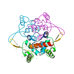
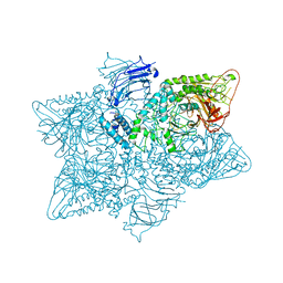
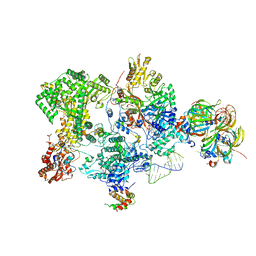 | | Structure of the human 20S U5 snRNP core | | 分子名称: | 116 kDa U5 small nuclear ribonucleoprotein component, CD2 antigen cytoplasmic tail-binding protein 2, GUANOSINE-5'-TRIPHOSPHATE, ... | | 著者 | Schneider, S, Galej, W.P. | | 登録日 | 2023-08-19 | | 公開日 | 2024-03-27 | | 実験手法 | ELECTRON MICROSCOPY (3.1 Å) | | 主引用文献 | Structure of the human 20S U5 snRNP.
Nat.Struct.Mol.Biol., 2024
|
|
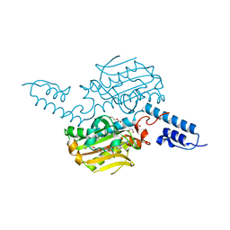
8RC0
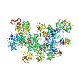 | | Structure of the human 20S U5 snRNP | | 分子名称: | 116 kDa U5 small nuclear ribonucleoprotein component, CD2 antigen cytoplasmic tail-binding protein 2, GUANOSINE-5'-TRIPHOSPHATE, ... | | 著者 | Schneider, S, Galej, W.P. | | 登録日 | 2023-12-05 | | 公開日 | 2024-03-27 | | 実験手法 | ELECTRON MICROSCOPY (3.2 Å) | | 主引用文献 | Structure of the human 20S U5 snRNP.
Nat.Struct.Mol.Biol., 2024
|
|
8R33
 | |
8R34
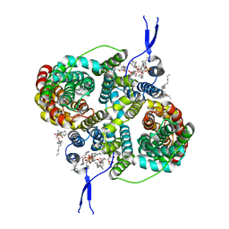
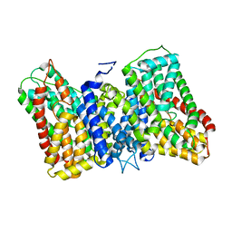
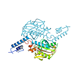
 | | CryoEM structure of the symmetric Pho90 dimer from yeast with substrates. | | 分子名称: | 1,2-DIACYL-GLYCEROL-3-SN-PHOSPHATE, Low-affinity phosphate transporter PHO90, PHOSPHATE ION, ... | | 著者 | Schneider, S, Kuehlbrandt, W, Yildiz, O. | | 登録日 | 2023-11-08 | | 公開日 | 2024-04-24 | | 実験手法 | ELECTRON MICROSCOPY (2.62 Å) | | 主引用文献 | Complementary structures of the yeast phosphate transporter Pho90 provide insights into its transport mechanism
Structure, 2024
|
|
8R35
 | |
3KOF
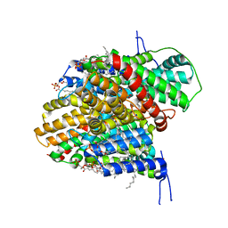
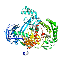
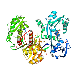
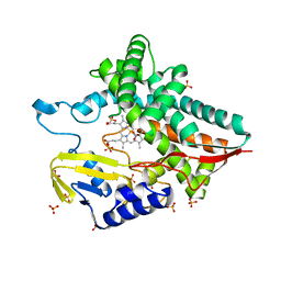
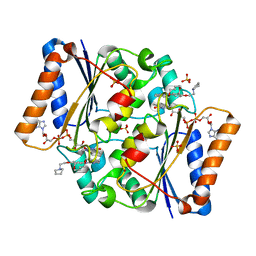
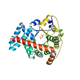 | | Crystal structure of the double mutant F178Y/R181E of E.coli transaldolase B | | 分子名称: | SULFATE ION, Transaldolase B | | 著者 | Schneider, S, Gutierrez, M, Sandalova, T, Schneider, G, Clapes, P, Sprenger, G.A, Samland, A.K. | | 登録日 | 2009-11-13 | | 公開日 | 2010-02-23 | | 最終更新日 | 2023-09-06 | | 実験手法 | X-RAY DIFFRACTION (1.9 Å) | | 主引用文献 | Redesigning the Active Site of Transaldolase TalB from Escherichia coli: New Variants with Improved Affinity towards Nonphosphorylated Substrates.
Chembiochem, 11, 2010
|
|
2J0R
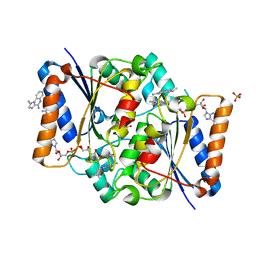
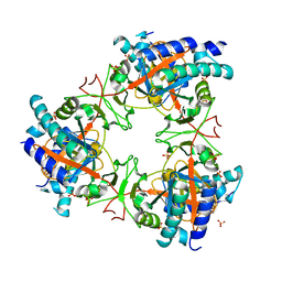
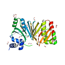 | | Structure of the haem-chaperone Proteobacteria-protein HemS | | 分子名称: | 1,2-ETHANEDIOL, DI(HYDROXYETHYL)ETHER, DODECAETHYLENE GLYCOL, ... | | 著者 | Schneider, S, Sharp, K.H, Barker, P.D, Paoli, M. | | 登録日 | 2006-08-04 | | 公開日 | 2006-08-29 | | 最終更新日 | 2023-12-13 | | 実験手法 | X-RAY DIFFRACTION (1.9 Å) | | 主引用文献 | An Induced Fit Conformational Change Underlies the Binding Mechanism of the Heme Transport Proteobacteria-Protein Hems.
J.Biol.Chem., 281, 2006
|
|
2J0P
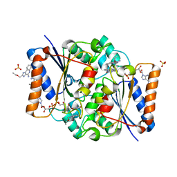
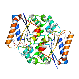
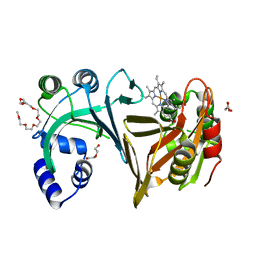 | | Structure of the haem-chaperone Proteobacteria-protein HemS | | 分子名称: | DI(HYDROXYETHYL)ETHER, DODECAETHYLENE GLYCOL, HEMIN TRANSPORT PROTEIN HEMS, ... | | 著者 | Schneider, S, Sharp, K.H, Barker, P.D, Paoli, M. | | 登録日 | 2006-08-04 | | 公開日 | 2006-08-29 | | 最終更新日 | 2023-12-13 | | 実験手法 | X-RAY DIFFRACTION (1.7 Å) | | 主引用文献 | An Induced Fit Conformational Change Underlies the Binding Mechanism of the Heme Transport Proteobacteria-Protein Hems.
J.Biol.Chem., 281, 2006
|
|
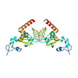
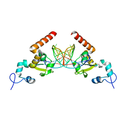
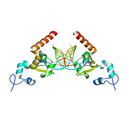
2V79
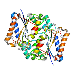
 | | Crystal Structure of the N-terminal domain of DnaD from Bacillus Subtilis | | 分子名称: | CHLORIDE ION, DNA REPLICATION PROTEIN DNAD, SODIUM ION | | 著者 | Schneider, S, Zhang, W, Soultanas, P, Paoli, M. | | 登録日 | 2007-07-27 | | 公開日 | 2008-01-15 | | 最終更新日 | 2017-06-28 | | 実験手法 | X-RAY DIFFRACTION (2 Å) | | 主引用文献 | Structure of the N-Terminal Oligomerization Domain of Dnad Reveals a Unique Tetramerization Motif and Provides Insights Into Scaffold Formation.
J.Mol.Biol., 376, 2008
|
|
4BWA
 | | PylRS Y306G, Y384F, I405R mutant in complex with adenylated norbornene | | 分子名称: | 1,2-ETHANEDIOL, Adenylated Norbornene, DI(HYDROXYETHYL)ETHER, ... | | 著者 | Schneider, S, Vrabel, M, Gattner, M.J, Fluegel, V, Lopez-Carillo, V, Carell, T. | | 登録日 | 2013-07-01 | | 公開日 | 2013-07-31 | | 最終更新日 | 2023-12-20 | | 実験手法 | X-RAY DIFFRACTION (2.45 Å) | | 主引用文献 | Structural Insights Into Incorporation of Norbornene Amino Acids for Click Modification of Proteins
Chem.Bio.Chem., 14, 2013
|
|
4BW9
 | | PylRS Y306G, Y384F, I405R mutant in complex with AMP-PNP | | 分子名称: | 1,2-ETHANEDIOL, MAGNESIUM ION, PENTAETHYLENE GLYCOL, ... | | 著者 | Schneider, S, Vrabel, M, Gattner, M.J, Fluegel, V, Lopez-Carillo, V, Carell, T. | | 登録日 | 2013-07-01 | | 公開日 | 2013-07-31 | | 最終更新日 | 2023-12-20 | | 実験手法 | X-RAY DIFFRACTION (2.35 Å) | | 主引用文献 | Structural Insights Into Incorporation of Norbornene Amino Acids for Click Modification of Proteins
Chem.Bio.Chem., 14, 2013
|
|
5G5S
 | | Structure of the Argonaute protein from Methanocaldcoccus janaschii | | 分子名称: | 2-AMINO-2-HYDROXYMETHYL-PROPANE-1,3-DIOL, ARGONAUTE, MAGNESIUM ION | | 著者 | Schneider, S, Oellig, C.A, Keegan, R, Grohmann, D, Zander, A, Willkomm, S. | | 登録日 | 2016-06-03 | | 公開日 | 2017-02-08 | | 最終更新日 | 2017-03-29 | | 実験手法 | X-RAY DIFFRACTION (2.29 Å) | | 主引用文献 | Structural and mechanistic insights into an archaeal DNA-guided Argonaute protein.
Nat Microbiol, 2, 2017
|
|
5G5T
 | | Structure of the Argonaute protein from Methanocaldcoccus janaschii in complex with guide DNA | | 分子名称: | ARGONAUTE, GUIDE DNA, MAGNESIUM ION, ... | | 著者 | Schneider, S, Oellig, C.A, Keegan, R, Grohmann, D, Zander, A, Willkomm, S. | | 登録日 | 2016-06-03 | | 公開日 | 2017-02-08 | | 最終更新日 | 2024-01-10 | | 実験手法 | X-RAY DIFFRACTION (2.85 Å) | | 主引用文献 | Structural and mechanistic insights into an archaeal DNA-guided Argonaute protein.
Nat Microbiol, 2, 2017
|
|
5LBT
 | |
5LBU
 | |
5JM0
 | | Structure of the S. cerevisiae alpha-mannosidase 1 | | 分子名称: | Alpha-mannosidase,Alpha-mannosidase,Alpha-mannosidase | | 著者 | Schneider, S, Kosinski, J, Jakobi, A.J, Hagen, W.J.H, Sachse, C. | | 登録日 | 2016-04-28 | | 公開日 | 2016-06-15 | | 最終更新日 | 2017-08-02 | | 実験手法 | ELECTRON MICROSCOPY (6.3 Å) | | 主引用文献 | Higher-order assemblies of oligomeric cargo receptor complexes form the membrane scaffold of the Cvt vesicle.
Embo Rep., 17, 2016
|
|
7AYX
 | |
7Z06
 | |
5LBW
 | | Structure of the human quinone reductase 2 (NQO2) in complex with volitinib | | 分子名称: | FLAVIN-ADENINE DINUCLEOTIDE, Ribosyldihydronicotinamide dehydrogenase [quinone], ZINC ION, ... | | 著者 | Schneider, S, Medard, G, Kuester, B. | | 登録日 | 2016-06-17 | | 公開日 | 2017-11-29 | | 最終更新日 | 2024-01-10 | | 実験手法 | X-RAY DIFFRACTION (1.9 Å) | | 主引用文献 | The target landscape of clinical kinase drugs.
Science, 358, 2017
|
|
5LBY
 | | Structure of the human quinone reductase 2 (NQO2) in complex with crenolanib | | 分子名称: | 1-(2-{5-[(3-Methyloxetan-3-yl)methoxy]-1H-benzimidazol-1-yl}quinolin-8-yl)piperidin-4-amine, FLAVIN-ADENINE DINUCLEOTIDE, Ribosyldihydronicotinamide dehydrogenase [quinone], ... | | 著者 | Schneider, S, Medard, G, Kuester, B. | | 登録日 | 2016-06-17 | | 公開日 | 2017-11-29 | | 最終更新日 | 2024-01-10 | | 実験手法 | X-RAY DIFFRACTION (1.4 Å) | | 主引用文献 | The target landscape of clinical kinase drugs.
Science, 358, 2017
|
|
5LBZ
 | | Structure of the human quinone reductase 2 (NQO2) in complex with pacritinib | | 分子名称: | 11-(2-pyrrolidin-1-yl-ethoxy)-14,19-dioxa-5,7,26-triaza-tetracyclo[19.3.1.1(2,6).1(8,12)]heptacosa-1(25),2(26),3,5,8,10,12(27),16,21,23-decaene, FLAVIN-ADENINE DINUCLEOTIDE, Ribosyldihydronicotinamide dehydrogenase [quinone], ... | | 著者 | Schneider, S, Medard, G, Kuster, B. | | 登録日 | 2016-06-17 | | 公開日 | 2017-11-29 | | 最終更新日 | 2024-01-10 | | 実験手法 | X-RAY DIFFRACTION (1.4 Å) | | 主引用文献 | The target landscape of clinical kinase drugs.
Science, 358, 2017
|
|
5G32
 | | Structure of Rad14 in complex with acetylaminophenyl-guanine containing DNA | | 分子名称: | 5'-D(*GP*CP*TP*CP*TP*AP*6FKP*TP*CP*AP*TP*CP*AP*CP)-3', 5'-D(*GP*TP*GP*AP*TP*GP*AP*CP*GP*TP*AP*GP*AP*GP)-3', RAD14, ... | | 著者 | Simon, N, Ebert, C, Schneider, S. | | 登録日 | 2016-04-18 | | 公開日 | 2016-06-01 | | 最終更新日 | 2019-05-15 | | 実験手法 | X-RAY DIFFRACTION (2.2 Å) | | 主引用文献 | Structural Basis for Bulky Adduct DNA Lesion Recognition by the Nucleotide Excision Repair Protein Rad14.
Chemistry, 22, 2016
|
|
5G35
 | | Structure of Rad14 in complex with acetylaminopyren-C8-guanine containing DNA | | 分子名称: | 5'-D(*GP*CP*TP*CP*TP*AP*8PYP*TP*CP*AP*TP*CP*AP*CP)-3', 5'-D(*GP*TP*GP*AP*TP*GP*AP*CP*GP*TP*AP*GP*AP*GP)-3', RAD14, ... | | 著者 | Simon, N, Ebert, C, Schneider, S. | | 登録日 | 2016-04-18 | | 公開日 | 2016-06-01 | | 最終更新日 | 2024-01-10 | | 実験手法 | X-RAY DIFFRACTION (2 Å) | | 主引用文献 | Structural Basis for Bulky Adduct DNA Lesion Recognition by the Nucleotide Excision Repair Protein Rad14.
Chemistry, 22, 2016
|
|
5G33
 | | Structure of Rad14 in complex with acetylnaphtyl-guanine containing DNA | | 分子名称: | 5'-D(*GP*CP*TP*CP*TP*AP*MFOP*TP*CP*AP*TP*CP*AP*CP)-3', 5'-D(*GP*TP*GP*AP*TP*GP*AP*CP*GP*TP*AP*GP*AP*GP)-3', RAD14, ... | | 著者 | Simon, N, Ebert, C, Schneider, S. | | 登録日 | 2016-04-18 | | 公開日 | 2016-06-01 | | 最終更新日 | 2024-01-10 | | 実験手法 | X-RAY DIFFRACTION (2.4 Å) | | 主引用文献 | Structural Basis for Bulky Adduct DNA Lesion Recognition by the Nucleotide Excision Repair Protein Rad14.
Chemistry, 22, 2016
|
|
5G34
 | | Structure of Rad14 in complex with acetylaminoanthracene-C8-guanine containing DNA | | 分子名称: | 5'-D(*GP*CP*TP*CP*TP*AP*6FKP*TP*CP*AP*TP*CP*AP*CP)-3', 5'-D(*GP*TP*GP*AP*TP*GP*AP*CP*GP*TP*AP*GP*AP*GP)-3', RAD14, ... | | 著者 | Simon, N, Ebert, C, Schneider, S. | | 登録日 | 2016-04-18 | | 公開日 | 2016-06-01 | | 最終更新日 | 2024-01-10 | | 実験手法 | X-RAY DIFFRACTION (1.9 Å) | | 主引用文献 | Structural Basis for Bulky Adduct DNA Lesion Recognition by the Nucleotide Excision Repair Protein Rad14.
Chemistry, 22, 2016
|
|


