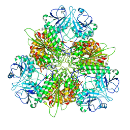
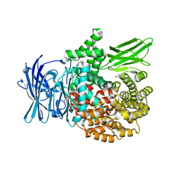
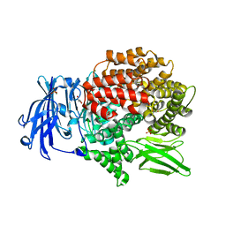
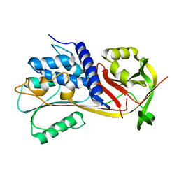
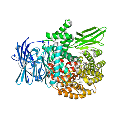
5KYO
 
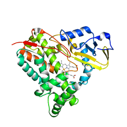 | | Crystal Structure of CYP101J2 | | 分子名称: | CYP101J2, PROTOPORPHYRIN IX CONTAINING FE | | 著者 | Unterweger, B, Drinkwater, N, Dumsday, G.J, McGowan, S. | | 登録日 | 2016-07-22 | | 公開日 | 2017-01-04 | | 最終更新日 | 2023-10-04 | | 実験手法 | X-RAY DIFFRACTION (1.8 Å) | | 主引用文献 | X-ray crystal structure of cytochrome P450 monooxygenase CYP101J2 from Sphingobium yanoikuyae strain B2.
Proteins, 85, 2017
|
|
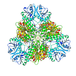
7KWW
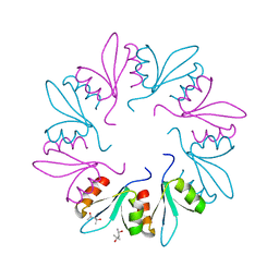 | | X-ray Crystal Structure of PlyCB Mutant K59H | | 分子名称: | (4S)-2-METHYL-2,4-PENTANEDIOL, PlyCB | | 著者 | Williams, D.E, Broendum, S.S, Hayes, B.K, Drinkwater, N, McGowan, S. | | 登録日 | 2020-12-02 | | 公開日 | 2021-04-07 | | 最終更新日 | 2023-10-18 | | 実験手法 | X-RAY DIFFRACTION (1.8 Å) | | 主引用文献 | High avidity drives the interaction between the streptococcal C1 phage endolysin, PlyC, with the cell surface carbohydrates of Group A Streptococcus.
Mol.Microbiol., 116, 2021
|
|
7KWT
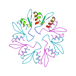 | | X-ray Crystal Structure of PlyCB Mutant Y28H | | 分子名称: | PlyCB | | 著者 | Williams, D.E, Broendum, S.S, Hayes, B.K, Drinkwater, N, McGowan, S. | | 登録日 | 2020-12-02 | | 公開日 | 2021-04-07 | | 最終更新日 | 2023-10-18 | | 実験手法 | X-RAY DIFFRACTION (1.79 Å) | | 主引用文献 | High avidity drives the interaction between the streptococcal C1 phage endolysin, PlyC, with the cell surface carbohydrates of Group A Streptococcus.
Mol.Microbiol., 116, 2021
|
|
7KWY
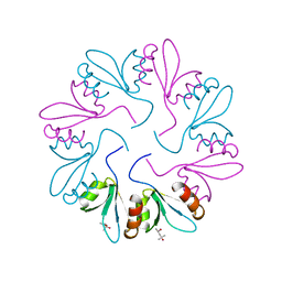 | | X-ray Crystal Structure of PlyCB Mutant R66K | | 分子名称: | (4S)-2-METHYL-2,4-PENTANEDIOL, PlyCB | | 著者 | Williams, D.E, Broendum, S.S, Hayes, B.K, Drinkwater, N, McGowan, S. | | 登録日 | 2020-12-02 | | 公開日 | 2021-04-07 | | 最終更新日 | 2023-10-18 | | 実験手法 | X-RAY DIFFRACTION (1.7 Å) | | 主引用文献 | High avidity drives the interaction between the streptococcal C1 phage endolysin, PlyC, with the cell surface carbohydrates of Group A Streptococcus.
Mol.Microbiol., 116, 2021
|
|
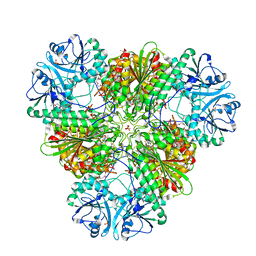
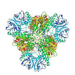
7K5K
 | | Plasmodium vivax M17 leucyl aminopeptidase Pv-M17 | | 分子名称: | CARBONATE ION, M17 leucyl aminopeptidase, putative, ... | | 著者 | Malcolm, T.R, McGowan, S, Belousoff, M.J. | | 登録日 | 2020-09-17 | | 公開日 | 2020-12-16 | | 最終更新日 | 2024-03-06 | | 実験手法 | ELECTRON MICROSCOPY (2.66 Å) | | 主引用文献 | Active site metals mediate an oligomeric equilibrium in Plasmodium M17 aminopeptidases.
J.Biol.Chem., 296, 2020
|
|
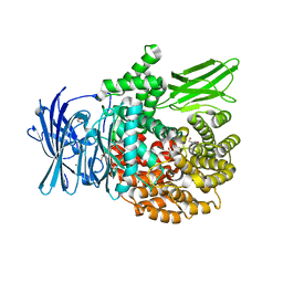
7RIE
 | | Plasmodium falciparum M17 in complex with inhibitor MIPS2571 | | 分子名称: | CARBONATE ION, M17 leucyl aminopeptidase, N-{(1R)-2-(hydroxyamino)-1-[4'-(hydroxymethyl)[1,1'-biphenyl]-4-yl]-2-oxoethyl}-2,2-dimethylpropanamide, ... | | 著者 | Webb, C.T, McGowan, S. | | 登録日 | 2021-07-19 | | 公開日 | 2022-09-14 | | 最終更新日 | 2023-10-25 | | 実験手法 | X-RAY DIFFRACTION (2.49 Å) | | 主引用文献 | Genetic and chemical validation of Plasmodium falciparum aminopeptidase Pf A-M17 as a drug target in the hemoglobin digestion pathway.
Elife, 11, 2022
|
|
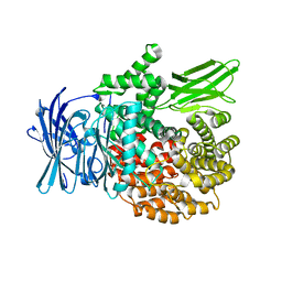
6EE3
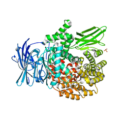 | | X-ray crystal structure of Pf-M1 in complex with inhibitor (6k) and catalytic zinc ion | | 分子名称: | (1R,2r,3S,5R,7R)-N-[(1R)-2-(hydroxyamino)-2-oxo-1-(3',4',5'-trifluoro[1,1'-biphenyl]-4-yl)ethyl]tricyclo[3.3.1.1~3,7~]decane-2-carboxamide, GLYCEROL, M1 family aminopeptidase, ... | | 著者 | Drinkwater, N, McGowan, S. | | 登録日 | 2018-08-13 | | 公開日 | 2018-12-26 | | 最終更新日 | 2024-03-13 | | 実験手法 | X-RAY DIFFRACTION (1.82 Å) | | 主引用文献 | Hydroxamic Acid Inhibitors Provide Cross-Species Inhibition of Plasmodium M1 and M17 Aminopeptidases.
J. Med. Chem., 62, 2019
|
|
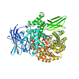
6EED
 | | X-ray crystal structure of Pf-M1 in complex with inhibitor (6p) and catalytic zinc ion | | 分子名称: | (2R)-2-[(cyclohexylacetyl)amino]-N-hydroxy-2-(3',4',5'-trifluoro[1,1'-biphenyl]-4-yl)acetamide, DIMETHYL SULFOXIDE, GLYCEROL, ... | | 著者 | Drinkwater, N, McGowan, S. | | 登録日 | 2018-08-13 | | 公開日 | 2018-12-26 | | 最終更新日 | 2024-03-13 | | 実験手法 | X-RAY DIFFRACTION (1.5 Å) | | 主引用文献 | Hydroxamic Acid Inhibitors Provide Cross-Species Inhibition of Plasmodium M1 and M17 Aminopeptidases.
J. Med. Chem., 62, 2019
|
|
6EA2
 | |
6EAB
 | |
6EAA
 | |
6EEE
 | | X-ray crystal structure of Pf-M17 in complex with inhibitor (6k) and regulatory zinc ion | | 分子名称: | (1R,2r,3S,5R,7R)-N-[(1R)-2-(hydroxyamino)-2-oxo-1-(3',4',5'-trifluoro[1,1'-biphenyl]-4-yl)ethyl]tricyclo[3.3.1.1~3,7~]decane-2-carboxamide, 1,2-ETHANEDIOL, CARBONATE ION, ... | | 著者 | Drinkwater, N, McGowan, S. | | 登録日 | 2018-08-13 | | 公開日 | 2018-12-26 | | 最終更新日 | 2024-03-13 | | 実験手法 | X-RAY DIFFRACTION (2.3 Å) | | 主引用文献 | Hydroxamic Acid Inhibitors Provide Cross-Species Inhibition of Plasmodium M1 and M17 Aminopeptidases.
J. Med. Chem., 62, 2019
|
|
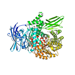
6EA1
 | | X-ray crystal structure of Pf-M1 in complex with inhibitor (6da) and catalytic zinc ion | | 分子名称: | (2R)-2,3,3,3-tetrafluoro-N-[(1R)-2-(hydroxyamino)-2-oxo-1-(3',4',5'-trifluoro[1,1'-biphenyl]-4-yl)ethyl]propanamide, GLYCEROL, M1 family aminopeptidase, ... | | 著者 | Drinkwater, N, McGowan, S. | | 登録日 | 2018-08-02 | | 公開日 | 2018-12-26 | | 最終更新日 | 2024-03-13 | | 実験手法 | X-RAY DIFFRACTION (1.815 Å) | | 主引用文献 | Hydroxamic Acid Inhibitors Provide Cross-Species Inhibition of Plasmodium M1 and M17 Aminopeptidases.
J. Med. Chem., 62, 2019
|
|
6EE4
 | | X-ray crystal structure of Pf-M1 in complex with inhibitor (6m) and catalytic zinc ion | | 分子名称: | (2R)-2-[(cyclopropylacetyl)amino]-N-hydroxy-2-(3',4',5'-trifluoro[1,1'-biphenyl]-4-yl)acetamide, GLYCEROL, M1 family aminopeptidase, ... | | 著者 | Drinkwater, N, McGowan, S. | | 登録日 | 2018-08-13 | | 公開日 | 2018-12-26 | | 最終更新日 | 2024-03-13 | | 実験手法 | X-RAY DIFFRACTION (1.58 Å) | | 主引用文献 | Hydroxamic Acid Inhibitors Provide Cross-Species Inhibition of Plasmodium M1 and M17 Aminopeptidases.
J. Med. Chem., 62, 2019
|
|
6EE2
 | |
6EE6
 | | X-ray crystal structure of Pf-M1 in complex with inhibitor (6o) and catalytic zinc ion | | 分子名称: | (2R)-2-[(cyclopentylacetyl)amino]-N-hydroxy-2-(3',4',5'-trifluoro[1,1'-biphenyl]-4-yl)acetamide, GLYCEROL, M1 family aminopeptidase, ... | | 著者 | Drinkwater, N, McGowan, S. | | 登録日 | 2018-08-13 | | 公開日 | 2018-12-26 | | 最終更新日 | 2024-03-13 | | 実験手法 | X-RAY DIFFRACTION (1.5 Å) | | 主引用文献 | Hydroxamic Acid Inhibitors Provide Cross-Species Inhibition of Plasmodium M1 and M17 Aminopeptidases.
J. Med. Chem., 62, 2019
|
|
6NVB
 | | Crystal structure of the inhibitor-free form of the serine protease kallikrein-4 | | 分子名称: | GLYCEROL, Kallikrein-4, SULFATE ION | | 著者 | Riley, B.T, Buckle, A.M, McGowan, S. | | 登録日 | 2019-02-04 | | 公開日 | 2019-07-17 | | 最終更新日 | 2023-10-11 | | 実験手法 | X-RAY DIFFRACTION (1.636 Å) | | 主引用文献 | Crystal structure of the inhibitor-free form of the serine protease kallikrein-4.
Acta Crystallogr.,Sect.F, 75, 2019
|
|
6EE5
 | |
4R7M
 | |
4R5V
 | |
4R5X
 | |
4R76
 | |
4R5T
 | |
4R6T
 | |
5CDZ
 | | Crystal structure of conserpin in the latent state | | 分子名称: | Conserpin in the latent state, GLYCEROL | | 著者 | Porebski, B.T, McGowan, S, Keleher, S, Buckle, A.M. | | 登録日 | 2015-07-06 | | 公開日 | 2016-07-20 | | 最終更新日 | 2023-09-27 | | 実験手法 | X-RAY DIFFRACTION (1.449 Å) | | 主引用文献 | Smoothing a rugged protein folding landscape by sequence-based redesign.
Sci Rep, 6, 2016
|
|


