Loading
PDBj
MenuPDBj@FacebookPDBj@TwitterPDBj@YouTubewwPDB FoundationwwPDB
RCSB PDBPDBeBMRBAdv. SearchSearch help
Search by PDB author
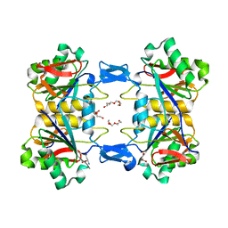
8UP6
DownloadVisualize
BU of 8up6 by Molmil
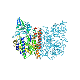
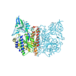
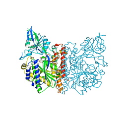
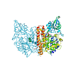
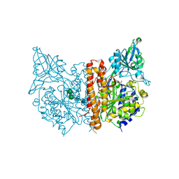
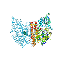
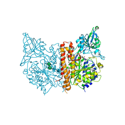
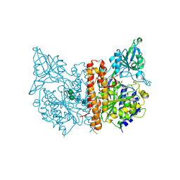
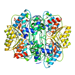
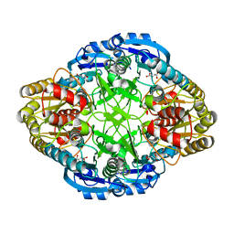
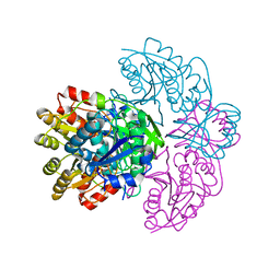
Structure of atypical asparaginase from Rhodospirillum rubrum (mutant K19A) in complex with L-Asp
Descriptor: ASPARTIC ACID, Asparaginase, TETRAETHYLENE GLYCOL
Authors:Lubkowski, J, Wlodawer, A, Zhang, D.
Deposit date:2023-10-21
Release date:2024-04-03
Method:X-RAY DIFFRACTION (1.7 Å)
Cite:RrA, an enzyme from Rhodospirillum rubrum, is a prototype of a new family of short-chain L-asparaginases.
Protein Sci., 33, 2024
1ZMH
DownloadVisualize
BU of 1zmh by Molmil
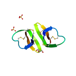
Crystal structure of human neutrophil peptide 2, HNP-2 (variant Gly16-> D-Ala)
Descriptor: GLYCEROL, HEXAETHYLENE GLYCOL, Neutrophil defensin 2, ...
Authors:Lubkowski, J, Prahl, A, Lu, W.
Deposit date:2005-05-10
Release date:2005-08-16
Last modified:2023-08-23
Method:X-RAY DIFFRACTION (1.5 Å)
Cite:Reconstruction of the conserved beta-bulge in mammalian defensins using D-amino acids.
J.Biol.Chem., 280, 2005
1ZMI
DownloadVisualize
BU of 1zmi by Molmil
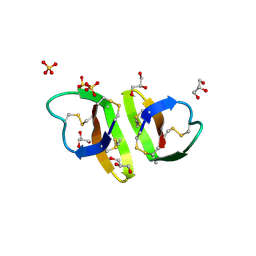
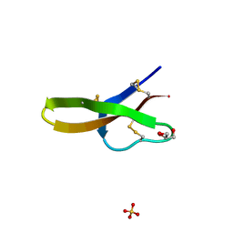
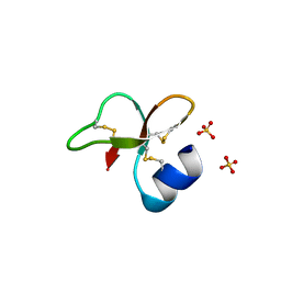
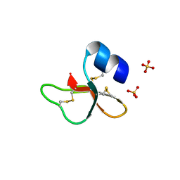
Crystal structure of human alpha_defensin-2 (variant GLY16->D-ALA), P 32 2 1 space group )
Descriptor: GLYCEROL, Neutrophil defensin 2, SULFATE ION
Authors:Lubkowski, J, Prahl, A, Lu, W.
Deposit date:2005-05-10
Release date:2005-08-16
Last modified:2023-08-23
Method:X-RAY DIFFRACTION (1.15 Å)
Cite:Reconstruction of the conserved beta-bulge in mammalian defensins using D-amino acids.
J.Biol.Chem., 280, 2005
1ZMM
DownloadVisualize
BU of 1zmm by Molmil
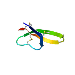
Crystal structure of human alpha-defensin-4
Descriptor: Neutrophil defensin 4
Authors:Lubkowski, J, Szyk, A, Lu, W.
Deposit date:2005-05-10
Release date:2006-05-30
Last modified:2023-08-23
Method:X-RAY DIFFRACTION (1.6 Å)
Cite:Crystal structures of human {alpha}-defensins HNP4, HD5, and HD6.
Protein Sci., 15, 2006
1ZMK
DownloadVisualize
BU of 1zmk by Molmil
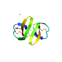
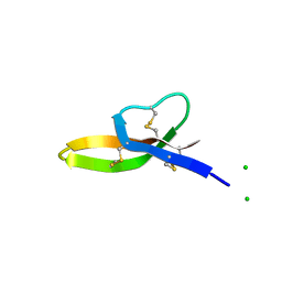
Crystal structure of human alpha-defensin-2 (variant Gly16-> D-ALA), P 42 21 2 space group
Descriptor: CHLORIDE ION, GLYCEROL, Neutrophil defensin 2
Authors:Lubkowski, J, Prahl, A, Lu, W.
Deposit date:2005-05-10
Release date:2005-08-16
Last modified:2023-08-23
Method:X-RAY DIFFRACTION (1.3 Å)
Cite:Reconstruction of the conserved beta-bulge in mammalian defensins using D-amino acids.
J.Biol.Chem., 280, 2005
1ZMP
DownloadVisualize
BU of 1zmp by Molmil
Crystal structure of human defensin-5
Descriptor: CHLORIDE ION, Defensin 5, GLYCEROL, ...
Authors:Lubkowski, J, Szyk, A, Lu, W.
Deposit date:2005-05-10
Release date:2006-05-30
Last modified:2024-04-03
Method:X-RAY DIFFRACTION (1.65 Å)
Cite:Crystal structures of human {alpha}-defensins HNP4, HD5, and HD6.
Protein Sci., 15, 2006
1ZMQ
DownloadVisualize
BU of 1zmq by Molmil
Crystal structure of human alpha-defensin-6
Descriptor: CHLORIDE ION, Defensin 6
Authors:Lubkowski, J, Szyk, A, Lu, W.
Deposit date:2005-05-10
Release date:2006-05-30
Last modified:2024-04-03
Method:X-RAY DIFFRACTION (2.1 Å)
Cite:Crystal structures of human {alpha}-defensins HNP4, HD5, and HD6.
Protein Sci., 15, 2006
2I9A
DownloadVisualize
BU of 2i9a by Molmil
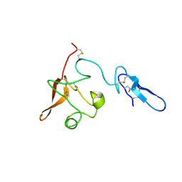
Crystal structure of the free aminoterminal fragment of urokinase type plasminogen activator (ATF)
Descriptor: PHOSPHATE ION, Urokinase-type plasminogen activator
Authors:Lubkowski, J, Barinka, C.
Deposit date:2006-09-05
Release date:2006-11-28
Last modified:2023-08-30
Method:X-RAY DIFFRACTION (1.9 Å)
Cite:Structural basis of interaction between urokinase-type plasminogen activator and its receptor.
J.Mol.Biol., 363, 2006
2I9B
DownloadVisualize
BU of 2i9b by Molmil
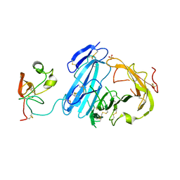
Crystal structure of ATF-urokinase receptor complex
Descriptor: 2-acetamido-2-deoxy-beta-D-glucopyranose-(1-4)-2-acetamido-2-deoxy-beta-D-glucopyranose, SULFATE ION, Urokinase plasminogen activator surface receptor, ...
Authors:Lubkowski, J, Barinka, C.
Deposit date:2006-09-05
Release date:2007-01-02
Last modified:2023-08-30
Method:X-RAY DIFFRACTION (2.8 Å)
Cite:Structural basis of interaction between urokinase-type plasminogen activator and its receptor.
J.Mol.Biol., 363, 2006
3FF3
DownloadVisualize
BU of 3ff3 by Molmil
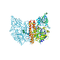
The high resolution structure of human glutamate carboxypeptidase III (GCPIII/NAALADase II) in complex with L-glutamate
Descriptor: 2-acetamido-2-deoxy-beta-D-glucopyranose, 2-acetamido-2-deoxy-beta-D-glucopyranose-(1-4)-2-acetamido-2-deoxy-beta-D-glucopyranose, CALCIUM ION, ...
Authors:Lubkowski, J, Barinka, C.
Deposit date:2008-12-01
Release date:2009-08-25
Last modified:2023-09-06
Method:X-RAY DIFFRACTION (1.37 Å)
Cite:Structural insight into the evolutionary and pharmacologic homology of glutamate carboxypeptidases II and III
Febs J., 276, 2009
3FEE
DownloadVisualize
BU of 3fee by Molmil
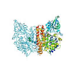
The high resolution structure of human glutamate carboxypeptidase III (GCPIII/NAALADase II) in complex with quisqualic acid
Descriptor: (S)-2-AMINO-3-(3,5-DIOXO-[1,2,4]OXADIAZOLIDIN-2-YL)-PROPIONIC ACID, 2-acetamido-2-deoxy-beta-D-glucopyranose, 2-acetamido-2-deoxy-beta-D-glucopyranose-(1-4)-2-acetamido-2-deoxy-beta-D-glucopyranose, ...
Authors:Lubkowski, J, Barinka, C, Hlouchova, K.
Deposit date:2008-11-28
Release date:2009-08-25
Last modified:2023-09-06
Method:X-RAY DIFFRACTION (1.56 Å)
Cite:Structural insight into the evolutionary and pharmacologic homology of glutamate carboxypeptidases II and III
Febs J., 276, 2009
3FED
DownloadVisualize
BU of 3fed by Molmil
The high resolution structure of human glutamate carboxypeptidase III (GCPIII/NAALADase II) in complex with a transition state analog of Glu-Glu
Descriptor: (2S)-2-{[(S)-[(3S)-3-amino-3-carboxypropyl](hydroxy)phosphoryl]methyl}pentanedioic acid, 2-acetamido-2-deoxy-beta-D-glucopyranose, 2-acetamido-2-deoxy-beta-D-glucopyranose-(1-4)-2-acetamido-2-deoxy-beta-D-glucopyranose, ...
Authors:Lubkowski, J, Barinka, C, Hlouchova, K.
Deposit date:2008-11-28
Release date:2009-08-25
Last modified:2023-09-06
Method:X-RAY DIFFRACTION (1.29 Å)
Cite:Structural insight into the evolutionary and pharmacologic homology of glutamate carboxypeptidases II and III
Febs J., 276, 2009
3BI0
DownloadVisualize
BU of 3bi0 by Molmil
X-ray structure of human glutamate carboxypeptidase II (GCPII) in complex with a transition state analog of Glu-Glu
Descriptor: (2S)-2-{[(S)-[(3S)-3-amino-3-carboxypropyl](hydroxy)phosphoryl]methyl}pentanedioic acid, 2-acetamido-2-deoxy-beta-D-glucopyranose, 2-acetamido-2-deoxy-beta-D-glucopyranose-(1-4)-2-acetamido-2-deoxy-beta-D-glucopyranose, ...
Authors:Lubkowski, J, Barinka, C.
Deposit date:2007-11-29
Release date:2008-01-08
Last modified:2023-08-30
Method:X-RAY DIFFRACTION (1.67 Å)
Cite:Structural basis of interactions between human glutamate carboxypeptidase II and its substrate analogs
J.Mol.Biol., 376, 2008
3BHX
DownloadVisualize
BU of 3bhx by Molmil
X-ray structure of human glutamate carboxypeptidase II (GCPII) in complex with a transition state analog of Asp-Glu
Descriptor: (2S)-2-{[(S)-(2-carboxyethyl)(hydroxy)phosphoryl]methyl}pentanedioic acid, 2-acetamido-2-deoxy-beta-D-glucopyranose, 2-acetamido-2-deoxy-beta-D-glucopyranose-(1-4)-2-acetamido-2-deoxy-beta-D-glucopyranose, ...
Authors:Lubkowski, J, Barinka, C.
Deposit date:2007-11-29
Release date:2008-01-08
Last modified:2023-08-30
Method:X-RAY DIFFRACTION (1.6 Å)
Cite:Structural basis of interactions between human glutamate carboxypeptidase II and its substrate analogs
J.Mol.Biol., 376, 2008
3BXM
DownloadVisualize
BU of 3bxm by Molmil
Structure of an inactive mutant of human glutamate carboxypeptidase II [GCPII(E424A)] in complex with N-acetyl-Asp-Glu (NAAG)
Descriptor: 2-acetamido-2-deoxy-beta-D-glucopyranose, 2-acetamido-2-deoxy-beta-D-glucopyranose-(1-4)-2-acetamido-2-deoxy-beta-D-glucopyranose, CALCIUM ION, ...
Authors:Lubkowski, J, Barinka, C.
Deposit date:2008-01-14
Release date:2009-01-27
Last modified:2023-08-30
Method:X-RAY DIFFRACTION (1.71 Å)
Cite:Reaction mechanism of glutamate carboxypeptidase II revealed by mutagenesis, X-ray crystallography, and computational methods.
Biochemistry, 48, 2009
3D7G
DownloadVisualize
BU of 3d7g by Molmil
A high resolution crystal structure of human glutamate carboxypeptidase II (GCPII) in a complex with DCMC, a urea-based inhibitor
Descriptor: 2-acetamido-2-deoxy-beta-D-glucopyranose, 2-acetamido-2-deoxy-beta-D-glucopyranose-(1-4)-2-acetamido-2-deoxy-beta-D-glucopyranose, CALCIUM ION, ...
Authors:Lubkowski, J, Barinka, C.
Deposit date:2008-05-21
Release date:2008-12-30
Last modified:2023-08-30
Method:X-RAY DIFFRACTION (1.75 Å)
Cite:Interactions between Human Glutamate Carboxypeptidase II and Urea-Based Inhibitors: Structural Characterization
J.Med.Chem., 51, 2008
3D7F
DownloadVisualize
BU of 3d7f by Molmil
A high resolution crystal structure of human glutamate carboxypeptidase II (GCPII) in a complex with DCIT, a urea-based inhibitor
Descriptor: 2-acetamido-2-deoxy-beta-D-glucopyranose, 2-acetamido-2-deoxy-beta-D-glucopyranose-(1-4)-2-acetamido-2-deoxy-beta-D-glucopyranose, CALCIUM ION, ...
Authors:Lubkowski, J, Barinka, C.
Deposit date:2008-05-21
Release date:2008-12-30
Last modified:2023-08-30
Method:X-RAY DIFFRACTION (1.54 Å)
Cite:Interactions between Human Glutamate Carboxypeptidase II and Urea-Based Inhibitors: Structural Characterization
J.Med.Chem., 51, 2008
3D7H
DownloadVisualize
BU of 3d7h by Molmil
A high resolution crystal structure of human glutamate carboxypeptidase II (GCPII) in a complex with DCIBzL, a urea-based inhibitor
Descriptor: 2-acetamido-2-deoxy-beta-D-glucopyranose, 2-acetamido-2-deoxy-beta-D-glucopyranose-(1-4)-2-acetamido-2-deoxy-beta-D-glucopyranose, CALCIUM ION, ...
Authors:Lubkowski, J, Barinka, C.
Deposit date:2008-05-21
Release date:2008-12-30
Last modified:2023-08-30
Method:X-RAY DIFFRACTION (1.55 Å)
Cite:Interactions between Human Glutamate Carboxypeptidase II and Urea-Based Inhibitors: Structural Characterization
J.Med.Chem., 51, 2008
3D7D
DownloadVisualize
BU of 3d7d by Molmil
A high resolution crystal structure of human glutamate carboxypeptidase II (GCPII) in a complex with DCFBD, a urea-based inhibitor
Descriptor: 2-acetamido-2-deoxy-beta-D-glucopyranose, 2-acetamido-2-deoxy-beta-D-glucopyranose-(1-4)-2-acetamido-2-deoxy-beta-D-glucopyranose, CALCIUM ION, ...
Authors:Lubkowski, J, Barinka, C.
Deposit date:2008-05-21
Release date:2008-12-30
Last modified:2023-08-30
Method:X-RAY DIFFRACTION (1.69 Å)
Cite:Interactions between Human Glutamate Carboxypeptidase II and Urea-Based Inhibitors: Structural Characterization
J.Med.Chem., 51, 2008
1HG1
DownloadVisualize
BU of 1hg1 by Molmil
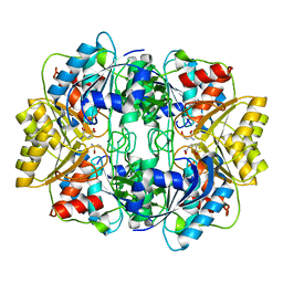
X-ray structure of the complex between Erwinia chrysanthemi L-asparaginase and D-aspartate
Descriptor: D-ASPARTIC ACID, L-ASPARAGINASE
Authors:Lubkowski, J, Wlodawer, A, Kolyani, K.A.
Deposit date:2000-12-08
Release date:2001-08-07
Last modified:2011-07-13
Method:X-RAY DIFFRACTION (1.8 Å)
Cite:Stuctural Basis for the Activity and Substrate Specificity of Erwinia Chrysanthemi L-Asparaginase
Biochemistry, 40, 2001
1HFW
DownloadVisualize
BU of 1hfw by Molmil
X-ray structure of the complex between Erwinia chrysanthemi L-asparaginase and L-Glutamate
Descriptor: GLUTAMIC ACID, L-ASPARAGINASE
Authors:Lubkowski, J, Wlodawer, A, Kolyani, K.A.
Deposit date:2000-12-08
Release date:2001-08-07
Last modified:2011-07-13
Method:X-RAY DIFFRACTION (1.8 Å)
Cite:Stuctural Basis for the Activity and Substrate Specificity of Erwinia Chrysanthemi L-Asparaginase
Biochemistry, 40, 2001
1HG0
DownloadVisualize
BU of 1hg0 by Molmil
X-ray structure of the complex between Erwinia chrysanthemi L-asparaginase and succinic acid
Descriptor: L-ASPARAGINASE, SUCCINIC ACID
Authors:Lubkowski, J, Wlodawer, A, Kolyani, K.A.
Deposit date:2000-12-08
Release date:2001-08-07
Last modified:2017-09-13
Method:X-RAY DIFFRACTION (1.9 Å)
Cite:Stuctural Basis for the Activity and Substrate Specificity of Erwinia Chrysanthemi L-Asparaginase
Biochemistry, 40, 2001
1HFK
DownloadVisualize
BU of 1hfk by Molmil
Asparaginase from Erwinia chrysanthemi, hexagonal form with weak sulfate
Descriptor: L-ASPARAGINE AMIDOHYDROLASE, SULFATE ION
Authors:Lubkowski, J, Palm, G.J, Kozak, M, Jaskolski, M, Wlodawer, A.
Deposit date:2000-12-05
Release date:2000-12-07
Last modified:2023-12-13
Method:X-RAY DIFFRACTION (2.17 Å)
Cite:Structures of Two Highly Homologous Bacterial L-Asparaginases: A Case of Enantiomorphic Space Groups
Acta Crystallogr.,Sect.D, 57, 2001
2NLF
DownloadVisualize
BU of 2nlf by Molmil
Human beta-defensin-1 (Mutant Leu13Glu)
Descriptor: Beta-defensin 1, SULFATE ION
Authors:Lubkowski, J, Pazgier, M.
Deposit date:2006-10-20
Release date:2006-10-31
Last modified:2023-08-30
Method:X-RAY DIFFRACTION (1.45 Å)
Cite:Studies of the Biological Properties of Human beta-Defensin 1.
J.Biol.Chem., 282, 2007
2NLD
DownloadVisualize
BU of 2nld by Molmil
Human beta-defensin-1 (Mutant Gln11Ala)
Descriptor: ACETATE ION, Beta-defensin 1, SULFATE ION
Authors:Lubkowski, J, Pazgier, M.
Deposit date:2006-10-20
Release date:2006-10-31
Last modified:2023-08-30
Method:X-RAY DIFFRACTION (1.49 Å)
Cite:Studies of the Biological Properties of Human beta-Defensin 1.
J.Biol.Chem., 282, 2007

218853

PDB entries from 2024-04-24

PDB statisticsPDBj update infoContact PDBjnumon