7YHL
 
 | |
7YGH
 | |
7YGL
 | |
8JBB
 | |
8JH1
 | |
8JBC
 | |
5XNP
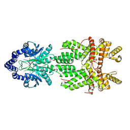
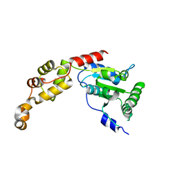
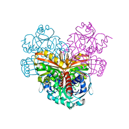
 | | Crystal structures of human SALM5 in complex with human PTPdelta | | 分子名称: | 2-acetamido-2-deoxy-beta-D-glucopyranose, CALCIUM ION, CHLORIDE ION, ... | | 著者 | Liu, H, Lin, Z, Xu, F. | | 登録日 | 2017-05-24 | | 公開日 | 2018-01-24 | | 最終更新日 | 2023-11-22 | | 実験手法 | X-RAY DIFFRACTION (3.729 Å) | | 主引用文献 | Structural basis of SALM5-induced PTP delta dimerization for synaptic differentiation
Nat Commun, 9, 2018
|
|
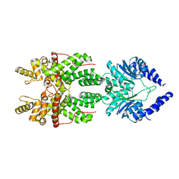
5XNQ
 | | Crystal structures of human SALM5 | | 分子名称: | 2-acetamido-2-deoxy-beta-D-glucopyranose, CALCIUM ION, CHLORIDE ION, ... | | 著者 | Liu, H, Lin, Z, Xu, F. | | 登録日 | 2017-05-24 | | 公開日 | 2018-01-24 | | 最終更新日 | 2020-07-29 | | 実験手法 | X-RAY DIFFRACTION (2.802 Å) | | 主引用文献 | Structural basis of SALM5-induced PTP delta dimerization for synaptic differentiation
Nat Commun, 9, 2018
|
|
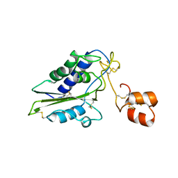
5XME
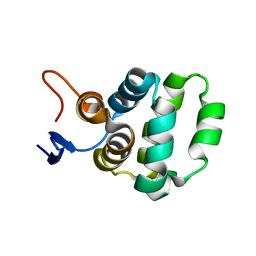 | | Solution structure of C-terminal domain of TRADD | | 分子名称: | Tumor necrosis factor receptor type 1-associated DEATH domain protein | | 著者 | Lin, Z, Zhang, N. | | 登録日 | 2017-05-15 | | 公開日 | 2017-09-06 | | 最終更新日 | 2024-05-01 | | 実験手法 | SOLUTION NMR | | 主引用文献 | Structure of the C-terminal domain of TRADD reveals a novel fold in the death domain superfamily.
Sci Rep, 7, 2017
|
|
5Z6Q
 | |
5ZGG
 | |
4XSK
 | | Structure of PAItrap, an uPA mutant | | 分子名称: | GLYCEROL, SULFATE ION, TRIETHYLENE GLYCOL, ... | | 著者 | Gong, L, Proulle, V, Hong, Z, Lin, Z, Liu, M, Yuan, C, Lin, L, Furie, B, Flaumenhaft, R, Andreasen, P, Furie, B, Huang, M. | | 登録日 | 2015-01-22 | | 公開日 | 2016-02-03 | | 最終更新日 | 2023-11-08 | | 実験手法 | X-RAY DIFFRACTION (1.5 Å) | | 主引用文献 | Structure of PAItrap, an uPA mutant
To Be Published
|
|
1GP7
 | |
1M8S
 | | Crystal Structures of Cadmium-binding Acidic Phospholipase A2 from the Venom of Agkistrodon halys pallas at 1.9 Resolution (crystal grown at pH 5.9) | | 分子名称: | 1,4-BUTANEDIOL, CADMIUM ION, phospholipase a2 | | 著者 | Xu, S, Gu, L, Zhou, Y, Lin, Z. | | 登録日 | 2002-07-25 | | 公開日 | 2003-02-11 | | 最終更新日 | 2023-10-25 | | 実験手法 | X-RAY DIFFRACTION (1.9 Å) | | 主引用文献 | Structures of cadmium-binding acidic phospholipase A(2) from the venom of Agkistrodon halys Pallas at 1.9A resolutio
Biochem.Biophys.Res.Commun., 300, 2003
|
|
1M8R
 | | Crystal Structures of Cadmium-binding Acidic Phospholipase A2 from the Venom of Agkistrodon halys pallas at 1.9 Resolution (crystal grown at pH 7.4) | | 分子名称: | 1,4-BUTANEDIOL, CADMIUM ION, phospholipase A2 | | 著者 | Xu, S, Gu, L, Zhou, Y, Lin, Z. | | 登録日 | 2002-07-25 | | 公開日 | 2003-02-11 | | 最終更新日 | 2023-10-25 | | 実験手法 | X-RAY DIFFRACTION (1.9 Å) | | 主引用文献 | Structures of cadmium-binding acidic phospholipase A(2) from the venom of Agkistrodon halys Pallas at 1.9A resolutio
Biochem.Biophys.Res.Commun., 300, 2003
|
|
1CRW
 | |
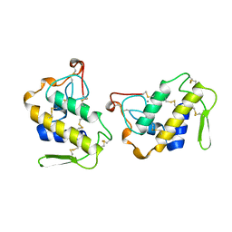
2GIZ
 | | Structural and functional analysis of Natrin, a member of crisp-3 family blocks a variety of ion channels | | 分子名称: | Natrin-1 | | 著者 | Jiang, T, Wang, F, Li, H, Yin, C, Zhou, Y, Shu, Y, Qi, Z, Lin, Z. | | 登録日 | 2006-03-30 | | 公開日 | 2006-11-28 | | 最終更新日 | 2023-10-25 | | 実験手法 | X-RAY DIFFRACTION (1.68 Å) | | 主引用文献 | Structural and functional analysis of natrin, a venom protein that targets various ion channels
Biochem.Biophys.Res.Commun., 351, 2006
|
|
1JIA
 | |
1M8T
 | | Structure of an acidic Phospholipase A2 from the venom of Ophiophagus hannah at 2.1 resolution from a hemihedrally twinned crystal form | | 分子名称: | CALCIUM ION, HEXANE-1,6-DIOL, Phospholipase a2 | | 著者 | Xu, S, Gu, L, Wang, Q, Shu, Y, Lin, Z. | | 登録日 | 2002-07-26 | | 公開日 | 2003-09-02 | | 最終更新日 | 2023-10-25 | | 実験手法 | X-RAY DIFFRACTION (2.1 Å) | | 主引用文献 | Structure of a king cobra phospholipase A2 determined from a hemihedrally twinned crystal.
Acta Crystallogr.,Sect.D, 59, 2003
|
|
1BK9
 | | PHOSPHOLIPASE A2 MODIFIED BY PBPB | | 分子名称: | 1,4-BUTANEDIOL, CALCIUM ION, PHOSPHOLIPASE A2, ... | | 著者 | Zhao, H, Tang, L, Wang, X, Lin, Z, Zhou, Y. | | 登録日 | 1998-07-16 | | 公開日 | 1999-03-02 | | 最終更新日 | 2023-08-02 | | 実験手法 | X-RAY DIFFRACTION (2 Å) | | 主引用文献 | Structure of a snake venom phospholipase A2 modified by p-bromo-phenacyl-bromide.
Toxicon, 36, 1998
|
|
4XKL
 | | Crystal structure of NDP52 ZF2 in complex with mono-ubiquitin | | 分子名称: | ACETATE ION, Calcium-binding and coiled-coil domain-containing protein 2, GLYCEROL, ... | | 著者 | Xie, X, Li, F, Wang, Y, Lin, Z, Chen, X, Liu, J, Pan, L. | | 登録日 | 2015-01-12 | | 公開日 | 2015-11-11 | | 最終更新日 | 2023-11-08 | | 実験手法 | X-RAY DIFFRACTION (2.1 Å) | | 主引用文献 | Molecular basis of ubiquitin recognition by the autophagy receptor CALCOCO2
Autophagy, 11, 2015
|
|
1HUO
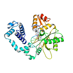 | | CRYSTAL STRUCTURE OF DNA POLYMERASE BETA COMPLEXED WITH DNA AND CR-TMPPCP | | 分子名称: | 5'-D(*AP*AP*TP*AP*GP*GP*CP*GP*TP*CP*G)-3', 5'-D(P*CP*GP*AP*CP*GP*CP*C)-3', CHROMIUM ION, ... | | 著者 | Arndt, J.W, Gong, W, Zhong, X, Showalter, A.K, Liu, J, Lin, Z, Paxson, C, Tsai, M.-D, Chan, M.K. | | 登録日 | 2001-01-04 | | 公開日 | 2001-04-23 | | 最終更新日 | 2024-02-07 | | 実験手法 | X-RAY DIFFRACTION (2.6 Å) | | 主引用文献 | Insight into the catalytic mechanism of DNA polymerase beta: structures of intermediate complexes.
Biochemistry, 40, 2001
|
|
1HUZ
 | | CRYSTAL STRUCTURE OF DNA POLYMERASE COMPLEXED WITH DNA AND CR-PCP | | 分子名称: | 5'-D(*AP*AP*TP*AP*GP*GP*CP*GP*TP*CP*G)-3', 5'-D(P*CP*GP*AP*CP*GP*CP*CP*T)-3', CHROMIUM ION, ... | | 著者 | Arndt, J.W, Gong, W, Zhong, X, Showalter, A.K, Liu, J, Lin, Z, Paxson, C, Tsai, M.-D, Chan, M.K. | | 登録日 | 2001-01-04 | | 公開日 | 2001-04-23 | | 最終更新日 | 2024-02-07 | | 実験手法 | X-RAY DIFFRACTION (2.6 Å) | | 主引用文献 | Insight into the catalytic mechanism of DNA polymerase beta: structures of intermediate complexes.
Biochemistry, 40, 2001
|
|
1IJL
 | | Crystal structure of acidic phospholipase A2 from deinagkistrodon acutus | | 分子名称: | CALCIUM ION, PHOSPHOLIPASE A2, ZINC ION | | 著者 | Gu, L, Zhang, H, Song, S, Zhou, Y, Lin, Z. | | 登録日 | 2001-04-27 | | 公開日 | 2001-12-28 | | 最終更新日 | 2023-10-25 | | 実験手法 | X-RAY DIFFRACTION (2.6 Å) | | 主引用文献 | Structure of an acidic phospholipase A2 from the venom of Deinagkistrodon acutus.
Acta Crystallogr.,Sect.D, 58, 2002
|
|
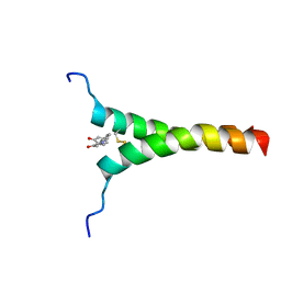
2M0M
 | | Structural Characterization of Minor Ampullate Spidroin Domains and their Distinct Roles in Fibroin Solubility and Fiber Formation | | 分子名称: | Minor ampullate fibroin 1 | | 著者 | Yang, D, Gao, Z, Lin, Z, Huang, W, Lai, C, Fan, J. | | 登録日 | 2012-10-30 | | 公開日 | 2013-03-27 | | 最終更新日 | 2024-05-01 | | 実験手法 | SOLUTION NMR | | 主引用文献 | Structural characterization of minor ampullate spidroin domains and their distinct roles in fibroin solubility and fiber formation
Plos One, 8, 2013
|
|


