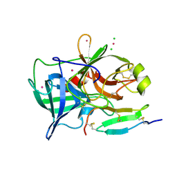
2QY0
 
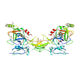 | | Active dimeric structure of the catalytic domain of C1r reveals enzyme-product like contacts | | 分子名称: | Complement C1r subcomponent, GLYCEROL | | 著者 | Kardos, J, Harmat, V, Pallo, A, Barabas, O, Szilagyi, K, Graf, L, Naray-Szabo, G, Goto, Y, Zavodszky, P, Gal, P. | | 登録日 | 2007-08-13 | | 公開日 | 2008-02-05 | | 最終更新日 | 2023-08-30 | | 実験手法 | X-RAY DIFFRACTION (2.6 Å) | | 主引用文献 | Revisiting the mechanism of the autoactivation of the complement protease C1r in the C1 complex: Structure of the active catalytic region of C1r.
Mol.Immunol., 45, 2008
|
|
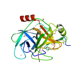
3IF7
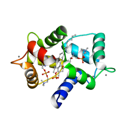 | | Structure of Calmodulin complexed with its first endogenous inhibitor, sphingosylphosphorylcholine | | 分子名称: | 2-{[(R)-{[(2S,3R,4E)-2-amino-3-hydroxyoctadec-4-en-1-yl]oxy}(hydroxy)phosphoryl]oxy}-N,N,N-trimethylethanaminium, CALCIUM ION, Calmodulin | | 著者 | Kovacs, E, Harmat, V, Toth, J, Vertessy, B.G, Modos, K, Kardos, J, Liliom, K. | | 登録日 | 2009-07-24 | | 公開日 | 2010-06-30 | | 最終更新日 | 2023-11-01 | | 実験手法 | X-RAY DIFFRACTION (1.6 Å) | | 主引用文献 | Structure and mechanism of calmodulin binding to a signaling sphingolipid reveal new aspects of lipid-protein interactions
Faseb J., 24, 2010
|
|
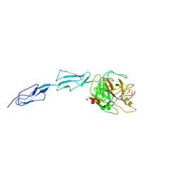
4BNR
 | | Extremely stable complex of crayfish trypsin with bovine trypsin inhibitor | | 分子名称: | CALCIUM ION, HEPATOPANCREAS TRYPSIN, PANCREATIC TRYPSIN INHIBITOR, ... | | 著者 | Molnar, T, Voros, J, Szeder, B, Takats, K, Kardos, J, Katona, G, Graf, L. | | 登録日 | 2013-05-17 | | 公開日 | 2013-09-04 | | 最終更新日 | 2023-12-20 | | 実験手法 | X-RAY DIFFRACTION (2 Å) | | 主引用文献 | Comparison of Complexes Formed by a Crustacean and a Vertebrate Trypsin with Bovine Pancreatic Trypsin Inhibitor - the Key to Achieving Extreme Stability?
FEBS J., 280, 2013
|
|
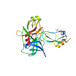
1Q3X
 | | Crystal structure of the catalytic region of human MASP-2 | | 分子名称: | GLYCEROL, Mannan-binding lectin serine protease 2, SODIUM ION | | 著者 | Harmat, V, Gal, P, Kardos, J, Szilagyi, K, Ambrus, G, Naray-Szabo, G, Zavodszky, P. | | 登録日 | 2003-08-01 | | 公開日 | 2004-08-03 | | 最終更新日 | 2023-08-16 | | 実験手法 | X-RAY DIFFRACTION (2.23 Å) | | 主引用文献 | The structure of MBL-associated serine protease-2 reveals that identical substrate specificities of C1s and MASP-2 are realized through different sets of enzyme-substrate interactions
J.Mol.Biol., 342, 2004
|
|
2F91
 | | 1.2A resolution structure of a crayfish trypsin complexed with a peptide inhibitor, SGTI | | 分子名称: | CADMIUM ION, CHLORIDE ION, Serine protease inhibitor I/II, ... | | 著者 | Fodor, K, Harmat, V, Hetenyi, C, Kardos, J, Antal, J, Perczel, A, Patthy, A, Katona, G, Graf, L. | | 登録日 | 2005-12-05 | | 公開日 | 2006-04-18 | | 最終更新日 | 2023-08-30 | | 実験手法 | X-RAY DIFFRACTION (1.2 Å) | | 主引用文献 | Enzyme:Substrate Hydrogen Bond Shortening during the Acylation Phase of Serine Protease Catalysis.
Biochemistry, 45, 2006
|
|
2XTT
 | | Bovine trypsin in complex with evolutionary enhanced Schistocerca gregaria protease inhibitor 1 (SGPI-1-P02) | | 分子名称: | ACETATE ION, CALCIUM ION, CATIONIC TRYPSIN, ... | | 著者 | Wahlgren, W.Y, Pal, G, Kardos, J, Porrogi, P, Szenthe, B, Patthy, A, Graf, L, Katona, G. | | 登録日 | 2010-10-12 | | 公開日 | 2010-11-10 | | 最終更新日 | 2023-12-20 | | 実験手法 | X-RAY DIFFRACTION (0.93 Å) | | 主引用文献 | The catalytic aspartate is protonated in the Michaelis complex formed between trypsin and an in vitro evolved substrate-like inhibitor: a refined mechanism of serine protease action.
J.Biol.Chem., 286, 2011
|
|
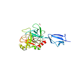
1ZJK
 | | Crystal structure of the zymogen catalytic region of human MASP-2 | | 分子名称: | Mannan-binding lectin serine protease 2 | | 著者 | Gal, P, Harmat, V, Kocsis, A, Bian, T, Barna, L, Ambrus, G, Vegh, B, Balczer, J, Sim, R.B, Naray-Szabo, G, Zavodszky, P. | | 登録日 | 2005-04-29 | | 公開日 | 2005-07-26 | | 最終更新日 | 2023-08-23 | | 実験手法 | X-RAY DIFFRACTION (2.18 Å) | | 主引用文献 | A True Autoactivating Enzyme: Structural insight into mannose-binding lectin-associated serine protease-2 activations
J.Biol.Chem., 280, 2005
|
|
3BAT
 | |
3BAS
 | |


