Loading
PDBj
MenuPDBj@FacebookPDBj@TwitterPDBj@YouTubewwPDB FoundationwwPDB
RCSB PDBPDBeBMRBAdv. SearchSearch help
Search by PDB author
3TG0
DownloadVisualize
BU of 3tg0 by Molmil
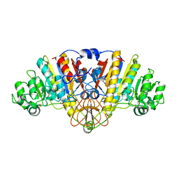
E. coli alkaline phosphatase with bound inorganic phosphate
Descriptor: Alkaline phosphatase, MAGNESIUM ION, PHOSPHATE ION, ...
Authors:Bobyr, E, Lassila, J.K, Wiersma-Koch, H.I, Fenn, T.D, Lee, J.J, Nikolic-Hughes, I, Hodgson, K.O, Rees, D.C, Hedman, B, Herschlag, D.
Deposit date:2011-08-16
Release date:2011-11-09
Last modified:2023-09-13
Method:X-RAY DIFFRACTION (1.2 Å)
Cite:High-resolution analysis of Zn(2+) coordination in the alkaline phosphatase superfamily by EXAFS and x-ray crystallography.
J.Mol.Biol., 415, 2012
2IIZ
DownloadVisualize
BU of 2iiz by Molmil
Crystal structure of putative melanin biosynthesis protein TyrA with bound heme (NP_716371.1) from Shewanella Oneidensis at 2.30 A resolution
Descriptor: 1,2-ETHANEDIOL, ISOPROPYL ALCOHOL, Melanin biosynthesis protein TyrA, ...
Authors:Joint Center for Structural Genomics (JCSG)
Deposit date:2006-09-28
Release date:2006-11-14
Last modified:2023-11-15
Method:X-RAY DIFFRACTION (2.3 Å)
Cite:Identification and structural characterization of heme binding in a novel dye-decolorizing peroxidase, TyrA.
Proteins, 69, 2007
3EQX
DownloadVisualize
BU of 3eqx by Molmil
CRYSTAL STRUCTURE OF A FIC FAMILY PROTEIN (SO_4266) FROM SHEWANELLA ONEIDENSIS AT 1.6 A RESOLUTION
Descriptor: FIC DOMAIN CONTAINING TRANSCRIPTIONAL REGULATOR, TRIETHYLENE GLYCOL
Authors:Joint Center for Structural Genomics (JCSG)
Deposit date:2008-10-01
Release date:2008-10-14
Last modified:2023-02-01
Method:X-RAY DIFFRACTION (1.6 Å)
Cite:Crystal structure of the Fic (Filamentation induced by cAMP) family protein SO4266 (gi|24375750) from Shewanella oneidensis MR-1 at 1.6 A resolution.
Proteins, 75, 2009
3F1Z
DownloadVisualize
BU of 3f1z by Molmil
Crystal structure of putative nucleic acid-binding lipoprotein (YP_001337197.1) from Klebsiella pneumoniae subsp. pneumoniae MGH 78578 at 2.46 A resolution
Descriptor: DI(HYDROXYETHYL)ETHER, putative nucleic acid-binding lipoprotein
Authors:Joint Center for Structural Genomics (JCSG)
Deposit date:2008-10-28
Release date:2008-11-18
Last modified:2023-02-01
Method:X-RAY DIFFRACTION (2.46 Å)
Cite:The structure of KPN03535 (gi|152972051), a novel putative lipoprotein from Klebsiella pneumoniae, reveals an OB-fold.
Acta Crystallogr.,Sect.F, 66, 2010
6MUJ
DownloadVisualize
BU of 6muj by Molmil
Formylglycine generating enzyme bound to copper
Descriptor: 2,3-DIHYDROXY-1,4-DITHIOBUTANE, CALCIUM ION, COPPER (II) ION, ...
Authors:Lafrance-Vanasse, J, Appel, M.J, Tsai, C.-L, Bertozzi, C, Tainer, J.A.
Deposit date:2018-10-23
Release date:2019-02-27
Last modified:2023-10-11
Method:X-RAY DIFFRACTION (2.249 Å)
Cite:Formylglycine-generating enzyme binds substrate directly at a mononuclear Cu(I) center to initiate O2activation.
Proc. Natl. Acad. Sci. U.S.A., 116, 2019
4PNJ
DownloadVisualize
BU of 4pnj by Molmil
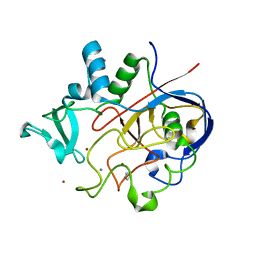
Recombinant Sperm Whale P6 Myoglobin Solved with Single Pulse Free Electron Laser Data
Descriptor: Myoglobin, PROTOPORPHYRIN IX CONTAINING FE, SULFATE ION
Authors:Cohen, A, Gonzalez, A, Lam, W, Lyubimov, A, Sauter, N, Tsai, Y, Uervirojnangkoorn, M, Brunger, A, Soltis, M.
Deposit date:2014-05-23
Release date:2014-11-05
Last modified:2023-09-27
Method:X-RAY DIFFRACTION (1.36 Å)
Cite:Goniometer-based femtosecond crystallography with X-ray free electron lasers.
Proc.Natl.Acad.Sci.USA, 111, 2014
3L5O
DownloadVisualize
BU of 3l5o by Molmil
Crystal structure of protein with unknown function from DUF364 family (ZP_00559375.1) from Desulfitobacterium hafniense DCB-2 at 2.01 A resolution
Descriptor: 1,2-ETHANEDIOL, CHLORIDE ION, IMIDAZOLE, ...
Authors:Joint Center for Structural Genomics, Joint Center for Structural Genomics (JCSG)
Deposit date:2009-12-22
Release date:2010-02-02
Last modified:2023-02-01
Method:X-RAY DIFFRACTION (2.01 Å)
Cite:Structure of the first representative of Pfam family PF04016 (DUF364) reveals enolase and Rossmann-like folds that combine to form a unique active site with a possible role in heavy-metal chelation.
Acta Crystallogr.,Sect.F, 66, 2010
3K5J
DownloadVisualize
BU of 3k5j by Molmil
Crystal structure of Putative SUFU (suppressor of fused protein) homolog (YP_208451.1) from Neisseria gonorrhoeae FA 1090 at 1.40 A resolution
Descriptor: GLYCEROL, SULFATE ION, Suppressor of fused family protein
Authors:Joint Center for Structural Genomics (JCSG)
Deposit date:2009-10-07
Release date:2010-01-26
Last modified:2019-07-17
Method:X-RAY DIFFRACTION (1.4 Å)
Cite:The crystal structure of a bacterial Sufu-like protein defines a novel group of bacterial proteins that are similar to the N-terminal domain of human Sufu.
Protein Sci., 19, 2010
3KK7
DownloadVisualize
BU of 3kk7 by Molmil
Crystal structure of Putative cell invasion protein with MAC/Perforin domain (NP_812351.1) from BACTERIODES THETAIOTAOMICRON VPI-5482 at 2.46 A resolution
Descriptor: (4S)-2-METHYL-2,4-PENTANEDIOL, 1,2-ETHANEDIOL, CHLORIDE ION, ...
Authors:Joint Center for Structural Genomics (JCSG)
Deposit date:2009-11-04
Release date:2009-11-24
Last modified:2023-02-01
Method:X-RAY DIFFRACTION (2.46 Å)
Cite:Structure of a membrane-attack complex/perforin (MACPF) family protein from the human gut symbiont Bacteroides thetaiotaomicron.
Acta Crystallogr.,Sect.F, 66, 2010
3NL9
DownloadVisualize
BU of 3nl9 by Molmil
Crystal structure of a putative NTP pyrophosphohydrolase (Exig_1061) from EXIGUOBACTERIUM SP. 255-15 at 1.78 A resolution
Descriptor: 1,2-ETHANEDIOL, putative NTP pyrophosphohydrolase
Authors:Joint Center for Structural Genomics (JCSG)
Deposit date:2010-06-21
Release date:2010-07-21
Last modified:2023-02-01
Method:X-RAY DIFFRACTION (1.78 Å)
Cite:Structure of a putative NTP pyrophosphohydrolase: YP_001813558.1 from Exiguobacterium sibiricum 255-15.
Acta Crystallogr.,Sect.F, 66, 2010
3H41
DownloadVisualize
BU of 3h41 by Molmil
CRYSTAL STRUCTURE OF A NLPC/P60 FAMILY PROTEIN (BCE_2878) FROM BACILLUS CEREUS ATCC 10987 AT 1.79 A RESOLUTION
Descriptor: ALANINE, D-GLUTAMIC ACID, NLP/P60 family protein, ...
Authors:Joint Center for Structural Genomics (JCSG)
Deposit date:2009-04-17
Release date:2009-05-05
Last modified:2023-02-01
Method:X-RAY DIFFRACTION (1.79 Å)
Cite:Structure of the gamma-D-glutamyl-L-diamino acid endopeptidase YkfC from Bacillus cereus in complex with L-Ala-[gamma]-D-Glu: insights into substrate recognition by NlpC/P60 cysteine peptidases
Acta Crystallogr.,Sect.F, 66, 2010
3H50
DownloadVisualize
BU of 3h50 by Molmil
CRYSTAL STRUCTURE OF A TETRACENOMYCIN POLYKETIDE SYNTHESIS PROTEIN (TCMJ) FROM XANTHOMONAS CAMPESTRIS PV. CAMPESTRIS AT 1.60 A RESOLUTION
Descriptor: ACETATE ION, Tetracenomycin polyketide synthesis protein, ZINC ION
Authors:Joint Center for Structural Genomics (JCSG)
Deposit date:2009-04-21
Release date:2009-05-05
Last modified:2023-02-01
Method:X-RAY DIFFRACTION (1.6 Å)
Cite:Conformational changes associated with the binding of zinc acetate at the putative active site of XcTcmJ, a cupin from Xanthomonas campestris pv. campestris.
Acta Crystallogr.,Sect.F, 66, 2010
3GF8
DownloadVisualize
BU of 3gf8 by Molmil
Crystal structure of putative polysaccharide binding proteins (DUF1812) (NP_809975.1) from BACTEROIDES THETAIOTAOMICRON VPI-5482 at 2.20 A resolution
Descriptor: 1,2-ETHANEDIOL, putative polysaccharide binding proteins (DUF1812)
Authors:Joint Center for Structural Genomics (JCSG)
Deposit date:2009-02-26
Release date:2009-03-24
Last modified:2023-02-01
Method:X-RAY DIFFRACTION (2.2 Å)
Cite:A conserved fold for fimbrial components revealed by the crystal structure of a putative fimbrial assembly protein (BT1062) from Bacteroides thetaiotaomicron at 2.2 A resolution
Acta Crystallogr.,Sect.F, 66, 2010
3HSA
DownloadVisualize
BU of 3hsa by Molmil
Crystal structure of pleckstrin homology domain (YP_926556.1) from SHEWANELLA AMAZONENSIS SB2B at 1.99 A resolution
Descriptor: DI(HYDROXYETHYL)ETHER, GLYCEROL, pleckstrin homology domain
Authors:Joint Center for Structural Genomics (JCSG)
Deposit date:2009-06-10
Release date:2009-06-23
Last modified:2023-02-01
Method:X-RAY DIFFRACTION (1.99 Å)
Cite:Bacterial pleckstrin homology domains: a prokaryotic origin for the PH domain.
J.Mol.Biol., 396, 2010
3HBZ
DownloadVisualize
BU of 3hbz by Molmil
Crystal structure of a putative glycoside hydrolase (bt_2081) from bacteroides thetaiotaomicron vpi-5482 at 2.05 A resolution
Descriptor: ACETATE ION, CACODYLATE ION, CALCIUM ION, ...
Authors:Joint Center for Structural Genomics (JCSG)
Deposit date:2009-05-05
Release date:2009-05-26
Last modified:2023-02-01
Method:X-RAY DIFFRACTION (2.05 Å)
Cite:Structure of Bacteroides thetaiotaomicron BT2081 at 2.05 A resolution: the first structural representative of a new protein family that may play a role in carbohydrate metabolism.
Acta Crystallogr.,Sect.F, 66, 2010
3IRB
DownloadVisualize
BU of 3irb by Molmil
Crystal structure of protein with unknown function from DUF35 family (13815350) from SULFOLOBUS SOLFATARICUS at 1.80 A resolution
Descriptor: ACETIC ACID, SULFATE ION, ZINC ION, ...
Authors:Joint Center for Structural Genomics (JCSG)
Deposit date:2009-08-21
Release date:2009-09-01
Last modified:2023-02-01
Method:X-RAY DIFFRACTION (1.8 Å)
Cite:The structure of SSO2064, the first representative of Pfam family PF01796, reveals a novel two-domain zinc-ribbon OB-fold architecture with a potential acyl-CoA-binding role.
Acta Crystallogr.,Sect.F, 66, 2010
3H0N
DownloadVisualize
BU of 3h0n by Molmil
Crystal structure of a duf1470 family protein (jann_2411) from jannaschia sp. ccs1 at 1.45 A resolution
Descriptor: ACETATE ION, GLYCEROL, NICKEL (II) ION, ...
Authors:Joint Center for Structural Genomics (JCSG)
Deposit date:2009-04-09
Release date:2009-04-21
Last modified:2023-02-01
Method:X-RAY DIFFRACTION (1.45 Å)
Cite:The structure of Jann_2411 (DUF1470) from Jannaschia sp. at 1.45  resolution reveals a new fold (the ABATE domain) and suggests its possible role as a transcription regulator.
Acta Crystallogr.,Sect.F, 66, 2010
5FD1
DownloadVisualize
BU of 5fd1 by Molmil
CRYSTAL STRUCTURES OF OXIDIZED AND REDUCED AZOTOBACTER VINELANDII FERREDOXIN AT PH 8 AND PH 6
Descriptor: FE3-S4 CLUSTER, FERREDOXIN, IRON/SULFUR CLUSTER
Authors:Stout, C.D.
Deposit date:1993-06-29
Release date:1993-10-31
Last modified:2024-03-13
Method:X-RAY DIFFRACTION (1.9 Å)
Cite:Crystal structures of oxidized and reduced Azotobacter vinelandii ferredoxin at pH 8 and 6.
J.Biol.Chem., 268, 1993
2OOK
DownloadVisualize
BU of 2ook by Molmil
Crystal structure of a protein with unknown function (YP_749275.1) from Shewanella Frigidimarina NCIMB 400 at 1.80 A resolution
Descriptor: 1,2-ETHANEDIOL, Hypothetical protein
Authors:Joint Center for Structural Genomics (JCSG)
Deposit date:2007-01-25
Release date:2007-02-06
Last modified:2023-01-25
Method:X-RAY DIFFRACTION (1.8 Å)
Cite:Open and closed conformations of two SpoIIAA-like proteins (YP_749275.1 and YP_001095227.1) provide insights into membrane association and ligand binding.
Acta Crystallogr.,Sect.F, 66, 2010
2OOC
DownloadVisualize
BU of 2ooc by Molmil
Crystal structure of Histidine Phosphotransferase ShpA (NP_419930.1) from Caulobacter crescentus at 1.52 A resolution
Descriptor: GLYCEROL, Histidine phosphotransferase, TETRAETHYLENE GLYCOL
Authors:Joint Center for Structural Genomics (JCSG)
Deposit date:2007-01-25
Release date:2007-02-06
Last modified:2023-01-25
Method:X-RAY DIFFRACTION (1.52 Å)
Cite:Crystal structure of histidine phosphotransfer protein ShpA, an essential regulator of stalk biogenesis in Caulobacter crescentus.
J.Mol.Biol., 390, 2009
2RA9
DownloadVisualize
BU of 2ra9 by Molmil
Crystal structure of a duf1285 family protein (sbal_2486) from shewanella baltica os155 at 1.40 A resolution
Descriptor: 1,2-ETHANEDIOL, SODIUM ION, Uncharacterized protein DUF1285
Authors:Joint Center for Structural Genomics (JCSG)
Deposit date:2007-09-14
Release date:2007-10-16
Last modified:2023-01-25
Method:X-RAY DIFFRACTION (1.4 Å)
Cite:Structures of the first representatives of Pfam family PF06938 (DUF1285) reveal a new fold with repeated structural motifs and possible involvement in signal transduction.
Acta Crystallogr.,Sect.F, 66, 2010
2PV7
DownloadVisualize
BU of 2pv7 by Molmil
Crystal structure of chorismate mutase / prephenate dehydrogenase (tyrA) (1574749) from Haemophilus influenzae RD at 2.00 A resolution
Descriptor: NICOTINAMIDE-ADENINE-DINUCLEOTIDE, T-protein [Includes: Chorismate mutase (EC 5.4.99.5) (CM) and Prephenate dehydrogenase (EC 1.3.1.12) (PDH)], TYROSINE
Authors:Joint Center for Structural Genomics (JCSG)
Deposit date:2007-05-09
Release date:2007-05-22
Last modified:2023-01-25
Method:X-RAY DIFFRACTION (2 Å)
Cite:The structure of Haemophilus influenzae prephenate dehydrogenase suggests unique features of bifunctional TyrA enzymes.
Acta Crystallogr.,Sect.F, 66, 2010
2Q3L
DownloadVisualize
BU of 2q3l by Molmil
CRYSTAL STRUCTURE OF AN UNCHARACTERIZED PROTEIN FROM DUF3478 FAMILY WITH A SPOIIAA-LIKE FOLD (SHEW_3102) FROM SHEWANELLA LOIHICA PV-4 AT 2.25 A RESOLUTION
Descriptor: (4S)-2-METHYL-2,4-PENTANEDIOL, CHLORIDE ION, SODIUM ION, ...
Authors:Joint Center for Structural Genomics (JCSG)
Deposit date:2007-05-30
Release date:2007-06-12
Last modified:2023-01-25
Method:X-RAY DIFFRACTION (2.25 Å)
Cite:Open and closed conformations of two SpoIIAA-like proteins (YP_749275.1 and YP_001095227.1) provide insights into membrane association and ligand binding.
Acta Crystallogr.,Sect.F, 66, 2010
2Q8U
DownloadVisualize
BU of 2q8u by Molmil
CRYSTAL STRUCTURE OF MRE11 FROM THERMOTOGA MARITIMA MSB8 (TM1635) AT 2.20 A RESOLUTION
Descriptor: Exonuclease, putative
Authors:Joint Center for Structural Genomics (JCSG)
Deposit date:2007-06-11
Release date:2007-06-26
Last modified:2023-01-25
Method:X-RAY DIFFRACTION (2.2 Å)
Cite:Crystal structure of the first eubacterial Mre11 nuclease reveals novel features that may discriminate substrates during DNA repair.
J.Mol.Biol., 397, 2010
1J5S
DownloadVisualize
BU of 1j5s by Molmil
Crystal structure of uronate isomerase (TM0064) from Thermotoga maritima at 2.85 A resolution
Descriptor: URONATE ISOMERASE
Authors:Joint Center for Structural Genomics (JCSG)
Deposit date:2002-07-02
Release date:2002-07-17
Last modified:2024-05-22
Method:X-RAY DIFFRACTION (2.85 Å)
Cite:Crystal structure of uronate isomerase (TM0064) from Thermotoga maritima at 2.85 A resolution.
Proteins, 53, 2003

221716

PDB entries from 2024-06-26

PDB statisticsPDBj update infoContact PDBjnumon