Loading
PDBj
MenuPDBj@FacebookPDBj@TwitterPDBj@YouTubewwPDB FoundationwwPDB
RCSB PDBPDBeBMRBAdv. SearchSearch help
Search by PDB author
2IL8
DownloadVisualize
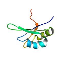
BU of 2il8 by Molmil
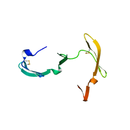
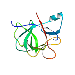
THREE-DIMENSIONAL STRUCTURE OF INTERLEUKIN 8 IN SOLUTION
Descriptor: INTERLEUKIN-8
Authors:Clore, G.M.
Deposit date:1990-03-08
Release date:1991-01-15
Last modified:2022-03-09
Method:SOLUTION NMR
Cite:Three-dimensional structure of interleukin 8 in solution.
Biochemistry, 29, 1990
1QCK
DownloadVisualize
BU of 1qck by Molmil
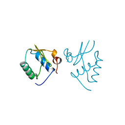
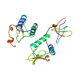
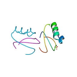
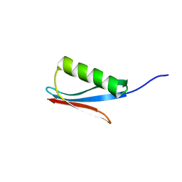
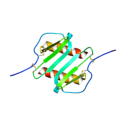
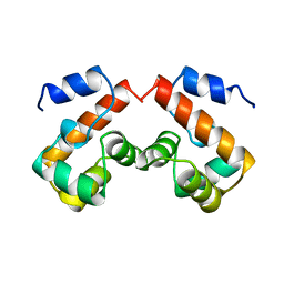
SOLUTION STRUCTURE OF HUMAN BARRIER-TO-AUTOINTEGRATION FACTOR BAF, NMR, REGULARIZED MEAN STRUCTURE PLUS 20 INDIVIDUAL SIMULATED ANNEALING STRUCTURES
Descriptor: PROTEIN (BARRIER-TO-AUTOINTEGRATION FACTOR)
Authors:Clore, G.M.
Deposit date:1999-05-06
Release date:1999-06-23
Last modified:2023-12-27
Method:SOLUTION NMR
Cite:IMPROVING THE PACKING AND ACCURACY OF NMR STRUCTURES WITH A PSEUDOPOTENTIAL FOR THE RADIUS OF GYRATION
J.Am.Chem.Soc., 121, 1999
1AVV
DownloadVisualize
BU of 1avv by Molmil
HIV-1 NEF PROTEIN, UNLIGANDED CORE DOMAIN
Descriptor: NEGATIVE FACTOR
Authors:Arold, S, Franken, P, Dumas, C.
Deposit date:1997-09-21
Release date:1998-03-25
Last modified:2023-08-02
Method:X-RAY DIFFRACTION (3 Å)
Cite:The crystal structure of HIV-1 Nef protein bound to the Fyn kinase SH3 domain suggests a role for this complex in altered T cell receptor signaling.
Structure, 5, 1997
1TWO
DownloadVisualize
BU of 1two by Molmil
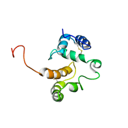
NMR structure of the pheromone binding protein from Antheraea polyphemus at acidic pH
Descriptor: Pheromone-binding protein
Authors:Mohanty, S, Zubkov, S.
Deposit date:2004-07-01
Release date:2005-10-25
Last modified:2022-03-02
Method:SOLUTION NMR
Cite:Structural Consequences of the pH-induced Conformational Switch in A.polyphemus Pheromone-binding Protein: Mechanisms of Ligand Release
J.Mol.Biol., 354, 2005
3EST
DownloadVisualize
BU of 3est by Molmil
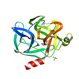
STRUCTURE OF NATIVE PORCINE PANCREATIC ELASTASE AT 1.65 ANGSTROMS RESOLUTION
Descriptor: CALCIUM ION, PORCINE PANCREATIC ELASTASE, SULFATE ION
Authors:Meyer, E.F, Cole, G, Radhakrishnan, R, Epp, O.
Deposit date:1987-09-17
Release date:1988-01-16
Last modified:2011-07-13
Method:X-RAY DIFFRACTION (1.65 Å)
Cite:Structure of native porcine pancreatic elastase at 1.65 A resolutions.
Acta Crystallogr.,Sect.B, 44, 1988
3EZM
DownloadVisualize
BU of 3ezm by Molmil
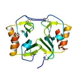
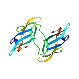
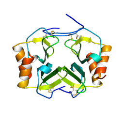
CYANOVIRIN-N
Descriptor: PROTEIN (CYANOVIRIN-N)
Authors:Yang, F, Wlodawer, A.
Deposit date:1998-12-15
Release date:1998-12-23
Last modified:2023-09-06
Method:X-RAY DIFFRACTION (1.5 Å)
Cite:Crystal structure of cyanovirin-N, a potent HIV-inactivating protein, shows unexpected domain swapping.
J.Mol.Biol., 288, 1999
2SXL
DownloadVisualize
BU of 2sxl by Molmil
SEX-LETHAL RBD1, NMR, MINIMIZED AVERAGE STRUCTURE
Descriptor: SEX-LETHAL PROTEIN
Authors:Inoue, M, Muto, Y, Sakamoto, H, Kigawa, T, Takio, K, Shimura, Y, Yokoyama, S, RIKEN Structural Genomics/Proteomics Initiative (RSGI)
Deposit date:1997-07-16
Release date:1998-07-22
Last modified:2021-11-03
Method:SOLUTION NMR
Cite:A characteristic arrangement of aromatic amino acid residues in the solution structure of the amino-terminal RNA-binding domain of Drosophila sex-lethal.
J.Mol.Biol., 272, 1997
3J3Y
DownloadVisualize
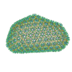
BU of 3j3y by Molmil
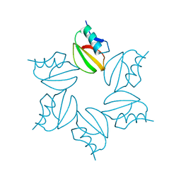
Atomic-level structure of the entire HIV-1 capsid (186 hexamers + 12 pentamers)
Descriptor: capsid protein
Authors:Perilla, J.R, Zhao, G, Zhang, P, Schulten, K.J.
Deposit date:2013-05-06
Release date:2013-05-29
Last modified:2018-07-18
Method:ELECTRON MICROSCOPY
Cite:Mature HIV-1 capsid structure by cryo-electron microscopy and all-atom molecular dynamics.
Nature, 497, 2013
3J4F
DownloadVisualize
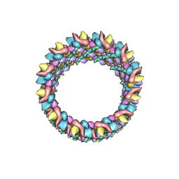
BU of 3j4f by Molmil
Structure of HIV-1 capsid protein by cryo-EM
Descriptor: capsid protein
Authors:Zhao, G, Perilla, J.R, Meng, X, Schulten, K, Zhang, P.
Deposit date:2013-07-11
Release date:2013-07-24
Last modified:2018-07-18
Method:ELECTRON MICROSCOPY (8.6 Å)
Cite:Mature HIV-1 capsid structure by cryo-electron microscopy and all-atom molecular dynamics.
Nature, 497, 2013
3J3Q
DownloadVisualize
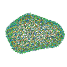
BU of 3j3q by Molmil
Atomic-level structure of the entire HIV-1 capsid
Descriptor: capsid protein
Authors:Perilla, J.R, Zhao, G, Zhang, P, Schulten, K.J.
Deposit date:2013-04-12
Release date:2013-05-29
Last modified:2018-07-18
Method:ELECTRON MICROSCOPY
Cite:Mature HIV-1 capsid structure by cryo-electron microscopy and all-atom molecular dynamics.
Nature, 497, 2013
1AVZ
DownloadVisualize
BU of 1avz by Molmil
V-1 NEF PROTEIN IN COMPLEX WITH WILD TYPE FYN SH3 DOMAIN
Descriptor: FYN TYROSINE KINASE, NEGATIVE FACTOR
Authors:Arold, S, Franken, P, Dumas, C.
Deposit date:1997-09-23
Release date:1998-03-25
Last modified:2023-08-02
Method:X-RAY DIFFRACTION (3 Å)
Cite:The crystal structure of HIV-1 Nef protein bound to the Fyn kinase SH3 domain suggests a role for this complex in altered T cell receptor signaling.
Structure, 5, 1997
1NAP
DownloadVisualize
BU of 1nap by Molmil
THE CRYSTAL STRUCTURE OF RECOMBINANT HUMAN NEUTROPHIL-ACTIVATING PEPTIDE-2 (M6L) AT 1.9-ANGSTROMS RESOLUTION
Descriptor: NEUTROPHIL ACTIVATING PEPTIDE-2
Authors:Malkowski, M.G, Edwards, B.F.P.
Deposit date:1994-12-19
Release date:1995-12-19
Last modified:2019-08-14
Method:X-RAY DIFFRACTION (1.9 Å)
Cite:The crystal structure of recombinant human neutrophil-activating peptide-2 (M6L) at 1.9-A resolution.
J.Biol.Chem., 270, 1995
1PGA
DownloadVisualize
BU of 1pga by Molmil
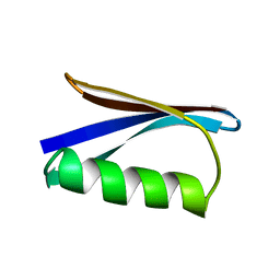
TWO CRYSTAL STRUCTURES OF THE B1 IMMUNOGLOBULIN-BINDING DOMAIN OF STREPTOCOCCAL PROTEIN G AND COMPARISON WITH NMR
Descriptor: PROTEIN G
Authors:Gallagher, T, Alexander, P, Bryan, P, Gilliland, G.L.
Deposit date:1993-11-23
Release date:1994-04-30
Last modified:2024-02-14
Method:X-RAY DIFFRACTION (2.07 Å)
Cite:Two crystal structures of the B1 immunoglobulin-binding domain of streptococcal protein G and comparison with NMR.
Biochemistry, 33, 1994
1PGB
DownloadVisualize
BU of 1pgb by Molmil
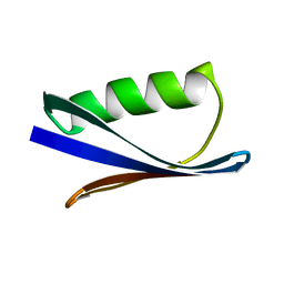
TWO CRYSTAL STRUCTURES OF THE B1 IMMUNOGLOBULIN-BINDING DOMAIN OF STREPTOCCOCAL PROTEIN G AND COMPARISON WITH NMR
Descriptor: PROTEIN G
Authors:Gallagher, T, Alexander, P, Bryan, P, Gilliland, G.L.
Deposit date:1993-11-23
Release date:1994-04-30
Last modified:2024-02-14
Method:X-RAY DIFFRACTION (1.92 Å)
Cite:Two crystal structures of the B1 immunoglobulin-binding domain of streptococcal protein G and comparison with NMR.
Biochemistry, 33, 1994
1CQ4
DownloadVisualize
BU of 1cq4 by Molmil
CI2 MUTANT WITH TETRAGLUTAMINE (MGQQQQGM) REPLACING MET59
Descriptor: PROTEIN (SERINE PROTEINASE INHIBITOR 2), SULFATE ION
Authors:Chen, Y.W, Stott, K.R.
Deposit date:1998-11-17
Release date:1998-11-25
Last modified:2023-08-09
Method:X-RAY DIFFRACTION (1.8 Å)
Cite:Crystal structure of a dimeric chymotrypsin inhibitor 2 mutant containing an inserted glutamine repeat.
Proc.Natl.Acad.Sci.USA, 96, 1999
1IOB
DownloadVisualize
BU of 1iob by Molmil
INTERLEUKIN-1 BETA FROM JOINT X-RAY AND NMR REFINEMENT
Descriptor: INTERLEUKIN-1 BETA
Authors:Shaanan, B, Clore, G.M.
Deposit date:1996-03-14
Release date:1996-08-17
Last modified:2011-07-13
Method:SOLUTION NMR (2 Å), X-RAY DIFFRACTION
Cite:Combining experimental information from crystal and solution studies: joint X-ray and NMR refinement.
Science, 257, 1992
1IGC
DownloadVisualize
BU of 1igc by Molmil
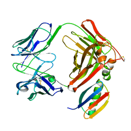
IGG1 FAB FRAGMENT (MOPC21) COMPLEX WITH DOMAIN III OF PROTEIN G FROM STREPTOCOCCUS
Descriptor: IGG1-KAPPA MOPC21 FAB (HEAVY CHAIN), IGG1-KAPPA MOPC21 FAB (LIGHT CHAIN), STREPTOCOCCAL PROTEIN G (DOMAIN III)
Authors:Derrick, J.P, Wigley, D.B.
Deposit date:1994-08-05
Release date:1995-06-03
Last modified:2011-07-13
Method:X-RAY DIFFRACTION (2.6 Å)
Cite:The third IgG-binding domain from streptococcal protein G. An analysis by X-ray crystallography of the structure alone and in a complex with Fab.
J.Mol.Biol., 243, 1994
1J4V
DownloadVisualize
BU of 1j4v by Molmil
CYANOVIRIN-N
Descriptor: CYANOVIRIN-N
Authors:Clore, G.M, Bewley, C.A.
Deposit date:2001-11-21
Release date:2002-03-06
Last modified:2023-12-27
Method:SOLUTION NMR
Cite:Using conjoined rigid body/torsion angle simulated annealing to determine the relative orientation of covalently linked protein domains from dipolar couplings.
J.Magn.Reson., 154, 2002
1IGD
DownloadVisualize
BU of 1igd by Molmil
THE THIRD IGG-BINDING DOMAIN FROM STREPTOCOCCAL PROTEIN G: AN ANALYSIS BY X-RAY CRYSTALLOGRAPHY OF THE STRUCTURE ALONE AND IN A COMPLEX WITH FAB
Descriptor: PROTEIN G
Authors:Derrick, J.P, Wigley, D.B.
Deposit date:1994-08-05
Release date:1994-11-01
Last modified:2024-02-07
Method:X-RAY DIFFRACTION (1.1 Å)
Cite:The third IgG-binding domain from streptococcal protein G. An analysis by X-ray crystallography of the structure alone and in a complex with Fab.
J.Mol.Biol., 243, 1994
2KLM
DownloadVisualize
BU of 2klm by Molmil
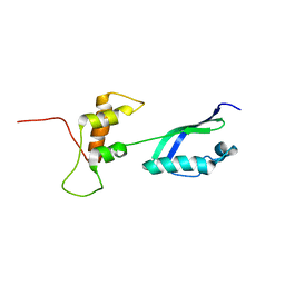
Solution Structure of L11 with SAXS and RDC
Descriptor: 50S ribosomal protein L11
Authors:Wang, J, Zuo, X, Yu, P, Schwieters, C.D, Wang, Y.
Deposit date:2009-07-06
Release date:2009-10-06
Last modified:2022-03-16
Method:SOLUTION NMR, SOLUTION SCATTERING
Cite:Determination of multicomponent protein structures in solution using global orientation and shape restraints.
J.Am.Chem.Soc., 131, 2009
2CI2
DownloadVisualize
BU of 2ci2 by Molmil
CRYSTAL AND MOLECULAR STRUCTURE OF THE SERINE PROTEINASE INHIBITOR CI-2 FROM BARLEY SEEDS
Descriptor: CHYMOTRYPSIN INHIBITOR 2
Authors:Mcphalen, C.A, James, M.N.G.
Deposit date:1988-09-05
Release date:1988-09-07
Last modified:2024-02-14
Method:X-RAY DIFFRACTION (2 Å)
Cite:Crystal and molecular structure of the serine proteinase inhibitor CI-2 from barley seeds.
Biochemistry, 26, 1987
1SXL
DownloadVisualize
BU of 1sxl by Molmil
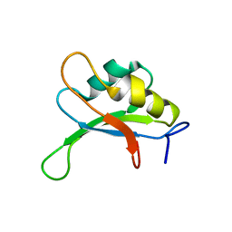
RESONANCE ASSIGNMENTS AND SOLUTION STRUCTURE OF THE SECOND RNA-BINDING DOMAIN OF SEX-LETHAL DETERMINED BY MULTIDIMENSIONAL HETERONUCLEAR MAGNETIC RESONANCE SPECTROSCOPY
Descriptor: SEX-LETHAL PROTEIN PROTEIN
Authors:Lee, A.L, Kanaar, R, Rio, D.C, Wemmer, D.E.
Deposit date:1994-07-01
Release date:1994-09-30
Last modified:2022-03-02
Method:SOLUTION NMR
Cite:Resonance assignments and solution structure of the second RNA-binding domain of sex-lethal determined by multidimensional heteronuclear magnetic resonance.
Biochemistry, 33, 1994
3BVB
DownloadVisualize
BU of 3bvb by Molmil
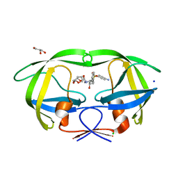
Cystal structure of HIV-1 Active Site Mutant D25N and inhibitor Darunavir
Descriptor: (3R,3AS,6AR)-HEXAHYDROFURO[2,3-B]FURAN-3-YL(1S,2R)-3-[[(4-AMINOPHENYL)SULFONYL](ISOBUTYL)AMINO]-1-BENZYL-2-HYDROXYPROPYLCARBAMATE, CHLORIDE ION, GLYCEROL, ...
Authors:Liu, F, Weber, I.T.
Deposit date:2008-01-05
Release date:2008-04-01
Last modified:2023-08-30
Method:X-RAY DIFFRACTION (1.3 Å)
Cite:Effect of the Active Site D25N Mutation on the Structure, Stability, and Ligand Binding of the Mature HIV-1 Protease.
J.Biol.Chem., 283, 2008
3BVA
DownloadVisualize
BU of 3bva by Molmil
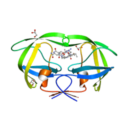
Cystal structure of HIV-1 Active Site Mutant D25N and p2-NC analog inhibitor
Descriptor: GLYCEROL, N-{(2S)-2-[(N-acetyl-L-threonyl-L-isoleucyl)amino]hexyl}-L-norleucyl-L-glutaminyl-N~5~-[amino(iminio)methyl]-L-ornithinamide, Protease (Retropepsin)
Authors:Liu, F, Weber, I.T.
Deposit date:2008-01-05
Release date:2008-04-01
Last modified:2023-08-30
Method:X-RAY DIFFRACTION (1.05 Å)
Cite:Effect of the Active Site D25N Mutation on the Structure, Stability, and Ligand Binding of the Mature HIV-1 Protease.
J.Biol.Chem., 283, 2008
1TVX
DownloadVisualize
BU of 1tvx by Molmil
NEUTROPHIL ACTIVATING PEPTIDE-2 VARIANT FORM M6L WITH FIVE ADDITIONAL AMINO TERMINAL RESIDUES (DSDLY)
Descriptor: NEUTROPHIL ACTIVATING PEPTIDE 2 VARIANT
Authors:Malkowski, M.G, Edwards, B.F.P.
Deposit date:1996-11-05
Release date:1997-01-11
Last modified:2023-08-09
Method:X-RAY DIFFRACTION (1.75 Å)
Cite:The amino-terminal residues in the crystal structure of connective tissue activating peptide-III (des10) block the ELR chemotactic sequence.
J.Mol.Biol., 266, 1997

218853

PDB entries from 2024-04-24

PDB statisticsPDBj update infoContact PDBjnumon