6EW8
 
 | |
6EW7
 | |
5WIA
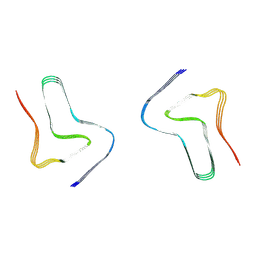
 | | Crystal structure of the segment, GNNSYS, from the low complexity domain of TDP-43, residues 370-375 | | 分子名称: | TAR DNA-binding protein 43 | | 著者 | Guenther, E.L, Trinh, H, Sawaya, M.R, Eisenberg, D.S. | | 登録日 | 2017-07-18 | | 公開日 | 2018-04-25 | | 最終更新日 | 2024-04-03 | | 実験手法 | X-RAY DIFFRACTION (1.002 Å) | | 主引用文献 | Atomic structures of TDP-43 LCD segments and insights into reversible or pathogenic aggregation.
Nat. Struct. Mol. Biol., 25, 2018
|
|
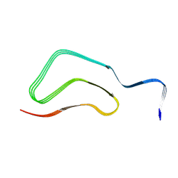
5WKD
 | | Crystal structure of the segment, GNNQGSN, from the low complexity domain of TDP-43, residues 300-306 | | 分子名称: | TAR DNA-binding protein 43 | | 著者 | Guenther, E.L, Trinh, H, Sawaya, M.R, Cascio, D, Eisenberg, D.S. | | 登録日 | 2017-07-25 | | 公開日 | 2018-04-18 | | 最終更新日 | 2024-04-03 | | 実験手法 | X-RAY DIFFRACTION (1.8 Å) | | 主引用文献 | Atomic structures of TDP-43 LCD segments and insights into reversible or pathogenic aggregation.
Nat. Struct. Mol. Biol., 25, 2018
|
|
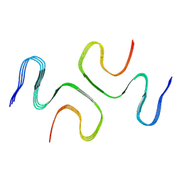
5WHN
 | | Crystal structure of the segment, NFGAFS, from the low complexity domain of TDP-43, residues 312-317 | | 分子名称: | Segment of TAR DNA-binding protein 43 | | 著者 | Guenther, E.L, Sawaya, M.R, Eisenberg, D.S. | | 登録日 | 2017-07-17 | | 公開日 | 2018-04-25 | | 最終更新日 | 2023-10-04 | | 実験手法 | X-RAY DIFFRACTION (1.1 Å) | | 主引用文献 | Atomic structures of TDP-43 LCD segments and insights into reversible or pathogenic aggregation.
Nat. Struct. Mol. Biol., 25, 2018
|
|
5WHP
 | | Crystal structure of the segment, NFGTFS, from the A315T familial variant of the low complexity domain of TDP-43, residues 312-317 | | 分子名称: | Segment of TAR DNA-binding protein 43 | | 著者 | Guenther, E.L, Sawaya, M.R, Eisenberg, D.S. | | 登録日 | 2017-07-17 | | 公開日 | 2018-05-23 | | 最終更新日 | 2024-03-13 | | 実験手法 | X-RAY DIFFRACTION (1 Å) | | 主引用文献 | Atomic structures of TDP-43 LCD segments and insights into reversible or pathogenic aggregation.
Nat. Struct. Mol. Biol., 25, 2018
|
|
5WIQ
 | | Crystal structure of the segment, GFNGGFG, from the low complexity domain of TDP-43, residues 396-402 | | 分子名称: | TAR DNA-binding protein 43 | | 著者 | Guenther, E.L, Sawaya, M.R, Eisenberg, D.S. | | 登録日 | 2017-07-19 | | 公開日 | 2018-04-18 | | 最終更新日 | 2023-10-04 | | 実験手法 | X-RAY DIFFRACTION (1.25 Å) | | 主引用文献 | Atomic structures of TDP-43 LCD segments and insights into reversible or pathogenic aggregation.
Nat. Struct. Mol. Biol., 25, 2018
|
|
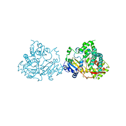
3I6D
 | | Crystal structure of PPO from bacillus subtilis with AF | | 分子名称: | 5-[2-CHLORO-4-(TRIFLUOROMETHYL)PHENOXY]-2-NITROBENZOIC ACID, FLAVIN-ADENINE DINUCLEOTIDE, PHOSPHATE ION, ... | | 著者 | Shen, Y. | | 登録日 | 2009-07-06 | | 公開日 | 2009-12-08 | | 最終更新日 | 2024-03-20 | | 実験手法 | X-RAY DIFFRACTION (2.9 Å) | | 主引用文献 | Structural insight into unique properties of protoporphyrinogen oxidase from Bacillus subtilis
J.Struct.Biol., 170, 2010
|
|
7SP1
 | | RNA-induced tau amyloid fibril | | 分子名称: | Isoform Tau-F of Microtubule-associated protein tau, RNA (5'-R(*AP*AP*AP*AP*AP*AP*AP*AP*AP*A)-3') | | 著者 | Abskharon, R, Sawaya, M.R, Boyer, D.R, Eisenberg, D.S. | | 登録日 | 2021-11-02 | | 公開日 | 2022-03-30 | | 最終更新日 | 2022-06-22 | | 実験手法 | ELECTRON MICROSCOPY (3.4 Å) | | 主引用文献 | Cryo-EM structure of RNA-induced tau fibrils reveals a small C-terminal core that may nucleate fibril formation.
Proc.Natl.Acad.Sci.USA, 119, 2022
|
|
3NR2
 | | Crystal structure of Caspase-6 zymogen | | 分子名称: | Caspase-6 | | 著者 | Su, X.-D, Wang, X.-J, Liu, X, Mi, W, Wang, K.-T. | | 登録日 | 2010-06-30 | | 公開日 | 2010-10-27 | | 最終更新日 | 2023-11-01 | | 実験手法 | X-RAY DIFFRACTION (2.9 Å) | | 主引用文献 | Crystal structures of human caspase 6 reveal a new mechanism for intramolecular cleavage self-activation
Embo Rep., 11, 2010
|
|
5G5J
 | |
8H03
 | |
8H04
 | |
8H05
 | |
5WKB
 | | MicroED structure of the segment, NFGEFS, from the A315E familial variant of the low complexity domain of TDP-43, residues 312-317 | | 分子名称: | TAR DNA-binding protein 43 | | 著者 | Guenther, E.L, Sawaya, M.R, Cascio, D, Eisenberg, D.S. | | 登録日 | 2017-07-24 | | 公開日 | 2018-05-23 | | 最終更新日 | 2024-03-13 | | 実験手法 | ELECTRON CRYSTALLOGRAPHY (1 Å) | | 主引用文献 | Atomic structures of TDP-43 LCD segments and insights into reversible or pathogenic aggregation.
Nat. Struct. Mol. Biol., 25, 2018
|
|
7E0F
 | |
7V47
 | |
7V49
 | |
7V48
 | |
7XO2
 | |
7XO3
 | |
7XO0
 | |
7XO1
 | |
7VQQ
 | |


