9BHR
 
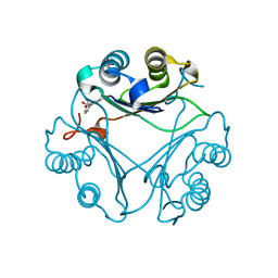
 | | Crystal structure of the WDR domain of human DCAF1 in complex with OICR-40155 compound | | 分子名称: | (4P)-N-[(1S)-3-amino-1-(3-chloro-4-fluorophenyl)-3-oxopropyl]-4-(4-chloro-2-fluorophenyl)-5-{(1E)-3-[(2-methoxyethyl)amino]-3-oxoprop-1-en-1-yl}-1H-pyrrole-3-carboxamide, DDB1- and CUL4-associated factor 1 | | 著者 | kimani, S, Dong, A, Li, Y, Seitova, A, Al-Awar, R, Krausser, C, Wilson, B, Ackloo, S, Arrowsmith, C.H, Edwards, A.M, Halabelian, L, Structural Genomics Consortium (SGC) | | 登録日 | 2024-04-21 | | 公開日 | 2024-05-08 | | 実験手法 | X-RAY DIFFRACTION (1.62 Å) | | 主引用文献 | Crystal structure of the WDR domain of human DCAF1 in complex with OICR-40155 compound
To be published
|
|
8VLB
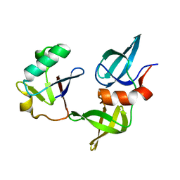
 | | Crystal structure of EloBC-VHL-CDO1 complex bound to compound 4 molecular glue | | 分子名称: | CITRIC ACID, Cysteine dioxygenase type 1, Elongin-B, ... | | 著者 | Shu, W, Ma, X, Tutter, A, Buckley, D, Golosov, A, Michaud, G. | | 登録日 | 2024-01-11 | | 公開日 | 2024-03-13 | | 実験手法 | X-RAY DIFFRACTION (2.9 Å) | | 主引用文献 | A small molecule VHL molecular glue degrader for cysteine dioxygenase 1
To Be Published
|
|
8VL9
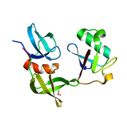
 | | Crystal structure of EloBC-VHL-CDO1 complex bound to compound 8 molecular glue | | 分子名称: | CITRIC ACID, Cysteine dioxygenase type 1, Elongin-B, ... | | 著者 | Shu, W, Ma, X, Tutter, A, Buckley, D, Golosov, A, Michaud, G. | | 登録日 | 2024-01-11 | | 公開日 | 2024-03-13 | | 実験手法 | X-RAY DIFFRACTION (2.5 Å) | | 主引用文献 | A small molecule VHL molecular glue degrader for cysteine dioxygenase 1
To Be Published
|
|
9BCZ
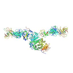
 | | Chicken 1-phosphatidylinositol 4,5-bisphosphate phosphodiesterase zeta-1 (PLCZ1) in complex with calcium and phosphorylated threonine | | 分子名称: | 1-phosphatidylinositol 4,5-bisphosphate phosphodiesterase zeta-1, 4-(2-HYDROXYETHYL)-1-PIPERAZINE ETHANESULFONIC ACID, CALCIUM ION, ... | | 著者 | Edwards, M.M, Dong, A, Theo-Emegano, N, Seitova, A, Loppnau, P, Leung, R, Li, H, Ilyassov, O, Edwards, A.M, Arrowsmith, C.H, Structural Genomics Consortium, Structural Genomics Consortium (SGC) | | 登録日 | 2024-04-10 | | 公開日 | 2024-05-15 | | 実験手法 | X-RAY DIFFRACTION (1.99 Å) | | 主引用文献 | Chicken 1-phosphatidylinositol 4,5-bisphosphate phosphodiesterase zeta-1 (PLCZ1) in complex with calcium and phosphorylated threonine
To be published
|
|
8VDY
 | | Crystal Structure of Delta 114-117 D-Dopachrome Tautomerase (D-DT) | | 分子名称: | D-dopachrome decarboxylase | | 著者 | Parkins, A, Pilien, A, Wolff, A, Thompson, M.C, Pantouris, G. | | 登録日 | 2023-12-18 | | 公開日 | 2024-05-01 | | 最終更新日 | 2024-05-22 | | 実験手法 | X-RAY DIFFRACTION (2.44 Å) | | 主引用文献 | The C-terminal Region of D-DT Regulates Molecular Recognition for Protein-Ligand Complexes.
J.Med.Chem., 67, 2024
|
|
8VFK
 | | Crystal Structure of Delta 109-117 D-Dopachrome Tautomerase (D-DT) | | 分子名称: | CITRATE ANION, D-dopachrome decarboxylase, SODIUM ION | | 著者 | Parkins, A, Pilien, A, Wolff, A, Thompson, M.C, Pantouris, G. | | 登録日 | 2023-12-21 | | 公開日 | 2024-05-01 | | 最終更新日 | 2024-05-22 | | 実験手法 | X-RAY DIFFRACTION (1.59 Å) | | 主引用文献 | The C-terminal Region of D-DT Regulates Molecular Recognition for Protein-Ligand Complexes.
J.Med.Chem., 67, 2024
|
|
8VFN
 | | Crystal Structure of WT D-Dopachrome Tautomerase (D-DT) at 310K | | 分子名称: | D-dopachrome decarboxylase | | 著者 | Parkins, A, Pilien, A, Wolff, A, Thompson, M.C, Pantouris, G. | | 登録日 | 2023-12-21 | | 公開日 | 2024-05-01 | | 最終更新日 | 2024-05-22 | | 実験手法 | X-RAY DIFFRACTION (1.29 Å) | | 主引用文献 | The C-terminal Region of D-DT Regulates Molecular Recognition for Protein-Ligand Complexes.
J.Med.Chem., 67, 2024
|
|
8VG7
 | | Crystal Structure of T115A Variant of D-Dopachrome Tautomerase (D-DT) | | 分子名称: | D-dopachrome decarboxylase | | 著者 | Parkins, A, Pilien, A, Wolff, A, Thompson, M.C, Pantouris, G. | | 登録日 | 2023-12-23 | | 公開日 | 2024-05-01 | | 最終更新日 | 2024-05-22 | | 実験手法 | X-RAY DIFFRACTION (1.24 Å) | | 主引用文献 | The C-terminal Region of D-DT Regulates Molecular Recognition for Protein-Ligand Complexes.
J.Med.Chem., 67, 2024
|
|
8VFL
 | | Crystal Structure of WT D-Dopachrome Tautomerase (D-DT) at 290K | | 分子名称: | D-dopachrome decarboxylase | | 著者 | Parkins, A, Pilien, A, Wolff, A, Thompson, M.C, Pantouris, G. | | 登録日 | 2023-12-21 | | 公開日 | 2024-05-01 | | 最終更新日 | 2024-05-22 | | 実験手法 | X-RAY DIFFRACTION (1.23 Å) | | 主引用文献 | The C-terminal Region of D-DT Regulates Molecular Recognition for Protein-Ligand Complexes.
J.Med.Chem., 67, 2024
|
|
8VG8
 | | Crystal Structure of T115A Variant of D-Dopachrome Tautomerase (D-DT) Bound to 4CPPC | | 分子名称: | 4-(3-carboxyphenyl)pyridine-2,5-dicarboxylic acid, D-dopachrome decarboxylase | | 著者 | Parkins, A, Pilien, A, Wolff, A, Thompson, M.C, Pantouris, G. | | 登録日 | 2023-12-23 | | 公開日 | 2024-05-01 | | 最終更新日 | 2024-05-22 | | 実験手法 | X-RAY DIFFRACTION (1.33 Å) | | 主引用文献 | The C-terminal Region of D-DT Regulates Molecular Recognition for Protein-Ligand Complexes.
J.Med.Chem., 67, 2024
|
|
8SVG
 | | Ubiquitin variant i53 in complex with 53BP1 Tudor domain | | 分子名称: | Tumor protein p53 binding protein 1, Ubiquitin variant i53 | | 著者 | Holden, J.K, Partridge, J.R, Wibowo, A.S, Mulichak, A. | | 登録日 | 2023-05-16 | | 公開日 | 2024-03-27 | | 最終更新日 | 2024-04-03 | | 実験手法 | X-RAY DIFFRACTION (1.21 Å) | | 主引用文献 | Functional screening in human HSPCs identifies optimized protein-based enhancers of Homology Directed Repair.
Nat Commun, 15, 2024
|
|
8SVI
 | | Ubiquitin variant i53:Mutant L67H with 53BP1 Tudor domain | | 分子名称: | GLYCEROL, Tumor protein p53 binding protein 1, Ubiquitin Variant i53: Mutant L67H | | 著者 | Partridge, J.R, Holden, J.K, Wibowo, A.S, Mulichak, A. | | 登録日 | 2023-05-16 | | 公開日 | 2024-03-27 | | 最終更新日 | 2024-04-03 | | 実験手法 | X-RAY DIFFRACTION (1.15 Å) | | 主引用文献 | Functional screening in human HSPCs identifies optimized protein-based enhancers of Homology Directed Repair.
Nat Commun, 15, 2024
|
|
8TNT
 | | Crystal structure of Epstein-Barr virus gH/gL/gp42 in complex with antibodies F-2-1 and 769C2 | | 分子名称: | 2-acetamido-2-deoxy-beta-D-glucopyranose, 769C2 heavy chain, 769C2 light chain, ... | | 著者 | Bu, W, Kumar, A, Board, N, Kim, J, Dowdell, K, Zhang, S, Lei, Y, Hostal, A, Krogmann, T, Wang, Y, Pittaluga, S, Marcotrigiano, J, Cohen, J.I. | | 登録日 | 2023-08-02 | | 公開日 | 2024-03-27 | | 実験手法 | X-RAY DIFFRACTION (3.15 Å) | | 主引用文献 | Epstein-Barr virus gp42 antibodies reveal sites of vulnerability for receptor binding and fusion to B cells.
Immunity, 57, 2024
|
|
8T2D
 | | Ubiquitin variant i53:Mutant T12Y.T14E.L67R with 53BP1 Tudor domain | | 分子名称: | Tumor protein p53 binding protein 1, Ubiquitin variant i53 | | 著者 | Partridge, J.R, Holden, J.K, Wibowo, A.S, Mulichak, A. | | 登録日 | 2023-06-05 | | 公開日 | 2024-03-27 | | 最終更新日 | 2024-04-03 | | 実験手法 | X-RAY DIFFRACTION (1.751 Å) | | 主引用文献 | Functional screening in human HSPCs identifies optimized protein-based enhancers of Homology Directed Repair.
Nat Commun, 15, 2024
|
|
8SVH
 | | Ubiquitin variant i53 mutant L67R bound to 53BP1 Tudor Domain | | 分子名称: | Tumor protein p53 binding protein 1, Ubiquitin variant i53: mutant L67R | | 著者 | Holden, J.K, Partridge, J.R, Wibowo, A.S, Mulichak, A. | | 登録日 | 2023-05-16 | | 公開日 | 2024-03-27 | | 最終更新日 | 2024-04-03 | | 実験手法 | X-RAY DIFFRACTION (1.16 Å) | | 主引用文献 | Functional screening in human HSPCs identifies optimized protein-based enhancers of Homology Directed Repair.
Nat Commun, 15, 2024
|
|
8TOO
 | | Crystal structure of Epstein-Barr virus gp42 in complex with antibody 4C12 | | 分子名称: | 4C12 heavy chain, 4C12 light chain, Glycoprotein 42 | | 著者 | Bu, W, Kumar, A, Board, N, Kim, J, Dowdell, K, Zhang, S, Lei, Y, Hostal, A, Krogmann, T, Wang, Y, Pittaluga, S, Marcotrigiano, J, Cohen, J.I. | | 登録日 | 2023-08-03 | | 公開日 | 2024-03-27 | | 実験手法 | X-RAY DIFFRACTION (2.6 Å) | | 主引用文献 | Epstein-Barr virus gp42 antibodies reveal sites of vulnerability for receptor binding and fusion to B cells.
Immunity, 57, 2024
|
|
8TXA
 | | Apo Structure of (N1G37) tRNA Methyltransferase from Mycobacterium marinum | | 分子名称: | tRNA (guanine-N(1)-)-methyltransferase | | 著者 | Balsamo, A, Bruno, C, Edele, C, Fabian, T, Jannotta, R, Lee, R, Schryver, D, Warsaw, J, Warsaw, L, Stojanoff, V, Battaile, K, Perez, A, Bolen, R. | | 登録日 | 2023-08-23 | | 公開日 | 2024-04-10 | | 実験手法 | X-RAY DIFFRACTION (1.591 Å) | | 主引用文献 | Apo Structure of (N1G37) tRNA Methyltransferase from Mycobacterium marinum
To Be Published
|
|
8TNN
 | | Crystal structure of Epstein-Barr virus gH/gL/gp42 in complex with gp42 antibody A10 | | 分子名称: | 2-acetamido-2-deoxy-beta-D-glucopyranose, A10 heavy chain, A10 light chain, ... | | 著者 | Bu, W, Kumar, A, Board, N, Kim, J, Dowdell, K, Zhang, S, Lei, Y, Hostal, A, Krogmann, T, Wang, Y, Pittaluga, S, Marcotrigiano, J, Cohen, J.I. | | 登録日 | 2023-08-02 | | 公開日 | 2024-03-27 | | 最終更新日 | 2024-05-22 | | 実験手法 | X-RAY DIFFRACTION (3.36 Å) | | 主引用文献 | Epstein-Barr virus gp42 antibodies reveal sites of vulnerability for receptor binding and fusion to B cells.
Immunity, 57, 2024
|
|
8SVJ
 | | Ubiquitin variant i53: mutant VHH with 53BP1 Tudor domain | | 分子名称: | GLYCEROL, Tumor protein p53 binding protein 1, Ubiquitin varient i53 mutant VHH | | 著者 | Holden, J, Partridge, J.R, Wibowo, A.S, Mulichak, A. | | 登録日 | 2023-05-16 | | 公開日 | 2024-03-27 | | 最終更新日 | 2024-04-03 | | 実験手法 | X-RAY DIFFRACTION (1.5 Å) | | 主引用文献 | Functional screening in human HSPCs identifies optimized protein-based enhancers of Homology Directed Repair.
Nat Commun, 15, 2024
|
|
8SUO
 | | BA.2/AZD1061/AZD3152 structure analysis | | 分子名称: | AZD1061 heavy chain, AZD1061 light chain, AZD3152 heavy chain, ... | | 著者 | Oganesyan, V, van Dyk, N, Dippel, A, Barnes, A, O'Connor, E. | | 登録日 | 2023-05-12 | | 公開日 | 2024-05-15 | | 実験手法 | X-RAY DIFFRACTION (3.3 Å) | | 主引用文献 | X-ray crystal structure of BA.2 RBD bound by two neutralizing antibodies
To Be Published
|
|
8WJY
 | | PKMYT1_Cocrystal_Cpd 4 | | 分子名称: | DIMETHYL SULFOXIDE, GLYCEROL, Membrane-associated tyrosine- and threonine-specific cdc2-inhibitory kinase, ... | | 著者 | Wang, Y, Wang, C, Liu, T, Qi, H, Chen, S, Cai, X, Zhang, M, Aliper, A, Ren, F, Ding, X, Zhavoronkov, A. | | 登録日 | 2023-09-26 | | 公開日 | 2023-11-22 | | 実験手法 | X-RAY DIFFRACTION (1.88 Å) | | 主引用文献 | PKMYT1_Cocrystal_Cpd 4
To Be Published
|
|
8WO8
 | | Crystal Structure of an RNA-binding protein, FAU-1, from Pyrococcus furiosus | | 分子名称: | Probable ribonuclease FAU-1, RNA (5'-R(P*AP*UP*A)-3') | | 著者 | Kawai, G, Okada, K, Baba, S, Sato, A, Sakamoto, T, Kanai, A. | | 登録日 | 2023-10-06 | | 公開日 | 2024-02-14 | | 実験手法 | X-RAY DIFFRACTION (2.78 Å) | | 主引用文献 | Homo-trimeric structure of the ribonuclease for rRNA processing, FAU-1, from Pyrococcus furiosus.
J.Biochem., 2024
|
|
8UPT
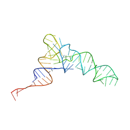 | | Candidatus Methanomethylophilus alvus tRNAPyl in A-site of ribosome | | 分子名称: | RNA (71-MER) | | 著者 | Krahn, N, Zhang, J, Melnikov, S.V, Tharp, J.M, Villa, A, Patel, A, Howard, R.J, Gabir, H, Patel, T.R, Stetefeld, J, Puglisi, J, Soll, D. | | 登録日 | 2023-10-23 | | 公開日 | 2024-01-10 | | 最終更新日 | 2024-02-07 | | 実験手法 | ELECTRON MICROSCOPY (2.8 Å) | | 主引用文献 | tRNA shape is an identity element for an archaeal pyrrolysyl-tRNA synthetase from the human gut.
Nucleic Acids Res., 52, 2024
|
|
8WAG
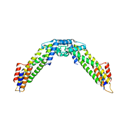 | | Crystal structure of the C-terminal fragment (residues 716-982) of Arabidopsis thaliana CHUP1 | | 分子名称: | Protein CHUP1, chloroplastic | | 著者 | Shimada, A, Nakamura, Y, Takano, A, Kohda, D. | | 登録日 | 2023-09-07 | | 公開日 | 2024-01-17 | | 最終更新日 | 2024-04-10 | | 実験手法 | X-RAY DIFFRACTION (3.003 Å) | | 主引用文献 | CHLOROPLAST UNUSUAL POSITIONING 1 is a plant-specific actin polymerization factor regulating chloroplast movement.
Plant Cell, 36, 2024
|
|
8WAF
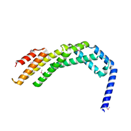 | | Crystal structure of the C-terminal fragment (residues 756-982 with the C864S mutation) of Arabidopsis thaliana CHUP1 | | 分子名称: | Protein CHUP1, chloroplastic | | 著者 | Shimada, A, Takano, A, Nakamura, Y, Kohda, D. | | 登録日 | 2023-09-07 | | 公開日 | 2024-01-17 | | 最終更新日 | 2024-04-10 | | 実験手法 | X-RAY DIFFRACTION (2.8 Å) | | 主引用文献 | CHLOROPLAST UNUSUAL POSITIONING 1 is a plant-specific actin polymerization factor regulating chloroplast movement.
Plant Cell, 36, 2024
|
|


