Loading
PDBj
MenuPDBj@FacebookPDBj@TwitterPDBj@YouTubewwPDB FoundationwwPDB
RCSB PDBPDBeBMRBAdv. SearchSearch help
Search by PDB author
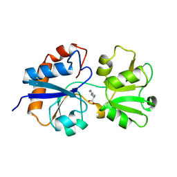
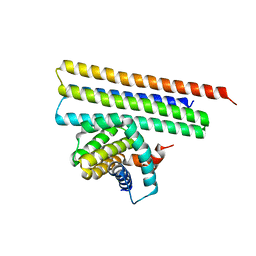
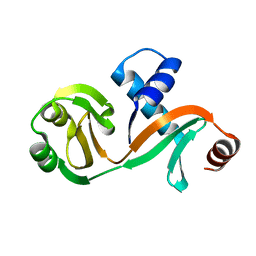
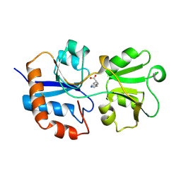
1LAG
DownloadVisualize
BU of 1lag by Molmil
STRUCTURAL BASES FOR MULTIPLE LIGAND SPECIFICITY OF THE PERIPLASMIC LYSINE-, ARGININE-, ORNITHINE-BINDING PROTEIN
Descriptor: HISTIDINE, LYSINE, ARGININE, ...
Authors:Kim, S.-H, Oh, B.-H.
Deposit date:1993-10-06
Release date:1995-07-10
Last modified:2017-11-29
Method:X-RAY DIFFRACTION (2.06 Å)
Cite:Structural basis for multiple ligand specificity of the periplasmic lysine-, arginine-, ornithine-binding protein.
J.Biol.Chem., 269, 1994
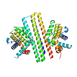
1LST
DownloadVisualize
BU of 1lst by Molmil
THREE-DIMENSIONAL STRUCTURES OF THE PERIPLASMIC LYSINE-, ARGININE-, ORNITHINE-BINDING PROTEIN WITH AND WITHOUT A LIGAND
Descriptor: LYSINE, ARGININE, ORNITHINE-BINDING PROTEIN
Authors:Kim, S.-H, Oh, B.-H.
Deposit date:1993-02-25
Release date:1994-06-22
Last modified:2017-11-29
Method:X-RAY DIFFRACTION (1.8 Å)
Cite:Three-dimensional structures of the periplasmic lysine/arginine/ornithine-binding protein with and without a ligand.
J.Biol.Chem., 268, 1993
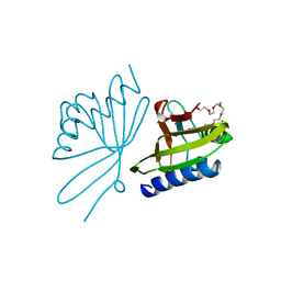
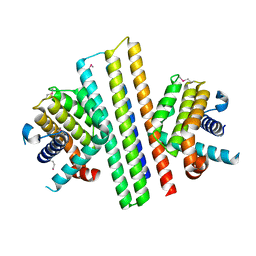
8CHO
DownloadVisualize
BU of 8cho by Molmil
CRYSTAL STRUCTURE OF DELTA5-3-KETOSTEROID ISOMERASE FROM PSEUDOMONAS TESTOSTERONI
Descriptor: O-ACETALDEHYDYL-HEXAETHYLENE GLYCOL, STEROID DELTA-ISOMERASE
Authors:Cho, H.-S, Oh, B.-H.
Deposit date:1998-01-06
Release date:1999-02-02
Last modified:2024-02-14
Method:X-RAY DIFFRACTION (2.3 Å)
Cite:Crystal structure and enzyme mechanism of Delta 5-3-ketosteroid isomerase from Pseudomonas testosteroni.
Biochemistry, 37, 1998
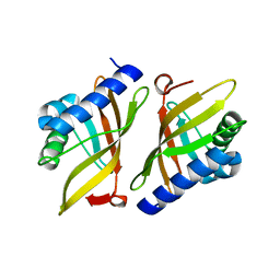
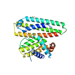
1W01
DownloadVisualize
BU of 1w01 by Molmil
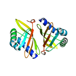
Crystal structure of mutant enzyme Y57F/D103L of ketosteroid isomerase from Pseudomonas putida biotype B
Descriptor: STEROID DELTA-ISOMERASE
Authors:Jang, D.S, Choi, K.Y.
Deposit date:2004-05-30
Release date:2004-07-08
Last modified:2014-02-19
Method:X-RAY DIFFRACTION (2.2 Å)
Cite:Structural Double-Mutant Cycle Analysis of a Hydrogen Bond Network in Ketosteroid Isomerase from Pseudomonas Putida Biotype B.
Biochem.J., 382, 2004
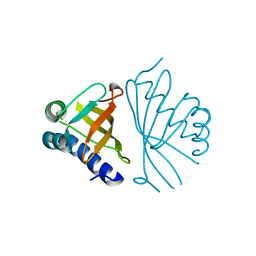
1W02
DownloadVisualize
BU of 1w02 by Molmil
Crystal structure of mutant enzyme Y16F/D103L of ketosteroid isomerase from Pseudomonas putida biotype B
Descriptor: STEROID DELTA-ISOMERASE
Authors:Jang, D.S, Choi, K.Y.
Deposit date:2004-05-30
Release date:2004-07-08
Last modified:2014-02-19
Method:X-RAY DIFFRACTION (2.3 Å)
Cite:Structural Double-Mutant Cycle Analysis of a Hydrogen Bond Network in Ketosteroid Isomerase from Pseudomonas Putida Biotype B.
Biochem.J., 382, 2004
4UW2
DownloadVisualize
BU of 4uw2 by Molmil
Crystal structure of Csm1 in T.onnurineus
Descriptor: CSM1
Authors:Jung, T.Y, An, Y, Park, K.H, Lee, M.H, Oh, B.H, Woo, E.J.
Deposit date:2014-08-08
Release date:2015-03-25
Last modified:2015-09-23
Method:X-RAY DIFFRACTION (2.632 Å)
Cite:Crystal Structure of the Csm1 Subunit of the Csm Complex and its Single-Stranded DNA-Specific Nuclease Activity.
Structure, 23, 2015
2POO
DownloadVisualize
BU of 2poo by Molmil
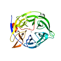
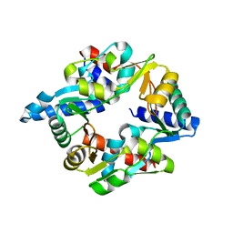
THERMOSTABLE PHYTASE IN FULLY CALCIUM LOADED STATE
Descriptor: CALCIUM ION, PROTEIN (PHYTASE)
Authors:Ha, N.-C, Oh, B.-H.
Deposit date:1999-04-16
Release date:2000-04-19
Last modified:2023-12-27
Method:X-RAY DIFFRACTION (2.05 Å)
Cite:Crystal structures of a novel, thermostable phytase in partially and fully calcium-loaded states.
Nat.Struct.Biol., 7, 2000
1CVM
DownloadVisualize
BU of 1cvm by Molmil
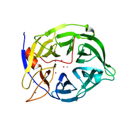
CADMIUM INHIBITED CRYSTAL STRUCTURE OF PHYTASE FROM BACILLUS AMYLOLIQUEFACIENS
Descriptor: CADMIUM ION, CALCIUM ION, PHYTASE
Authors:Shin, S, Ha, N.-C, Oh, B.-H.
Deposit date:1999-08-24
Release date:2000-02-24
Last modified:2024-02-07
Method:X-RAY DIFFRACTION (2.4 Å)
Cite:Crystal structures of a novel, thermostable phytase in partially and fully calcium-loaded states.
Nat.Struct.Biol., 7, 2000
7VT9
DownloadVisualize
BU of 7vt9 by Molmil
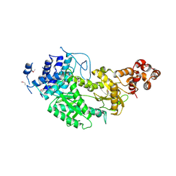
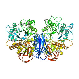
CRYSTAL STRUCTURE AT 3.4 ANGSTROMS RESOLUTION OF Maltodextrin glucosidase, MalZ, FROM Escherichia coli
Descriptor: Maltodextrin glucosidase
Authors:Ahn, W.-C, Ahn, Y, Woo, E.-J.
Deposit date:2021-10-28
Release date:2022-10-26
Last modified:2023-11-29
Method:X-RAY DIFFRACTION (3.3 Å)
Cite:Dimeric architecture of maltodextrin glucosidase (MalZ) provides insights into the substrate recognition and hydrolysis mechanism.
Biochem.Biophys.Res.Commun., 586, 2022
7VYT
DownloadVisualize
BU of 7vyt by Molmil
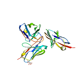
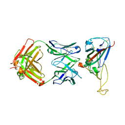
Crystal structure of human TIGIT(23-129) in complex with the scFv fragment of anti-TIGIT antibody MG1131
Descriptor: CITRATE ANION, MG1131 heavy chain variable region, MG1131 light chain variable region, ...
Authors:Jeong, B.-S, Nam, H, Kim, M, Oh, B.-H.
Deposit date:2021-11-15
Release date:2022-03-02
Last modified:2023-11-29
Method:X-RAY DIFFRACTION (1.53 Å)
Cite:Structural and functional characterization of a monoclonal antibody blocking TIGIT.
Mabs, 14, 2022
7CBC
DownloadVisualize
BU of 7cbc by Molmil
Crystal structure of a de novo designed switch protein caging a hemagglutinin binder
Descriptor: De novo designed switch protein caging a hemagglutinin binder (sCageHA267_1S), ETHANOL
Authors:Lee, H, Oh, B.-H, Baker, D.
Deposit date:2020-06-11
Release date:2020-12-23
Last modified:2021-03-31
Method:X-RAY DIFFRACTION (1.99 Å)
Cite:De novo design of modular and tunable protein biosensors.
Nature, 591, 2021
7DG5
DownloadVisualize
BU of 7dg5 by Molmil
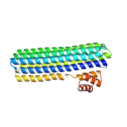
Crystal structure of mouse Smc1-Smc3 hinge domain containing a D574Y mutation
Descriptor: Structural maintenance of chromosomes protein 1A, Structural maintenance of chromosomes protein 3
Authors:Seo, H, Noh, H, Oh, B.-H.
Deposit date:2020-11-11
Release date:2021-11-17
Last modified:2023-11-29
Method:X-RAY DIFFRACTION (2 Å)
Cite:Folding of cohesin's coiled coil is important for Scc2/4-induced association with chromosomes.
Elife, 10, 2021
4YJ4
DownloadVisualize
BU of 4yj4 by Molmil
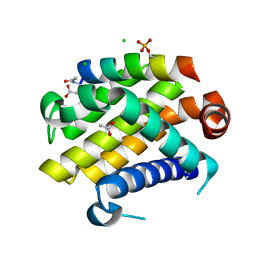
Crystal structure of Bcl-xL in complex with the BIM BH3 domain containing Ile155-to-Arg and Glu158-to-phosphoserine mutations
Descriptor: ACETATE ION, Bcl-2-like protein 1, BIM BH3 domain, ...
Authors:Ku, B, Ha, N.-C, Oh, B.-H.
Deposit date:2015-03-03
Release date:2015-07-29
Last modified:2017-09-27
Method:X-RAY DIFFRACTION (2.1 Å)
Cite:Conversion of cell-survival activity of Akt into apoptotic death of cancer cells by two mutations on the BIM BH3 domain.
Cell Death Dis, 6, 2015
1VZZ
DownloadVisualize
BU of 1vzz by Molmil
CRYSTAL STRUCTURE OF MUTANT ENZYME Y32F/D103L OF KETOSTEROID ISOMERASE FROM PSEUDOMONAS PUTIDA BIOTYPE B
Descriptor: STEROID DELTA-ISOMERASE
Authors:Jang, D.S, Cha, H.J, Choi, K.Y.
Deposit date:2004-05-30
Release date:2004-07-08
Last modified:2023-12-13
Method:X-RAY DIFFRACTION (2.3 Å)
Cite:Structural Double-Mutant Cycle Analysis of a Hydrogen Bond Network in Ketosteroid Isomerase from Pseudomonas Putida Biotype B.
Biochem.J., 382, 2004
7VYR
DownloadVisualize
BU of 7vyr by Molmil
Crystal structure of SARS-CoV-2 Spike RBD in complex with the D27 neutralizing antibody Fab fragment
Descriptor: D27 heavy chain, D27 light chain, Spike protein S1
Authors:Jeong, B.-S, Oh, B.-H.
Deposit date:2021-11-15
Release date:2022-03-02
Last modified:2023-11-29
Method:X-RAY DIFFRACTION (2.2 Å)
Cite:Computational design of a neutralizing antibody with picomolar binding affinity for all concerning SARS-CoV-2 variants.
Mabs, 14, 2022
7OGT
DownloadVisualize
BU of 7ogt by Molmil
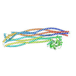
Folded elbow of cohesin
Descriptor: Structural maintenance of chromosomes protein 1, Structural maintenance of chromosomes protein 3
Authors:Lee, B.-G, Gonzalez Llamazares, A, Collier, J, Patele, N.J, Nasmyth, K.A, Lowe, J.
Deposit date:2021-05-07
Release date:2021-07-28
Method:ELECTRON MICROSCOPY (5.5 Å)
Cite:Folding of cohesin's coiled coil is important for Scc2/4-induced association with chromosomes.
Elife, 10, 2021
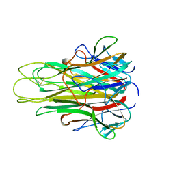
4TSV
DownloadVisualize
BU of 4tsv by Molmil
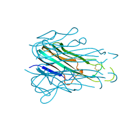
HIGH RESOLUTION CRYSTAL STRUCTURE OF A HUMAN TNF-ALPHA MUTANT
Descriptor: TUMOR NECROSIS FACTOR-ALPHA
Authors:Cha, S.-S, Kim, J.-S, Cho, H.-S, Oh, B.-H.
Deposit date:1997-10-29
Release date:1998-12-30
Last modified:2021-11-03
Method:X-RAY DIFFRACTION (1.8 Å)
Cite:High resolution crystal structure of a human tumor necrosis factor-alpha mutant with low systemic toxicity.
J.Biol.Chem., 273, 1998
7XGE
DownloadVisualize
BU of 7xge by Molmil
Crystal structure of MCL-1 in complex with computationally designed inhibitor protein
Descriptor: BCL-xL and MCL-1 dual binder 2, Induced myeloid leukemia cell differentiation protein Mcl-1
Authors:Oh, B.-H, Kim, S.
Deposit date:2022-04-04
Release date:2022-07-20
Last modified:2024-04-03
Method:X-RAY DIFFRACTION (2.38 Å)
Cite:Computational design of an apoptogenic protein that binds BCL-xL and MCL-1 simultaneously and potently.
Comput Struct Biotechnol J, 20, 2022
7XGF
DownloadVisualize
BU of 7xgf by Molmil
Crystal structure of BCL-xL in complex with computationally designed inhibitor protein
Descriptor: BCL-xL, BCL-xL and MCL-1 dual inhibitor 2
Authors:Oh, B.-H, Kim, S.
Deposit date:2022-04-04
Release date:2022-07-20
Last modified:2024-04-03
Method:X-RAY DIFFRACTION (1.9 Å)
Cite:Computational design of an apoptogenic protein that binds BCL-xL and MCL-1 simultaneously and potently.
Comput Struct Biotechnol J, 20, 2022
7XGG
DownloadVisualize
BU of 7xgg by Molmil
Crystal structure of BCL-xL in complex with computationally designed inhibitor protein
Descriptor: BCL-xL and MCL-1 dual inhibitor, Bcl-2-like protein 1
Authors:Oh, B.-H, Kim, S.
Deposit date:2022-04-04
Release date:2022-07-20
Method:X-RAY DIFFRACTION (1.9 Å)
Cite:Computational design of an apoptogenic protein that binds BCL-xL and MCL-1 simultaneously and potently.
Comput Struct Biotechnol J, 20, 2022
6IWB
DownloadVisualize
BU of 6iwb by Molmil
Crystal structure of a computationally designed protein (LD3) in complex with BCL-2
Descriptor: Apolipoprotein E, Apoptosis regulator Bcl-2,Apoptosis regulator Bcl-2, SULFATE ION
Authors:Kim, S, Kwak, M.J, Oh, B.-H, Correia, B.E, Gainza, P.
Deposit date:2018-12-05
Release date:2019-12-11
Last modified:2024-03-27
Method:X-RAY DIFFRACTION (2.5 Å)
Cite:A computationally designed chimeric antigen receptor provides a small-molecule safety switch for T-cell therapy.
Nat.Biotechnol., 38, 2020
6L7Q
DownloadVisualize
BU of 6l7q by Molmil
Crystal structure of a hypothetical protein PYCH_01220 derived from Pyrococcus yayanosii
Descriptor: hypothetical protein
Authors:Jeon, J.-H, Noh, H, Oh, B.-H.
Deposit date:2019-11-02
Release date:2020-11-04
Last modified:2024-03-27
Method:X-RAY DIFFRACTION (1.16 Å)
Cite:Crystal structure of PYCH_01220 from Pyrococcus yayanosii potentially involved in binding nucleic acid.
Proteins, 89, 2021
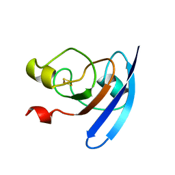
1FXA
DownloadVisualize
BU of 1fxa by Molmil
CRYSTALLIZATION AND STRUCTURE DETERMINATION TO 2.5-ANGSTROMS RESOLUTION OF THE OXIDIZED [2FE-2S] FERREDOXIN ISOLATED FROM ANABAENA 7120
Descriptor: FE2/S2 (INORGANIC) CLUSTER, [2FE-2S] FERREDOXIN
Authors:Rypniewski, W.R, Breiter, D.R, Benning, M.M, Wesenberg, G, Oh, B.-H, Markley, J.L, Rayment, I, Holden, H.M.
Deposit date:1991-01-09
Release date:1992-07-15
Last modified:2024-02-07
Method:X-RAY DIFFRACTION (2.5 Å)
Cite:Crystallization and structure determination to 2.5-A resolution of the oxidized [2Fe-2S] ferredoxin isolated from Anabaena 7120.
Biochemistry, 30, 1991
5TSW
DownloadVisualize
BU of 5tsw by Molmil
HIGH RESOLUTION CRYSTAL STRUCTURE OF A HUMAN TNF-ALPHA MUTANT
Descriptor: PROTEIN (TUMOR NECROSIS FACTOR-ALPHA)
Authors:Cha, S.-S, Kim, J.-S, Cho, H.-S, Oh, B.-H.
Deposit date:1999-04-22
Release date:1999-05-07
Last modified:2023-12-27
Method:X-RAY DIFFRACTION (2.5 Å)
Cite:High resolution crystal structure of a human tumor necrosis factor-alpha mutant with low systemic toxicity.
J.Biol.Chem., 273, 1998
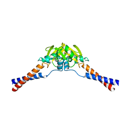
2WMM
DownloadVisualize
BU of 2wmm by Molmil
Crystal structure of the hinge domain of MukB
Descriptor: Chromosome partition protein MukB, D-MALATE
Authors:Ku, B, Oh, B.-H.
Deposit date:2009-07-01
Release date:2010-01-12
Last modified:2018-09-19
Method:X-RAY DIFFRACTION (2.3 Å)
Cite:Crystal structure of the MukB hinge domain with coiled-coil stretches and its functional implications.
Proteins, 78, 2010

218853

PDB entries from 2024-04-24

PDB statisticsPDBj update infoContact PDBjnumon