
Loading
PDBj
MenuPDBj@FacebookPDBj@TwitterPDBj@YouTubewwPDB FoundationwwPDB
RCSB PDBPDBeBMRBAdv. SearchSearch help
Search by PDB author
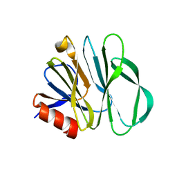
5YMT
DownloadVisualize
BU of 5ymt by Molmil
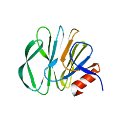
Functional and structural characterization of P[19] rotavirus VP8* interaction with histo-blood group antigens
Descriptor: Outer capsid protein VP4, beta-D-galactopyranose-(1-3)-2-acetamido-2-deoxy-beta-D-glucopyranose-(1-3)-beta-D-galactopyranose-(1-4)-alpha-D-glucopyranose
Authors:Sun, X, Duan, Z.
Deposit date:2017-10-22
Release date:2018-05-02
Last modified:2023-11-22
Method:X-RAY DIFFRACTION (2.199 Å)
Cite:Glycan Binding Specificity and Mechanism of Human and Porcine P[6]/P[19] Rotavirus VP8*s.
J. Virol., 92, 2018
7KXT
DownloadVisualize
BU of 7kxt by Molmil
Crystal structure of human EED
Descriptor: 1-[(4-fluorophenyl)methyl]-N-{1-[2-(4-methoxyphenyl)ethyl]piperidin-4-yl}-1H-benzimidazol-2-amine, Polycomb protein EED, UNKNOWN ATOM OR ION
Authors:Zhu, L, Dong, A, Du, D, Liu, Y, Luo, C, Arrowsmith, C.H, Edwards, A.M, Min, J, Structural Genomics Consortium (SGC)
Deposit date:2020-12-04
Release date:2021-02-24
Last modified:2023-10-18
Method:X-RAY DIFFRACTION (2.15 Å)
Cite:Structure-Guided Development of Small-Molecule PRC2 Inhibitors Targeting EZH2-EED Interaction.
J.Med.Chem., 64, 2021
5YMS
DownloadVisualize
BU of 5yms by Molmil
Structural basis of glycan specificity and identification of a novel glycan binding cavity in human P[19] rotavirus
Descriptor: 2-acetamido-2-deoxy-beta-D-glucopyranose, Outer capsid protein VP4, beta-D-galactopyranose-(1-3)-[2-acetamido-2-deoxy-beta-D-glucopyranose-(1-6)]2-acetamido-2-deoxy-alpha-D-galactopyranose
Authors:Sun, X, Dandi, L, Duan, Z.
Deposit date:2017-10-22
Release date:2018-05-02
Last modified:2023-11-22
Method:X-RAY DIFFRACTION (2.1 Å)
Cite:Glycan Binding Specificity and Mechanism of Human and Porcine P[6]/P[19] Rotavirus VP8*s.
J. Virol., 92, 2018
5GUT
DownloadVisualize
BU of 5gut by Molmil
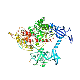
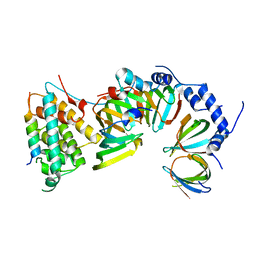
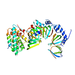
The crystal structure of mouse DNMT1 (731-1602) mutant - N1248A
Descriptor: DNA (cytosine-5)-methyltransferase 1, S-ADENOSYL-L-HOMOCYSTEINE, SULFATE ION, ...
Authors:Chen, S.J, Ye, F.
Deposit date:2016-08-31
Release date:2017-09-06
Last modified:2023-11-08
Method:X-RAY DIFFRACTION (2.099 Å)
Cite:Biochemical Studies and Molecular Dynamic Simulations Reveal the Molecular Basis of Conformational Changes in DNA Methyltransferase-1.
ACS Chem. Biol., 13, 2018
5HSI
DownloadVisualize
BU of 5hsi by Molmil
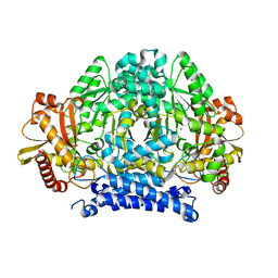
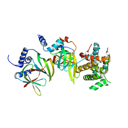
Crystal structure of tyrosine decarboxylase at 1.73 Angstroms resolution
Descriptor: MAGNESIUM ION, Putative decarboxylase
Authors:Ni, Y, Zhou, J, Zhu, H, Zhang, K.
Deposit date:2016-01-25
Release date:2016-09-21
Last modified:2024-03-20
Method:X-RAY DIFFRACTION (1.732 Å)
Cite:Crystal structure of tyrosine decarboxylase and identification of key residues involved in conformational swing and substrate binding
Sci Rep, 6, 2016
7EVJ
DownloadVisualize
BU of 7evj by Molmil
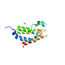
Crystal structure of CBP bromodomain liganded with 9c
Descriptor: 3-acetyl-1-((3-(1-cyclopropyl-1H-pyrazol-4-yl)-2-fluoro-5-(hydroxymethyl)phenyl)carbamoyl)indolizin-7-yl dimethylcarbamate, CREB-binding protein, GLYCEROL, ...
Authors:Xiang, Q, Wang, C, Wu, T, Zhang, Y, Zhang, C, Luo, G, Wu, X, Shen, H, Xu, Y.
Deposit date:2021-05-21
Release date:2022-02-16
Last modified:2023-11-29
Method:X-RAY DIFFRACTION (2.57 Å)
Cite:Design, Synthesis, and Biological Evaluation of 1-(Indolizin-3-yl)ethan-1-ones as CBP Bromodomain Inhibitors for the Treatment of Prostate Cancer.
J.Med.Chem., 65, 2022
2M30
DownloadVisualize
BU of 2m30 by Molmil
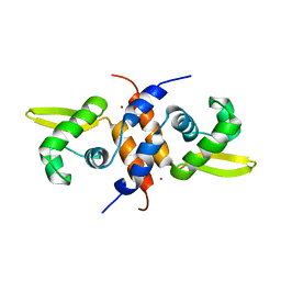
Solution NMR refinement of a metal ion bound protein using quantum mechanical/molecular mechanical and molecular dynamics methods
Descriptor: Repressor protein, ZINC ION
Authors:Chakravorty, D.K, Wang, B.I, Lee, C.I, Guerra, A.J, Giedroc, D.P, Merz Jr, K.M, Arunkumar, A.I, Pennella, M, Kong, X.
Deposit date:2013-01-04
Release date:2013-05-08
Last modified:2024-05-01
Method:SOLUTION NMR
Cite:Solution NMR refinement of a metal ion bound protein using metal ion inclusive restrained molecular dynamics methods.
J.Biomol.Nmr, 56, 2013
5XJR
DownloadVisualize
BU of 5xjr by Molmil
Crystal Structure of the Gemin2-binding domain of SMN, Gemin2dN39 in Complex with SmD1(1-82)/D2/F/E/G from Human
Descriptor: Gem-associated protein 2, Small nuclear ribonucleoprotein E, Small nuclear ribonucleoprotein F, ...
Authors:Yi, H, Zhang, R.
Deposit date:2017-05-04
Release date:2018-07-04
Last modified:2023-11-22
Method:X-RAY DIFFRACTION (3.12 Å)
Cite:Negative cooperativity between Gemin2 and RNA provides insights into RNA selection and the SMN complex's release in snRNP assembly.
Nucleic Acids Res., 2019
5XJQ
DownloadVisualize
BU of 5xjq by Molmil
Crystal Structure of the Gemin2-binding domain of SMN, Gemin2 in Complex with SmD1(1-82)/D2/F/E/G from Human
Descriptor: Gem-associated protein 2, Small nuclear ribonucleoprotein E, Small nuclear ribonucleoprotein F, ...
Authors:Yi, H, Zhang, R.
Deposit date:2017-05-04
Release date:2018-07-04
Last modified:2023-11-22
Method:X-RAY DIFFRACTION (3.28 Å)
Cite:Negative cooperativity between Gemin2 and RNA provides insights into RNA selection and the SMN complex's release in snRNP assembly.
Nucleic Acids Res., 2019
5XJS
DownloadVisualize
BU of 5xjs by Molmil
Crystal Structure of the Gemin2-binding domain of SMN, Gemin2dN39 in Complex with SmD1(1-82)/D2/F/E from Human
Descriptor: Gem-associated protein 2, Small nuclear ribonucleoprotein E, Small nuclear ribonucleoprotein F, ...
Authors:Yi, H, Zhang, R.
Deposit date:2017-05-04
Release date:2018-07-04
Last modified:2023-11-22
Method:X-RAY DIFFRACTION (3.38 Å)
Cite:Negative cooperativity between Gemin2 and RNA provides insights into RNA selection and the SMN complex's release in snRNP assembly.
Nucleic Acids Res., 2019
<12


226707

PDB entries from 2024-10-30

PDB statisticsPDBj update infoContact PDBjnumon