Loading
PDBj
MenuPDBj@FacebookPDBj@TwitterPDBj@YouTubewwPDB FoundationwwPDB
RCSB PDBPDBeBMRBAdv. SearchSearch help
Search by PDB author
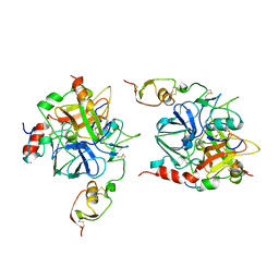
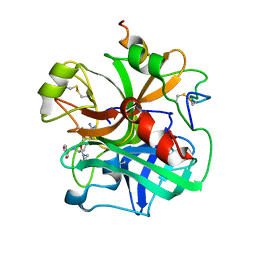
1CSB
DownloadVisualize
BU of 1csb by Molmil
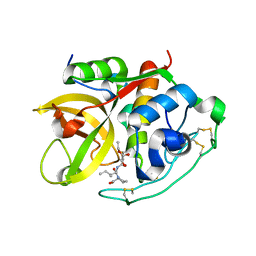
Crystal structure of cathepsin b inhibited with CA030 at 2.1 angstroms resolution: A basis for the design of specific epoxysuccinyl inhibitors
Descriptor: CATHEPSIN B heavy chain, CATHEPSIN B light chain, N-[(3R)-4-ethoxy-3-hydroxy-4-oxobutanoyl]-L-isoleucyl-L-proline
Authors:Turk, D, Bode, W.
Deposit date:1994-12-09
Release date:1996-04-03
Last modified:2012-12-12
Method:X-RAY DIFFRACTION (2 Å)
Cite:Crystal structure of cathepsin B inhibited with CA030 at 2.0-A resolution: A basis for the design of specific epoxysuccinyl inhibitors.
Biochemistry, 34, 1995
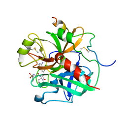
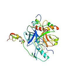
1SAV
DownloadVisualize
BU of 1sav by Molmil
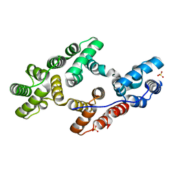
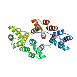
HUMAN ANNEXIN V WITH PROLINE SUBSTITUTION BY THIOPROLINE
Descriptor: ANNEXIN V, CALCIUM ION
Authors:Medrano, F.J, Minks, C, Budisa, N, Huber, R.
Deposit date:1997-11-24
Release date:1998-05-27
Last modified:2023-08-09
Method:X-RAY DIFFRACTION (2.5 Å)
Cite:Crystal and molecular structure of human annexin V after refinement. Implications for structure, membrane binding and ion channel formation of the annexin family of proteins.
J.Mol.Biol., 223, 1992
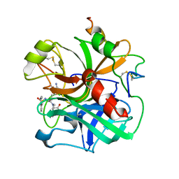
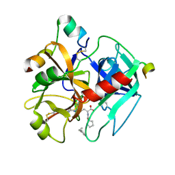
1AVR
DownloadVisualize
BU of 1avr by Molmil
CRYSTAL AND MOLECULAR STRUCTURE OF HUMAN ANNEXIN V AFTER REFINEMENT. IMPLICATIONS FOR STRUCTURE, MEMBRANE BINDING AND ION CHANNEL FORMATION OF THE ANNEXIN FAMILY OF PROTEINS
Descriptor: ANNEXIN V, CALCIUM ION, SULFATE ION
Authors:Huber, R, Berendes, R, Burger, A, Schneider, M, Karshikov, A, Luecke, H, Roemisch, J, Paques, E.
Deposit date:1991-10-17
Release date:1994-01-31
Last modified:2024-02-07
Method:X-RAY DIFFRACTION (2.3 Å)
Cite:Crystal and molecular structure of human annexin V after refinement. Implications for structure, membrane binding and ion channel formation of the annexin family of proteins.
J.Mol.Biol., 223, 1992
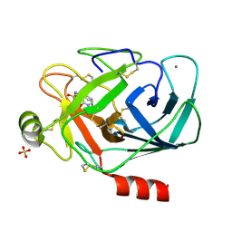
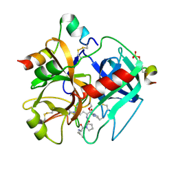
1PPF
DownloadVisualize
BU of 1ppf by Molmil
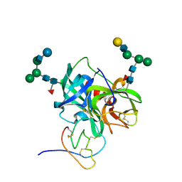
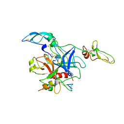
X-RAY CRYSTAL STRUCTURE OF THE COMPLEX OF HUMAN LEUKOCYTE ELASTASE (PMN ELASTASE) AND THE THIRD DOMAIN OF THE TURKEY OVOMUCOID INHIBITOR
Descriptor: HUMAN LEUKOCYTE ELASTASE, TURKEY OVOMUCOID INHIBITOR (OMTKY3), alpha-D-glucopyranose-(1-4)-2-acetamido-2-deoxy-beta-D-glucopyranose-(1-2)-alpha-D-mannopyranose-(1-6)-[alpha-D-mannopyranose-(1-3)]beta-D-mannopyranose-(1-4)-2-acetamido-2-deoxy-beta-D-glucopyranose-(1-4)-[alpha-L-fucopyranose-(1-6)]2-acetamido-2-deoxy-beta-D-glucopyranose, ...
Authors:Bode, W, Wei, A-Z.
Deposit date:1991-10-24
Release date:1994-01-31
Last modified:2020-07-29
Method:X-RAY DIFFRACTION (1.8 Å)
Cite:X-ray crystal structure of the complex of human leukocyte elastase (PMN elastase) and the third domain of the turkey ovomucoid inhibitor.
EMBO J., 5, 1986
1T32
DownloadVisualize
BU of 1t32 by Molmil
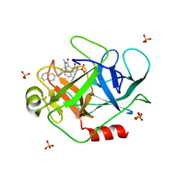
A Dual Inhibitor of the Leukocyte Proteases Cathepsin G and Chymase with Therapeutic Efficacy in Animals Models of Inflammation
Descriptor: 2-[3-({METHYL[1-(2-NAPHTHOYL)PIPERIDIN-4-YL]AMINO}CARBONYL)-2-NAPHTHYL]-1-(1-NAPHTHYL)-2-OXOETHYLPHOSPHONIC ACID, Cathepsin G, SULFATE ION
Authors:de Garavilla, L, Greco, M.N, Giardino, E.C, Wells, G.I, Haertlein, B.J, Kauffman, J.A, Corcoran, T.W, Derian, C.K, Eckardt, A.J, Abraham, W.M, Sukumar, N, Chen, Z, Pineda, A.O, Mathews, F.S, Di Cera, E, Andrade-Gordon, P, Damiano, B.P, Maryanoff, B.E.
Deposit date:2004-04-23
Release date:2005-03-01
Last modified:2023-08-23
Method:X-RAY DIFFRACTION (1.85 Å)
Cite:A novel, potent dual inhibitor of the leukocyte proteases cathepsin G and chymase: molecular mechanisms and anti-inflammatory activity in vivo.
J.Biol.Chem., 280, 2005
1BTH
DownloadVisualize
BU of 1bth by Molmil
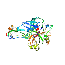
STRUCTURE OF THROMBIN COMPLEXED WITH BOVINE PANCREATIC TRYPSIN INHIBITOR
Descriptor: BOVINE PANCREATIC TRYPSIN INHIBITOR, THROMBIN
Authors:Van De Locht, A, Bode, W, Stubbs, M.T.
Deposit date:1996-12-03
Release date:1997-12-24
Last modified:2021-11-03
Method:X-RAY DIFFRACTION (2.3 Å)
Cite:The thrombin E192Q-BPTI complex reveals gross structural rearrangements: implications for the interaction with antithrombin and thrombomodulin.
EMBO J., 16, 1997
1TBR
DownloadVisualize
BU of 1tbr by Molmil
CRYSTAL STRUCTURE OF INSECT DERIVED DOUBLE DOMAIN KAZAL INHIBITOR RHODNIIN IN COMPLEX WITH THROMBIN
Descriptor: RHODNIIN, THROMBIN
Authors:Van De Locht, A, Lamba, D, Bode, W.
Deposit date:1995-03-03
Release date:1996-10-14
Last modified:2011-07-13
Method:X-RAY DIFFRACTION (2.6 Å)
Cite:Two heads are better than one: crystal structure of the insect derived double domain Kazal inhibitor rhodniin in complex with thrombin.
EMBO J., 14, 1995
1ETS
DownloadVisualize
BU of 1ets by Molmil
REFINED 2.3 ANGSTROMS X-RAY CRYSTAL STRUCTURE OF BOVINE THROMBIN COMPLEXES FORMED WITH THE BENZAMIDINE AND ARGININE-BASED THROMBIN INHIBITORS NAPAP, 4-TAPAP AND MQPA: A STARTING POINT FOR IMPROVING ANTITHROMBOTICS
Descriptor: 1-[N-(naphthalen-2-ylsulfonyl)glycyl-4-carbamimidoyl-D-phenylalanyl]piperidine, EPSILON-THROMBIN
Authors:Bode, W, Brandstetter, H.
Deposit date:1992-07-06
Release date:1994-01-31
Last modified:2024-06-05
Method:X-RAY DIFFRACTION (2.3 Å)
Cite:Refined 2.3 A X-ray crystal structure of bovine thrombin complexes formed with the benzamidine and arginine-based thrombin inhibitors NAPAP, 4-TAPAP and MQPA. A starting point for improving antithrombotics.
J.Mol.Biol., 226, 1992
1ETR
DownloadVisualize
BU of 1etr by Molmil
REFINED 2.3 ANGSTROMS X-RAY CRYSTAL STRUCTURE OF BOVINE THROMBIN COMPLEXES FORMED WITH THE BENZAMIDINE AND ARGININE-BASED THROMBIN INHIBITORS NAPAP, 4-TAPAP AND MQPA: A STARTING POINT FOR IMPROVING ANTITHROMBOTICS
Descriptor: EPSILON-THROMBIN, amino{[(4S)-5-[(2R,4R)-2-carboxy-4-methylpiperidin-1-yl]-4-({[(3R)-3-methyl-1,2,3,4-tetrahydroquinolin-8-yl]sulfonyl}amino)-5-oxopentyl]amino}methaniminium
Authors:Bode, W, Brandstetter, H.
Deposit date:1992-07-06
Release date:1994-01-31
Last modified:2024-06-05
Method:X-RAY DIFFRACTION (2.2 Å)
Cite:Refined 2.3 A X-ray crystal structure of bovine thrombin complexes formed with the benzamidine and arginine-based thrombin inhibitors NAPAP, 4-TAPAP and MQPA. A starting point for improving antithrombotics.
J.Mol.Biol., 226, 1992
1PPH
DownloadVisualize
BU of 1pph by Molmil
GEOMETRY OF BINDING OF THE NALPHA-TOSYLATED PIPERIDIDES OF M-AMIDINO-, P-AMIDINO-AND P-GUANIDINO PHENYLALANINE TO THROMBIN AND TRYPSIN: X-RAY CRYSTAL STRUCTURES OF THEIR TRYPSIN COMPLEXES AND MODELING OF THEIR THROMBIN COMPLEXES
Descriptor: 3-[(2S)-2-{[(4-methylphenyl)sulfonyl]amino}-3-oxo-3-piperidin-1-ylpropyl]benzenecarboximidamide, CALCIUM ION, SULFATE ION, ...
Authors:Bode, W, Turk, D.
Deposit date:1991-10-24
Release date:1994-01-31
Last modified:2024-06-05
Method:X-RAY DIFFRACTION (1.9 Å)
Cite:Geometry of binding of the N alpha-tosylated piperidides of m-amidino-, p-amidino- and p-guanidino phenylalanine to thrombin and trypsin. X-ray crystal structures of their trypsin complexes and modeling of their thrombin complexes.
FEBS Lett., 287, 1991
1ETT
DownloadVisualize
BU of 1ett by Molmil
REFINED 2.3 ANGSTROMS X-RAY CRYSTAL STRUCTURE OF BOVINE THROMBIN COMPLEXES FORMED WITH THE BENZAMIDINE AND ARGININE-BASED THROMBIN INHIBITORS NAPAP, 4-TAPAP AND MQPA: A STARTING POINT FOR IMPROVING ANTITHROMBOTICS
Descriptor: 4-[(2S)-2-{[(4-methylphenyl)sulfonyl]amino}-3-oxo-3-(piperidin-1-yl)propyl]benzene-1-carboximidamide, EPSILON-THROMBIN
Authors:Bode, W, Brandstetter, H.
Deposit date:1992-07-06
Release date:1994-01-31
Last modified:2018-09-12
Method:X-RAY DIFFRACTION (2.5 Å)
Cite:Refined 2.3 A X-ray crystal structure of bovine thrombin complexes formed with the benzamidine and arginine-based thrombin inhibitors NAPAP, 4-TAPAP and MQPA. A starting point for improving antithrombotics.
J.Mol.Biol., 226, 1992
1TBQ
DownloadVisualize
BU of 1tbq by Molmil
CRYSTAL STRUCTURE OF INSECT DERIVED DOUBLE DOMAIN KAZAL INHIBITOR RHODNIIN IN COMPLEX WITH THROMBIN
Descriptor: RHODNIIN, THROMBIN
Authors:Van De Locht, A, Lamba, D, Bode, W.
Deposit date:1995-03-02
Release date:1996-10-14
Last modified:2011-07-13
Method:X-RAY DIFFRACTION (3.1 Å)
Cite:Two heads are better than one: crystal structure of the insect derived double domain Kazal inhibitor rhodniin in complex with thrombin.
EMBO J., 14, 1995
1TMT
DownloadVisualize
BU of 1tmt by Molmil
CHANGES IN INTERACTIONS IN COMPLEXES OF HIRUDIN DERIVATIVES AND HUMAN ALPHA-THROMBIN DUE TO DIFFERENT CRYSTAL FORMS
Descriptor: 2-acetamido-2-deoxy-beta-D-glucopyranose, ALPHA-THROMBIN (LARGE SUBUNIT), ALPHA-THROMBIN (SMALL SUBUNIT), ...
Authors:Priestle, J.P, Gruetter, M.G.
Deposit date:1994-05-26
Release date:1994-09-30
Last modified:2022-12-21
Method:X-RAY DIFFRACTION (2.2 Å)
Cite:Changes in interactions in complexes of hirudin derivatives and human alpha-thrombin due to different crystal forms.
Protein Sci., 2, 1993
1TMU
DownloadVisualize
BU of 1tmu by Molmil
Changes in interactions in complexes of hirudin derivatives and human alpha-thrombin due to different crystal forms
Descriptor: 2-acetamido-2-deoxy-beta-D-glucopyranose, D-phenylalanyl-N-[(2S,3S)-6-{[amino(iminio)methyl]amino}-1-chloro-2-hydroxyhexan-3-yl]-L-prolinamide, Hirudin variant-2, ...
Authors:Priestle, J.P, Gruetter, M.G.
Deposit date:1994-05-26
Release date:1994-09-30
Last modified:2020-07-29
Method:X-RAY DIFFRACTION (2.5 Å)
Cite:Changes in interactions in complexes of hirudin derivatives and human alpha-thrombin due to different crystal forms.
Protein Sci., 2, 1993
1TOC
DownloadVisualize
BU of 1toc by Molmil
STRUCTURE OF SERINE PROTEINASE
Descriptor: ORNITHODORIN, THROMBIN
Authors:Van De Locht, A, Huber, R, Bode, W.
Deposit date:1996-07-20
Release date:1997-07-23
Last modified:2011-07-13
Method:X-RAY DIFFRACTION (3.1 Å)
Cite:The ornithodorin-thrombin crystal structure, a key to the TAP enigma?
EMBO J., 15, 1996
<12


224201

數據於2024-08-28公開中

PDB statisticsPDBj update infoContact PDBjnumon