Loading
PDBj
MenuPDBj@FacebookPDBj@TwitterPDBj@YouTubewwPDB FoundationwwPDB
RCSB PDBPDBeBMRBAdv. SearchSearch help
Search by PDB author
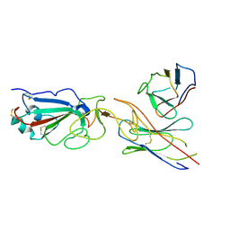
4D90
DownloadVisualize
BU of 4d90 by Molmil
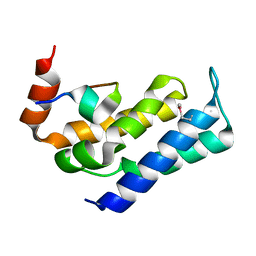
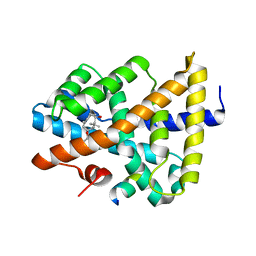
Crystal Structure of Del-1 EGF domains
Descriptor: 2-acetamido-2-deoxy-beta-D-galactopyranose, 2-acetamido-2-deoxy-beta-D-glucopyranose, CALCIUM ION, ...
Authors:Chen, Q, Schurpf, T, Springer, T, Wang, J.
Deposit date:2012-01-11
Release date:2012-05-30
Last modified:2023-09-13
Method:X-RAY DIFFRACTION (2.601 Å)
Cite:The RGD finger of Del-1 is a unique structural feature critical for integrin binding.
Faseb J., 26, 2012
4X3V
DownloadVisualize
BU of 4x3v by Molmil
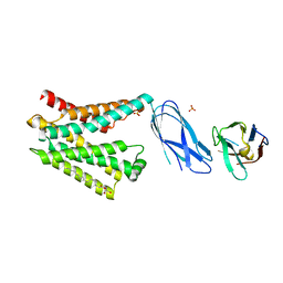
Crystal structure of human ribonucleotide reductase 1 bound to inhibitor
Descriptor: N~6~-{N-[(1,3-dioxo-1,3-dihydro-2H-isoindol-2-yl)acetyl]-2-methyl-D-alanyl}-D-lysine, Ribonucleoside-diphosphate reductase large subunit, THYMIDINE-5'-TRIPHOSPHATE
Authors:Dealwis, C.G, Ahmad, M.F, Alam, I.
Deposit date:2014-12-01
Release date:2015-11-11
Last modified:2023-09-27
Method:X-RAY DIFFRACTION (3.7 Å)
Cite:Identification of Non-nucleoside Human Ribonucleotide Reductase Modulators.
J.Med.Chem., 58, 2015
8DYP
DownloadVisualize
BU of 8dyp by Molmil
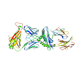
Crystal structure of human cystine transporter cystinosin
Descriptor: (2R)-2,3-dihydroxypropyl (9Z)-octadec-9-enoate, Cystinosin, Nanobody P10, ...
Authors:Guo, X, Feng, L.
Deposit date:2022-08-04
Release date:2022-11-16
Last modified:2023-10-18
Method:X-RAY DIFFRACTION (3.4 Å)
Cite:Structure and mechanism of human cystine exporter cystinosin.
Cell, 185, 2022
7E23
DownloadVisualize
BU of 7e23 by Molmil
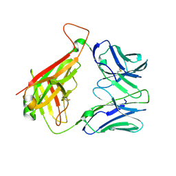
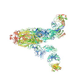
SARS-CoV-2 spike in complex with the CA521 neutralizing antibody Fab (focused refinement on Fab-RBD)
Descriptor: 2-acetamido-2-deoxy-beta-D-glucopyranose, CA521 Heavy Chain, CA521 Light Chain, ...
Authors:Liu, C, Song, D, Dou, C.
Deposit date:2021-02-04
Release date:2021-05-05
Method:ELECTRON MICROSCOPY (3.3 Å)
Cite:Structure and function analysis of a potent human neutralizing antibody CA521 FALA against SARS-CoV-2.
Commun Biol, 4, 2021
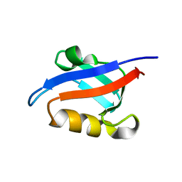
1U38
DownloadVisualize
BU of 1u38 by Molmil
Auto-inhibition Mechanism of X11s/Mints Family Scaffold Proteins Revealed by the Closed Conformation of the Tandem PDZ Domains
Descriptor: PVYI, amyloid beta A4 precursor protein-binding, family A, ...
Authors:Feng, W, Long, J.-F, Chan, L.-N, He, C, Fu, A, Xia, J, Ip, N.Y, Zhang, M.
Deposit date:2004-07-21
Release date:2005-07-26
Last modified:2024-05-29
Method:SOLUTION NMR
Cite:Autoinhibition of X11/Mint scaffold proteins revealed by the closed conformation of the PDZ tandem
Nat.Struct.Mol.Biol., 12, 2005
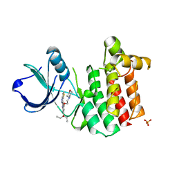
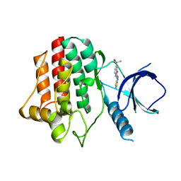
3V5L
DownloadVisualize
BU of 3v5l by Molmil
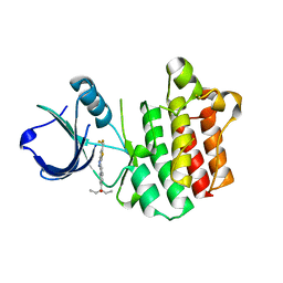
Crystal Structure of Interleukin-2 Inducible T-cell Kinase Itk Catalytic Domain with Thienopyrazolylindole Inhibitor 542
Descriptor: 3-[3-(4-methoxyphenyl)-2-(1H-thieno[3,2-c]pyrazol-3-yl)-1H-indol-6-yl]pentan-3-ol, SULFATE ION, Tyrosine-protein kinase ITK/TSK
Authors:McLean, L.R, Zhang, Y.
Deposit date:2011-12-16
Release date:2012-05-02
Last modified:2023-09-13
Method:X-RAY DIFFRACTION (1.86 Å)
Cite:X-ray crystallographic structure-based design of selective thienopyrazole inhibitors for interleukin-2-inducible tyrosine kinase.
Bioorg.Med.Chem.Lett., 22, 2012
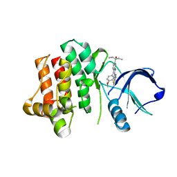
3VF8
DownloadVisualize
BU of 3vf8 by Molmil
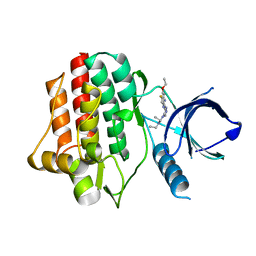
Crystal Structure of Spleen Tyrosine Kinase Syk Catalytic Domain with Pyrazolylbenzimidazole Inhibitor 416
Descriptor: 3-[5-(5-ethoxy-6-fluoro-1H-benzimidazol-2-yl)-1H-pyrazol-4-yl]-1,1-diethylurea, Tyrosine-protein kinase SYK
Authors:McLean, L.R, Zhang, Y.
Deposit date:2012-01-09
Release date:2012-05-02
Last modified:2024-02-28
Method:X-RAY DIFFRACTION (2.08 Å)
Cite:X-ray crystallographic structure-based design of selective thienopyrazole inhibitors for interleukin-2-inducible tyrosine kinase.
Bioorg.Med.Chem.Lett., 22, 2012
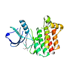
3V8W
DownloadVisualize
BU of 3v8w by Molmil
Crystal Structure of Interleukin-2 Inducible T-cell Kinase Itk Catalytic Domain with Thienopyrazolylindole Inhibitor 469
Descriptor: 3-[2-(5-phenyl-2H-thieno[3,2-c]pyrazol-3-yl)-1H-indol-6-yl]pentan-3-ol, SULFATE ION, Tyrosine-protein kinase ITK/TSK
Authors:McLean, L.R, Zhang, Y.
Deposit date:2011-12-23
Release date:2012-05-02
Last modified:2023-09-13
Method:X-RAY DIFFRACTION (2.27 Å)
Cite:X-ray crystallographic structure-based design of selective thienopyrazole inhibitors for interleukin-2-inducible tyrosine kinase.
Bioorg.Med.Chem.Lett., 22, 2012
3V5J
DownloadVisualize
BU of 3v5j by Molmil
Crystal Structure of Interleukin-2 Inducible T-cell Kinase Itk Catalytic Domain with Thienopyrazolylindole Inhibitor 090
Descriptor: 3-[4-(2-morpholin-4-ylethoxy)-2-(1~{H}-thieno[3,2-c]pyrazol-3-yl)-1~{H}-indol-6-yl]pentan-3-ol, SULFATE ION, Tyrosine-protein kinase ITK/TSK
Authors:McLean, L.R, Zhang, Y.
Deposit date:2011-12-16
Release date:2012-05-02
Last modified:2023-09-13
Method:X-RAY DIFFRACTION (2.59 Å)
Cite:X-ray crystallographic structure-based design of selective thienopyrazole inhibitors for interleukin-2-inducible tyrosine kinase.
Bioorg.Med.Chem.Lett., 22, 2012
3VF9
DownloadVisualize
BU of 3vf9 by Molmil
Crystal Structure of Spleen Tyrosine Kinase Syk Catalytic Domain with Thienopyrazolylindole Inhibitor 027
Descriptor: 3-{2-[5-(difluoromethyl)-2H-thieno[3,2-c]pyrazol-3-yl]-1H-indol-6-yl}pentan-3-ol, Tyrosine-protein kinase SYK
Authors:McLean, L.R, Zhang, Y.
Deposit date:2012-01-09
Release date:2012-05-02
Last modified:2024-02-28
Method:X-RAY DIFFRACTION (2.3 Å)
Cite:X-ray crystallographic structure-based design of selective thienopyrazole inhibitors for interleukin-2-inducible tyrosine kinase.
Bioorg.Med.Chem.Lett., 22, 2012
3V8T
DownloadVisualize
BU of 3v8t by Molmil
Crystal Structure of Interleukin-2 Inducible T-cell Kinase Itk Catalytic Domain with Thienopyrazolylindole Inhibitor 477
Descriptor: 3-{2-[5-(difluoromethyl)-2H-thieno[3,2-c]pyrazol-3-yl]-1H-indol-6-yl}pentan-3-ol, SULFATE ION, Tyrosine-protein kinase ITK/TSK
Authors:McLean, L.R, Zhang, Y.
Deposit date:2011-12-23
Release date:2012-05-02
Last modified:2023-09-13
Method:X-RAY DIFFRACTION (2 Å)
Cite:X-ray crystallographic structure-based design of selective thienopyrazole inhibitors for interleukin-2-inducible tyrosine kinase.
Bioorg.Med.Chem.Lett., 22, 2012
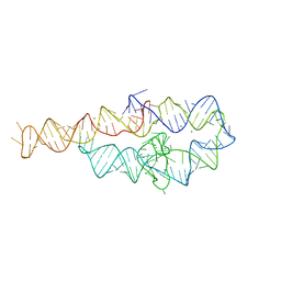
6D8M
DownloadVisualize
BU of 6d8m by Molmil
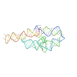
Group I self-splicing intron P4-P6 domain mutant A125U/G126U
Descriptor: Group I self-splicing intron, MAGNESIUM ION
Authors:Shoffner, G.M.
Deposit date:2018-04-26
Release date:2018-07-04
Last modified:2023-10-04
Method:X-RAY DIFFRACTION (3.7 Å)
Cite:In Crystallo Selection to Establish New RNA Crystal Contacts.
Structure, 26, 2018
6D8O
DownloadVisualize
BU of 6d8o by Molmil
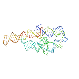
Group I self-splicing intron P4-P6 domain mutant A230U
Descriptor: Group I self-splicing intron, MAGNESIUM ION
Authors:Shoffner, G.M.
Deposit date:2018-04-26
Release date:2018-07-04
Last modified:2023-10-04
Method:X-RAY DIFFRACTION (2.80112481 Å)
Cite:In Crystallo Selection to Establish New RNA Crystal Contacts.
Structure, 26, 2018
6D8N
DownloadVisualize
BU of 6d8n by Molmil
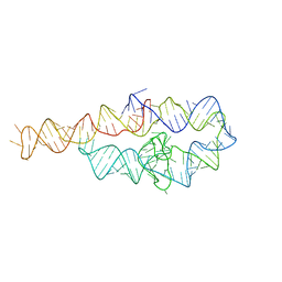
Group I self-splicing intron P4-P6 domain mutant G134A/U185AA
Descriptor: Group I self-splicing intron, MAGNESIUM ION
Authors:Shoffner, G.M.
Deposit date:2018-04-26
Release date:2018-07-18
Last modified:2023-10-04
Method:X-RAY DIFFRACTION (3.95427132 Å)
Cite:In Crystallo Selection to Establish New RNA Crystal Contacts.
Structure, 26, 2018
8GU7
DownloadVisualize
BU of 8gu7 by Molmil
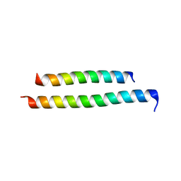
Selective targeting of the Beclin 2-Atg14L coiled coil complex by stapled peptides promotes autophagy and endolysosomal trafficking of GPCRs
Descriptor: Beclin 1-associated autophagy-related key regulator, Beclin-2
Authors:Qiu, X, Zhao, Y.
Deposit date:2022-09-11
Release date:2023-07-26
Last modified:2023-10-11
Method:X-RAY DIFFRACTION (2.6 Å)
Cite:The potent BECN2-ATG14 coiled-coil interaction is selectively critical for endolysosomal degradation of GPRASP1/GASP1-associated GPCRs.
Autophagy, 19, 2023
8GT9
DownloadVisualize
BU of 8gt9 by Molmil
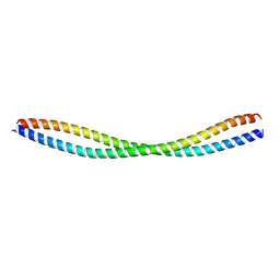
Selective targeting of the Beclin 2-Atg14L coiled coil complex by stapled peptides promotes autophagy and endolysosomal trafficking of GPCRs
Descriptor: Beclin-2
Authors:Qiu, X, Zhao, Y.
Deposit date:2022-09-07
Release date:2023-07-26
Last modified:2023-10-11
Method:X-RAY DIFFRACTION (2 Å)
Cite:The potent BECN2-ATG14 coiled-coil interaction is selectively critical for endolysosomal degradation of GPRASP1/GASP1-associated GPCRs.
Autophagy, 19, 2023
6D8L
DownloadVisualize
BU of 6d8l by Molmil
Group I self-splicing intron P4-P6 domain mutant U131A (without isopropanol soaking)
Descriptor: Group I self-splicing intron, MAGNESIUM ION
Authors:Shoffner, G.M.
Deposit date:2018-04-26
Release date:2018-07-18
Last modified:2023-10-04
Method:X-RAY DIFFRACTION (3.1402154 Å)
Cite:In Crystallo Selection to Establish New RNA Crystal Contacts.
Structure, 26, 2018
7KL9
DownloadVisualize
BU of 7kl9 by Molmil
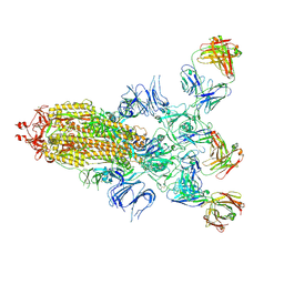
Structure of the SARS-CoV-2 S 6P trimer in complex with the ACE2 protein decoy, CTC-445.2 (State 4)
Descriptor: 2-acetamido-2-deoxy-beta-D-glucopyranose, 2-acetamido-2-deoxy-beta-D-glucopyranose-(1-4)-2-acetamido-2-deoxy-beta-D-glucopyranose, CTC-445.2 inhibitor, ...
Authors:Barnes, C.O, Bjorkman, P.J.
Deposit date:2020-10-29
Release date:2020-11-11
Last modified:2020-12-16
Method:ELECTRON MICROSCOPY (4.1 Å)
Cite:De novo design of potent and resilient hACE2 decoys to neutralize SARS-CoV-2.
Science, 370, 2020
7KJS
DownloadVisualize
BU of 7kjs by Molmil
Crystal structure of CDK2/cyclin E in complex with PF-06873600
Descriptor: 6-(difluoromethyl)-8-[(1R,2R)-2-hydroxy-2-methylcyclopentyl]-2-{[1-(methylsulfonyl)piperidin-4-yl]amino}pyrido[2,3-d]pyrimidin-7(8H)-one, Cyclin-dependent kinase 2, G1/S-specific cyclin-E1
Authors:McTigue, M.A, He, Y, Ferre, R.A.
Deposit date:2020-10-26
Release date:2021-06-23
Last modified:2023-10-18
Method:X-RAY DIFFRACTION (2.187 Å)
Cite:Discovery of PF-06873600, a CDK2/4/6 Inhibitor for the Treatment of Cancer.
J.Med.Chem., 64, 2021
5GL6
DownloadVisualize
BU of 5gl6 by Molmil
Msmeg rimP
Descriptor: Ribosome maturation factor RimP
Authors:Chu, T, Lau, T.C.K.
Deposit date:2016-07-08
Release date:2017-07-12
Last modified:2024-03-20
Method:X-RAY DIFFRACTION (2.2 Å)
Cite:The ribosomal maturation factor P fromMycobacterium smegmatisfacilitates the ribosomal biogenesis by binding to the small ribosomal protein S12.
J. Biol. Chem., 294, 2019
4YH3
DownloadVisualize
BU of 4yh3 by Molmil
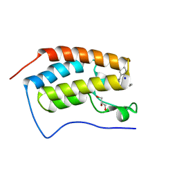
Crystal structure of human BRD4(1) in complex with 4-[(2E)-3-(4-methoxyphenyl)-2-phenylprop-2-enoyl]-3,4-dihydroquinoxalin-2(1H)-one (compound 19a)
Descriptor: 4-[(2E)-3-(4-methoxyphenyl)-2-phenylprop-2-enoyl]-3,4-dihydroquinoxalin-2(1H)-one, Bromodomain-containing protein 4
Authors:White, A, Lakshminarasimhan, D, Suto, R.K.
Deposit date:2015-02-26
Release date:2016-01-13
Last modified:2024-02-28
Method:X-RAY DIFFRACTION (1.6 Å)
Cite:Discovery of a new chemical series of BRD4(1) inhibitors using protein-ligand docking and structure-guided design.
Bioorg.Med.Chem.Lett., 25, 2015
5GO9
DownloadVisualize
BU of 5go9 by Molmil
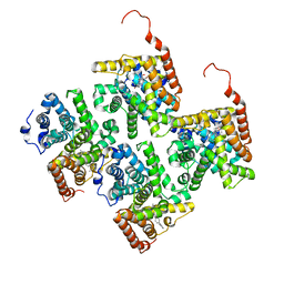
Cryo-EM structure of RyR2 in closed state
Descriptor: RyR2, ZINC ION
Authors:Peng, W, Wu, J.P, Yan, N.
Deposit date:2016-07-26
Release date:2016-10-05
Last modified:2024-03-27
Method:ELECTRON MICROSCOPY (4.4 Å)
Cite:Structural basis for the gating mechanism of the type 2 ryanodine receptor RyR2
Science, 354, 2016
5GOA
DownloadVisualize
BU of 5goa by Molmil
Cryo-EM structure of RyR2 in open state
Descriptor: RyR2, ZINC ION
Authors:Peng, W, Wu, J.P, Yan, N.
Deposit date:2016-07-26
Release date:2016-10-05
Last modified:2024-03-27
Method:ELECTRON MICROSCOPY (4.2 Å)
Cite:Structural basis for the gating mechanism of the type 2 ryanodine receptor RyR2
Science, 354, 2016
6A85
DownloadVisualize
BU of 6a85 by Molmil
Crystal structure of a novel DNA quadruplex
Descriptor: AMMONIUM ION, DNA (5'-D(*AP*GP*AP*GP*AP*GP*AP*TP*GP*GP*GP*TP*GP*CP*GP*TP*T)-3'), LEAD (II) ION, ...
Authors:Liu, H.H, Gan, J.H.
Deposit date:2018-07-06
Release date:2019-03-06
Last modified:2024-03-27
Method:X-RAY DIFFRACTION (1.45 Å)
Cite:High-resolution DNA quadruplex structure containing all the A-, G-, C-, T-tetrads.
Nucleic Acids Res., 46, 2018
4YH4
DownloadVisualize
BU of 4yh4 by Molmil
Crystal structure of human BRD4(1) in complex with 4-[(5-phenylpyridin-3-yl)carbonyl]-3,4-dihydroquinoxalin-2(1H)-one (compound 19d)
Descriptor: 4-[(5-phenylpyridin-3-yl)carbonyl]-3,4-dihydroquinoxalin-2(1H)-one, Bromodomain-containing protein 4, GLYCEROL, ...
Authors:Lakshminarasimhan, D, White, A, Suto, R.K.
Deposit date:2015-02-26
Release date:2016-01-13
Last modified:2024-02-28
Method:X-RAY DIFFRACTION (1.33 Å)
Cite:Discovery of a new chemical series of BRD4(1) inhibitors using protein-ligand docking and structure-guided design.
Bioorg.Med.Chem.Lett., 25, 2015

221371

數據於2024-06-19公開中

PDB statisticsPDBj update infoContact PDBjnumon