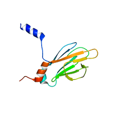
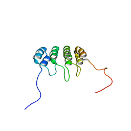
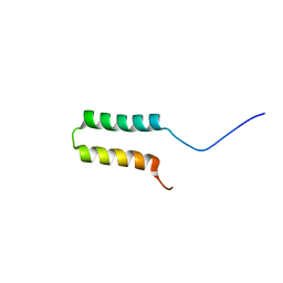
1J4Q
 
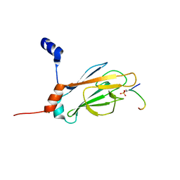 | | NMR STRUCTURE OF THE FHA1 DOMAIN OF RAD53 IN COMPLEX WITH A RAD9-DERIVED PHOSPHOTHREONINE (AT T192) PEPTIDE | | 分子名称: | DNA REPAIR PROTEIN RAD9, PROTEIN KINASE SPK1 | | 著者 | Yuan, C, Yongkiettrakul, S, Byeon, I.-J.L, Zhou, S, Tsai, M.-D. | | 登録日 | 2001-10-22 | | 公開日 | 2001-12-05 | | 最終更新日 | 2023-12-27 | | 実験手法 | SOLUTION NMR | | 主引用文献 | Solution structures of two FHA1-phosphothreonine peptide complexes provide insight into the structural basis of the ligand specificity of FHA1 from yeast Rad53.
J.Mol.Biol., 314, 2001
|
|
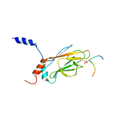
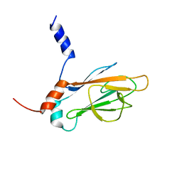
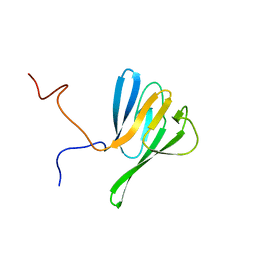
1K3Q
 | | NMR structure of the FHA1 Domain of Rad53 in Complex with a Rad9-derived Phosphothreonine (at T192) Peptide | | 分子名称: | DNA repair protein Rad9, Protein Kinase SPK1 | | 著者 | Yuan, C, Yongkiettrakul, S, Byeon, I.-J.L, Zhou, S, Tsai, M.-D. | | 登録日 | 2001-10-03 | | 公開日 | 2001-12-05 | | 最終更新日 | 2022-02-23 | | 実験手法 | SOLUTION NMR | | 主引用文献 | Solution structures of two FHA1-phosphothreonine peptide complexes provide insight into the structural basis of the ligand specificity of FHA1 from yeast Rad53.
J.Mol.Biol., 314, 2001
|
|
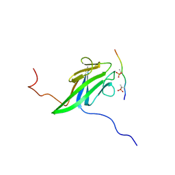
1K3N
 | | NMR Structure of the FHA1 Domain of Rad53 in Complex with a Rad9-derived Phosphothreonine (at T155) Peptide | | 分子名称: | DNA repair protein Rad9, Protein Kinase SPK1 | | 著者 | Yuan, C, Yongkiettrakul, S, Byeon, I.-J.L, Zhou, S, Tsai, M.-D. | | 登録日 | 2001-10-03 | | 公開日 | 2001-12-05 | | 最終更新日 | 2022-02-23 | | 実験手法 | SOLUTION NMR | | 主引用文献 | Solution structures of two FHA1-phosphothreonine peptide complexes provide insight into the structural basis of the ligand specificity of FHA1 from yeast Rad53.
J.Mol.Biol., 314, 2001
|
|
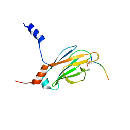
1J4P
 | | NMR STRUCTURE OF THE FHA1 DOMAIN OF RAD53 IN COMPLEX WITH A RAD9-DERIVED PHOSPHOTHREONINE (AT T155) PEPTIDE | | 分子名称: | DNA REPAIR PROTEIN RAD9, PROTEIN KINASE SPK1 | | 著者 | Yuan, C, Yongkiettrakul, S, Byeon, I.-J.L, Zhou, S, Tsai, M.-D. | | 登録日 | 2001-10-22 | | 公開日 | 2001-12-05 | | 最終更新日 | 2023-12-27 | | 実験手法 | SOLUTION NMR | | 主引用文献 | Solution structures of two FHA1-phosphothreonine peptide complexes provide insight into the structural basis of the ligand specificity of FHA1 from yeast Rad53.
J.Mol.Biol., 314, 2001
|
|
1TR4
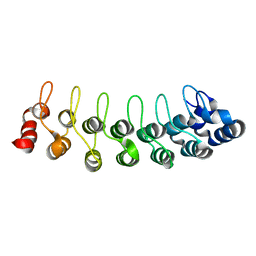 | | Solution structure of human oncogenic protein gankyrin | | 分子名称: | 26S proteasome non-ATPase regulatory subunit 10 | | 著者 | Yuan, C, Li, J, Mahajan, A, Poi, M.J, Byeon, I.J, Tsai, M.D. | | 登録日 | 2004-06-19 | | 公開日 | 2004-11-16 | | 最終更新日 | 2024-05-22 | | 実験手法 | SOLUTION NMR | | 主引用文献 | Solution structure of the human oncogenic protein gankyrin containing seven ankyrin repeats and analysis of its structure--function relationship.
Biochemistry, 43, 2004
|
|
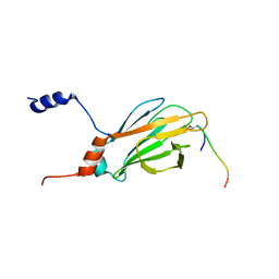
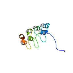
1K3J
 | | Refined NMR Structure of the FHA1 Domain of Yeast Rad53 | | 分子名称: | Protein Kinase SPK1 | | 著者 | Yuan, C, Yongkiettrakul, S, Byeon, I.-J.L, Zhou, S, Tsai, M.-D. | | 登録日 | 2001-10-03 | | 公開日 | 2001-12-05 | | 最終更新日 | 2024-05-22 | | 実験手法 | SOLUTION NMR | | 主引用文献 | Solution structures of two FHA1-phosphothreonine peptide complexes provide insight into the structural basis of the ligand specificity of FHA1 from yeast Rad53.
J.Mol.Biol., 314, 2001
|
|
1J4O
 | | REFINED NMR STRUCTURE OF THE FHA1 DOMAIN OF YEAST RAD53 | | 分子名称: | PROTEIN KINASE SPK1 | | 著者 | Yuan, C, Yongkiettrakul, S, Byeon, I.-J.L, Zhou, S, Tsai, M.-D. | | 登録日 | 2001-10-03 | | 公開日 | 2001-12-05 | | 最終更新日 | 2023-12-27 | | 実験手法 | SOLUTION NMR | | 主引用文献 | Solution structures of two FHA1-phosphothreonine peptide complexes provide insight into the structural basis of the ligand specificity of FHA1 from yeast Rad53.
J.Mol.Biol., 314, 2001
|
|
1D9S
 | | TUMOR SUPPRESSOR P15(INK4B) STRUCTURE BY COMPARATIVE MODELING AND NMR DATA | | 分子名称: | CYCLIN-DEPENDENT KINASE 4 INHIBITOR B | | 著者 | Yuan, C, Ji, L, Selby, T.L, Byeon, I.J.L, Tsai, M.D. | | 登録日 | 1999-10-29 | | 公開日 | 2000-07-28 | | 最終更新日 | 2024-05-22 | | 実験手法 | SOLUTION NMR | | 主引用文献 | Tumor suppressor INK4: comparisons of conformational properties between p16(INK4A) and p18(INK4C).
J.Mol.Biol., 294, 1999
|
|
5EW6
 | | Structure of ligand binding region of uPARAP at pH 7.4 without calcium | | 分子名称: | 2-acetamido-2-deoxy-beta-D-glucopyranose, 2-acetamido-2-deoxy-beta-D-glucopyranose-(1-4)-2-acetamido-2-deoxy-beta-D-glucopyranose-(1-4)-2-acetamido-2-deoxy-beta-D-glucopyranose, C-type mannose receptor 2, ... | | 著者 | Yuan, C, Huang, M. | | 登録日 | 2015-11-20 | | 公開日 | 2016-08-10 | | 最終更新日 | 2023-11-08 | | 実験手法 | X-RAY DIFFRACTION (2.29 Å) | | 主引用文献 | Crystal structures of the ligand-binding region of uPARAP: effect of calcium ion binding
Biochem.J., 473, 2016
|
|
5E4K
 | | Structure of ligand binding region of uPARAP at pH 7.4 | | 分子名称: | 2-acetamido-2-deoxy-beta-D-glucopyranose, 3,6,9,12,15,18,21,24-OCTAOXAHEXACOSAN-1-OL, C-type mannose receptor 2, ... | | 著者 | Yuan, C, Huang, M. | | 登録日 | 2015-10-06 | | 公開日 | 2016-08-10 | | 最終更新日 | 2023-11-08 | | 実験手法 | X-RAY DIFFRACTION (2.58 Å) | | 主引用文献 | Crystal structures of the ligand-binding region of uPARAP: effect of calcium ion binding
Biochem.J., 473, 2016
|
|
5E4L
 | |
3P8G
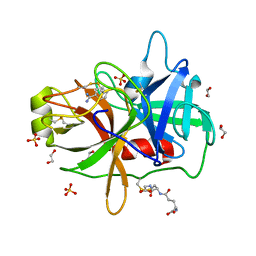 | | Crystal Structure of MT-SP1 in complex with benzamidine | | 分子名称: | 1,2-ETHANEDIOL, BENZAMIDINE, GLUTATHIONE, ... | | 著者 | Yuan, C, Huang, M, Chen, L. | | 登録日 | 2010-10-13 | | 公開日 | 2011-08-03 | | 最終更新日 | 2023-11-01 | | 実験手法 | X-RAY DIFFRACTION (1.2 Å) | | 主引用文献 | Structure of catalytic domain of Matriptase in complex with Sunflower trypsin inhibitor-1.
Bmc Struct.Biol., 11, 2011
|
|
3P8F
 | |
8Y6F
 | |
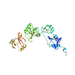
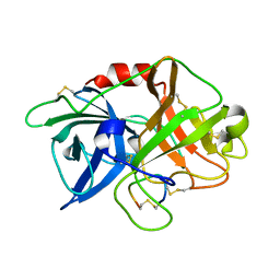
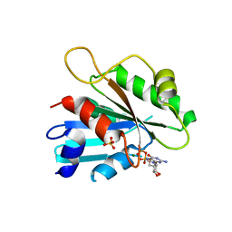
1G3G
 | | NMR STRUCTURE OF THE FHA1 DOMAIN OF YEAST RAD53 | | 分子名称: | PROTEIN KINASE SPK1 | | 著者 | Yuan, C, Liao, H, Su, M, Yongkiettrakul, S, Byeon, I.-J.L, Tsai, M.-D. | | 登録日 | 2000-10-24 | | 公開日 | 2001-01-10 | | 最終更新日 | 2024-05-22 | | 実験手法 | SOLUTION NMR | | 主引用文献 | Structure of the FHA1 domain of yeast Rad53 and identification of binding sites for both FHA1 and its target protein Rad9
J.Mol.Biol., 304, 2000
|
|
1DC2
 | | SOLUTION NMR STRUCTURE OF TUMOR SUPPRESSOR P16INK4A, 20 STRUCTURES | | 分子名称: | CYCLIN-DEPENDENT KINASE 4 INHIBITOR A (P16INK4A) | | 著者 | Byeon, I.-J.L, Li, J, Yuan, C, Tsai, M.-D. | | 登録日 | 1999-11-04 | | 公開日 | 1999-12-23 | | 最終更新日 | 2024-05-22 | | 実験手法 | SOLUTION NMR | | 主引用文献 | Tumor suppressor INK4: refinement of p16INK4A structure and determination of p15INK4B structure by comparative modeling and NMR data.
Protein Sci., 9, 2000
|
|
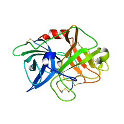
2JQJ
 | | NMR structure of yeast Dun1 FHA domain | | 分子名称: | DNA damage response protein kinase DUN1 | | 著者 | Yuan, C, Lee, H, Chang, C, Heierhorst, J, Tsai, M. | | 登録日 | 2007-06-02 | | 公開日 | 2008-06-24 | | 最終更新日 | 2023-12-20 | | 実験手法 | SOLUTION NMR | | 主引用文献 | Diphosphothreonine-specific interaction between an SQ/TQ cluster and an FHA domain in the Rad53-Dun1 kinase cascade.
Mol.Cell, 30, 2008
|
|
2JQL
 | | NMR structure of the yeast Dun1 FHA domain in complex with a doubly phosphorylated (pT) peptide derived from Rad53 SCD1 | | 分子名称: | DNA damage response protein kinase DUN1, Serine/threonine-protein kinase RAD53 | | 著者 | Yuan, C, Lee, H, Chang, C, Heierhorst, J, Tsai, M. | | 登録日 | 2007-06-02 | | 公開日 | 2008-06-24 | | 最終更新日 | 2023-12-20 | | 実験手法 | SOLUTION NMR | | 主引用文献 | Diphosphothreonine-specific interaction between an SQ/TQ cluster and an FHA domain in the Rad53-Dun1 kinase cascade.
Mol.Cell, 30, 2008
|
|
2JQI
 | |
6ITE
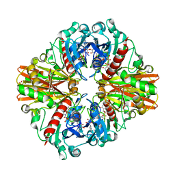 | | Crystal structure of group A Streptococcal surface dehydrogenase (SDH) | | 分子名称: | Glyceraldehyde-3-phosphate dehydrogenase, NICOTINAMIDE-ADENINE-DINUCLEOTIDE, SULFATE ION | | 著者 | Yuan, C, Li, R, Huang, M.D. | | 登録日 | 2018-11-21 | | 公開日 | 2019-09-25 | | 最終更新日 | 2023-11-22 | | 実験手法 | X-RAY DIFFRACTION (1.739 Å) | | 主引用文献 | Structural determination of group A Streptococcal surface dehydrogenase and characterization of its interaction with urokinase-type plasminogen activator receptor.
Biochem.Biophys.Res.Commun., 510, 2019
|
|
7V63
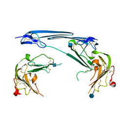 | | Structure of dimeric uPAR at low pH | | 分子名称: | 2-acetamido-2-deoxy-beta-D-glucopyranose, Urokinase plasminogen activator surface receptor | | 著者 | Yuan, C, Huang, M. | | 登録日 | 2021-08-19 | | 公開日 | 2021-12-22 | | 最終更新日 | 2022-04-06 | | 実験手法 | X-RAY DIFFRACTION (2.906 Å) | | 主引用文献 | Crystal structure and cellular functions of uPAR dimer
Nat Commun, 13, 2022
|
|
6XVD
 | | Crystal structure of complex of urokinase and a upain-1 variant(W3F) in pH7.4 condition | | 分子名称: | Urokinase-type plasminogen activator, upain-1-W3F | | 著者 | Xue, G.P, Xie, X, Zhou, Y, Yuan, C, Huang, M.D, Jiang, L.G. | | 登録日 | 2020-01-21 | | 公開日 | 2020-02-19 | | 最終更新日 | 2024-01-24 | | 実験手法 | X-RAY DIFFRACTION (1.4 Å) | | 主引用文献 | Insight to the residue in P2 position prevents the peptide inhibitor from being hydrolyzed by serine proteases.
Biosci.Biotechnol.Biochem., 84, 2020
|
|
3M61
 | | Crystal structure of complex of urokinase and a upain-1 variant(W3A) in pH4.6 condition | | 分子名称: | Urokinase-type plasminogen activator, upain-1 W3A | | 著者 | Jiang, L, Yuan, C, Wind, T, Andreasen, P.A, Chen, L, Meehan, E.J, Huang, M. | | 登録日 | 2010-03-15 | | 公開日 | 2010-04-07 | | 最終更新日 | 2023-11-01 | | 実験手法 | X-RAY DIFFRACTION (1.68 Å) | | 主引用文献 | Crystal structure of complex of urokinase and a upain-1 variant(W3A) in pH4.6 condition
TO BE PUBLISHED
|
|
6BV7
 | |
2FA9
 | | The crystal structure of Sar1[H79G]-GDP provides insight into the coat-controlled GTP hydrolysis in the disassembly of COP II | | 分子名称: | GTP-binding protein SAR1b, GUANOSINE-5'-DIPHOSPHATE, MAGNESIUM ION, ... | | 著者 | Rao, Y, Huang, M, Yuan, C, Bian, C, Hou, X. | | 登録日 | 2005-12-07 | | 公開日 | 2006-09-05 | | 最終更新日 | 2023-10-25 | | 実験手法 | X-RAY DIFFRACTION (2.5 Å) | | 主引用文献 | Crystal Structure of Sar1[H79G]-GDP Which Provides Insight into the Coat-controlled GTP Hydrolysis in the Disassembly of COP II
Chin.J.Struct.Chem., 25, 2006
|
|


