1CBI
 
 | |
2GGM
 | |
1YBA
 | |
1LSH
 | |
2Q1E
 | |
2Q20
 | |
4K07
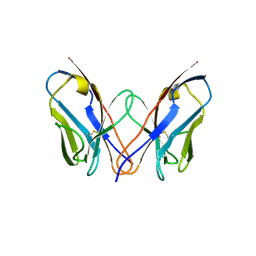
 | | Crystal structure of the amyloid-forming immunoglobulin AL-103 cis-proline 95 mutant | | 分子名称: | Amyloidogenic immunoglobulin light chain protein AL-103, SULFATE ION | | 著者 | Thompson, J.R, Berkholz, D.S, Mahlum, E.W, Ramirez-Alvarado, M. | | 登録日 | 2013-04-03 | | 公開日 | 2013-10-30 | | 最終更新日 | 2014-01-22 | | 実験手法 | X-RAY DIFFRACTION (2.83 Å) | | 主引用文献 | Kinetic control in protein folding for light chain amyloidosis and the differential effects of somatic mutations.
J.Mol.Biol., 426, 2014
|
|
3DVI
 | |
1IE3
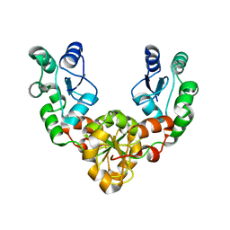
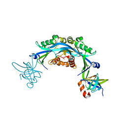
 | | CRYSTAL STRUCTURE OF R153C E. COLI MALATE DEHYDROGENASE | | 分子名称: | MALATE DEHYDROGENASE, NICOTINAMIDE-ADENINE-DINUCLEOTIDE, PYRUVIC ACID | | 著者 | Bell, J.K, Yennawar, H.P, Wright, S.K, Thompson, J.R, Viola, R.E, Banaszak, L.J. | | 登録日 | 2001-04-05 | | 公開日 | 2001-09-19 | | 最終更新日 | 2023-11-15 | | 実験手法 | X-RAY DIFFRACTION (2.5 Å) | | 主引用文献 | Structural Analyses of a Malate Dehydrogenase with a Variable Active Site
J.Biol.Chem., 276, 2001
|
|
4W8R
 | |
4W8S
 | |
4W8T
 | |
5KZ5
 | | Architecture of the Human Mitochondrial Iron-Sulfur Cluster Assembly Machinery: the Complex Formed by the Iron Donor, the Sulfur Donor, and the Scaffold | | 分子名称: | Cysteine desulfurase, mitochondrial, Frataxin, ... | | 著者 | Gakh, O, Ranatunga, W, Smith, D.Y, Ahlgren, E.C, Al-Karadaghi, S, Thompson, J.R, Isaya, G. | | 登録日 | 2016-07-22 | | 公開日 | 2016-08-31 | | 最終更新日 | 2019-12-18 | | 実験手法 | ELECTRON MICROSCOPY (14.3 Å) | | 主引用文献 | Architecture of the Human Mitochondrial Iron-Sulfur Cluster Assembly Machinery.
J.Biol.Chem., 291, 2016
|
|
7RR9
 | | Cryo-EM Structure of Nanodisc reconstituted ABCD1 in nucleotide bound outward open conformation | | 分子名称: | ATP-binding cassette sub-family D member 1, CHOLESTEROL, MAGNESIUM ION, ... | | 著者 | Alam, A, Le, L.T.M, Thompson, J.R. | | 登録日 | 2021-08-09 | | 公開日 | 2022-01-19 | | 最終更新日 | 2024-06-05 | | 実験手法 | ELECTRON MICROSCOPY (3.5 Å) | | 主引用文献 | Structures of the human peroxisomal fatty acid transporter ABCD1 in a lipid environment
Commun Biol, 5, 2022
|
|
7RRA
 | |
2FHD
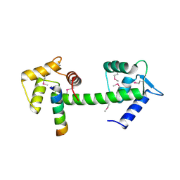
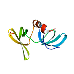
 | | Crystal structure of Crb2 tandem tudor domains | | 分子名称: | DNA repair protein rhp9/CRB2, PHOSPHATE ION | | 著者 | Lee, J, Botuyan, M.V, Thompson, J.R, Mer, G. | | 登録日 | 2005-12-23 | | 公開日 | 2007-01-02 | | 最終更新日 | 2011-07-13 | | 実験手法 | X-RAY DIFFRACTION (2.4 Å) | | 主引用文献 | Structural basis for the methylation state-specific recognition of histone H4-K20 by 53BP1 and Crb2 in DNA repair.
Cell(Cambridge,Mass.), 127, 2006
|
|
2ETX
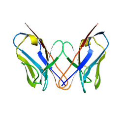
 | | Crystal Structure of MDC1 Tandem BRCT Domains | | 分子名称: | Mediator of DNA damage checkpoint protein 1 | | 著者 | Wasielewski, E, Kim, Y, Joachimiak, A, Thompson, J.R, Mer, G. | | 登録日 | 2005-10-27 | | 公開日 | 2005-11-15 | | 最終更新日 | 2024-02-14 | | 実験手法 | X-RAY DIFFRACTION (1.33 Å) | | 主引用文献 | Molecular Basis for the Association of Microcephalin (MCPH1) Protein with the Cell Division Cycle Protein 27 (Cdc27) Subunit of the Anaphase-promoting Complex.
J.Biol.Chem., 287, 2012
|
|
2G3R
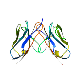
 | | Crystal Structure of 53BP1 tandem tudor domains at 1.2 A resolution | | 分子名称: | SULFATE ION, Tumor suppressor p53-binding protein 1 | | 著者 | Lee, J, Botuyan, M.V, Thompson, J.R, Mer, G. | | 登録日 | 2006-02-20 | | 公開日 | 2007-01-02 | | 最終更新日 | 2023-08-30 | | 実験手法 | X-RAY DIFFRACTION (1.25 Å) | | 主引用文献 | Structural Basis for the Methylation State-Specific Recognition of Histone H4-K20 by 53BP1 and Crb2 in DNA Repair.
Cell(Cambridge,Mass.), 127, 2006
|
|
3CDC
 | |
3CDY
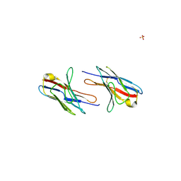
 | | AL-09 H87Y, immunoglobulin light chain variable domain | | 分子名称: | IMMUNOGLOBULIN LIGHT CHAIN | | 著者 | Baden, E.M, Randles, E.G, Aboagye, A.K, Thompson, J.R, Ramirez-Alvarado, M. | | 登録日 | 2008-02-27 | | 公開日 | 2008-09-02 | | 最終更新日 | 2023-08-30 | | 実験手法 | X-RAY DIFFRACTION (2.43 Å) | | 主引用文献 | Structural insights into the role of mutations in amyloidogenesis.
J.Biol.Chem., 283, 2008
|
|
3CDF
 | |
3PA6
 | |
6CO2
 | | Structure of an engineered protein (NUDT16TI) in complex with 53BP1 Tudor domains | | 分子名称: | NUDT16-Tudor-interacting (NUDT16TI), TP53-binding protein 1 | | 著者 | Botuyan, M.V, Thompson, J.R, Cui, G, Mer, G. | | 登録日 | 2018-03-10 | | 公開日 | 2018-06-06 | | 最終更新日 | 2023-10-04 | | 実験手法 | X-RAY DIFFRACTION (2.49 Å) | | 主引用文献 | Mechanism of 53BP1 activity regulation by RNA-binding TIRR and a designer protein.
Nat. Struct. Mol. Biol., 25, 2018
|
|
3P8D
 | | Crystal structure of the second Tudor domain of human PHF20 (homodimer form) | | 分子名称: | Medulloblastoma antigen MU-MB-50.72 | | 著者 | Cui, G, Lee, J, Thompson, J.R, Botuyan, M.V, Mer, G. | | 登録日 | 2010-10-13 | | 公開日 | 2011-06-22 | | 最終更新日 | 2024-10-16 | | 実験手法 | X-RAY DIFFRACTION (2 Å) | | 主引用文献 | PHF20 is an effector protein of p53 double lysine methylation that stabilizes and activates p53.
Nat.Struct.Mol.Biol., 19, 2012
|
|
3PD7
 | |


