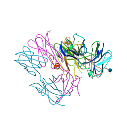
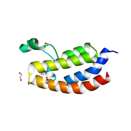
2YW6
 
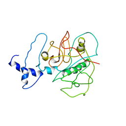
 | | Structural studies of N terminal deletion mutant of Dps from Mycobacterium smegmatis | | 分子名称: | DNA protection during starvation protein | | 著者 | Roy, S, Saraswathi, R, Gupta, S, Sekar, K, Chatterji, D, Vijayan, M. | | 登録日 | 2007-04-19 | | 公開日 | 2007-07-17 | | 最終更新日 | 2023-10-25 | | 実験手法 | X-RAY DIFFRACTION (2.53 Å) | | 主引用文献 | Role of N and C-terminal Tails in DNA Binding and Assembly in Dps: Structural Studies of Mycobacterium smegmatis Dps Deletion Mutants
J.Mol.Biol., 370, 2007
|
|
3USV
 | |
7M1S
 | |
3TNX
 | | Structure of the precursor of a thermostable variant of papain at 2.6 Angstroem resolution | | 分子名称: | CHLORIDE ION, Papain | | 著者 | Roy, S, Choudhury, D, Dattagupta, J.K, Biswas, S. | | 登録日 | 2011-09-02 | | 公開日 | 2012-09-12 | | 最終更新日 | 2023-11-01 | | 実験手法 | X-RAY DIFFRACTION (2.62 Å) | | 主引用文献 | The structure of a thermostable mutant of pro-papain reveals its activation mechanism
Acta Crystallogr.,Sect.D, 68, 2012
|
|
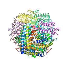
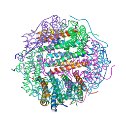
1VEQ
 | | Mycobacterium smegmatis Dps Hexagonal form | | 分子名称: | FE (III) ION, starvation-induced DNA protecting protein | | 著者 | Roy, S, Gupta, S, Das, S, Sekar, K, Chatterji, D, Vijayan, M. | | 登録日 | 2004-04-03 | | 公開日 | 2004-06-29 | | 最終更新日 | 2024-04-03 | | 実験手法 | X-RAY DIFFRACTION (3.98 Å) | | 主引用文献 | X-ray analysis of Mycobacterium smegmatis Dps and a comparative study involving other Dps and Dps-like molecules
J.Mol.Biol., 339, 2004
|
|
1VEL
 | | Mycobacterium smegmatis Dps tetragonal form | | 分子名称: | CADMIUM ION, SODIUM ION, SULFATE ION, ... | | 著者 | Roy, S, Gupta, S, Das, S, Sekar, K, Chatterji, D, Vijayan, M. | | 登録日 | 2004-04-01 | | 公開日 | 2004-06-29 | | 最終更新日 | 2024-04-03 | | 実験手法 | X-RAY DIFFRACTION (2.99 Å) | | 主引用文献 | X-ray analysis of Mycobacterium smegmatis Dps and a comparative study involving other Dps and Dps-like molecules
J.Mol.Biol., 339, 2004
|
|
1VEI
 | | Mycobacterium smegmatis Dps | | 分子名称: | FE (III) ION, SULFATE ION, starvation-induced DNA protecting protein | | 著者 | Roy, S, Gupta, S, Das, S, Sekar, K, Chatterji, D, Vijayan, M. | | 登録日 | 2004-03-31 | | 公開日 | 2004-06-29 | | 最終更新日 | 2023-12-27 | | 実験手法 | X-RAY DIFFRACTION (2.85 Å) | | 主引用文献 | X-ray Analysis of Mycobacterium smegmatis Dps and a Comparative Study Involving Other Dps and Dps-like Molecules
J.Mol.Biol., 339, 2004
|
|
2YW7
 | | Crystal structure of C-terminal deletion mutant of Mycobacterium smegmatis Dps | | 分子名称: | Starvation-induced DNA protecting protein | | 著者 | Roy, S, Saraswathi, R, Gupta, S, Sekar, K, Chatterji, D, Vijayan, M. | | 登録日 | 2007-04-19 | | 公開日 | 2007-07-17 | | 最終更新日 | 2023-10-25 | | 実験手法 | X-RAY DIFFRACTION (3.3 Å) | | 主引用文献 | Role of N and C-terminal Tails in DNA Binding and Assembly in Dps: Structural Studies of Mycobacterium smegmatis Dps Deletion Mutants
J.Mol.Biol., 370, 2007
|
|
6VKJ
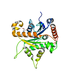 | |
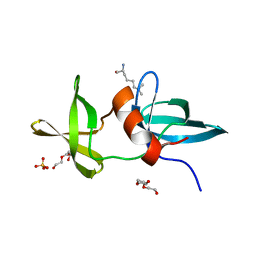
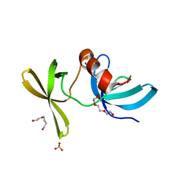
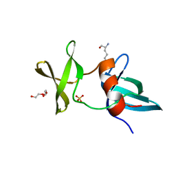
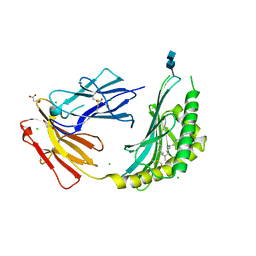
3BT4
 | | Crystal Structure Analysis of AmFPI-1, fungal protease inhibitor from Antheraea mylitta | | 分子名称: | Fungal protease inhibitor-1, GLYCEROL | | 著者 | Roy, S, Aravind, P, Madhurantakam, C, Ghosh, A.K, Sankarananarayanan, R, Das, A.K. | | 登録日 | 2007-12-27 | | 公開日 | 2008-12-30 | | 最終更新日 | 2017-10-25 | | 実験手法 | X-RAY DIFFRACTION (2.1 Å) | | 主引用文献 | Crystal structure of a fungal protease inhibitor from Antheraea mylitta
J.Struct.Biol., 166, 2009
|
|
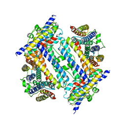
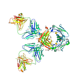
2Z90
 | | Crystal Structure of the Second Dps from Mycobacterium smegmatis | | 分子名称: | CHLORIDE ION, FE (II) ION, MAGNESIUM ION, ... | | 著者 | Roy, S, Saraswathi, R, Chatterji, D, Vijayan, M. | | 登録日 | 2007-09-13 | | 公開日 | 2008-04-22 | | 最終更新日 | 2023-11-01 | | 実験手法 | X-RAY DIFFRACTION (2.4 Å) | | 主引用文献 | Structural studies on the second Mycobacterium smegmatis Dps: invariant and variable features of structure, assembly and function.
J.Mol.Biol., 375, 2008
|
|
3LH0
 | |
3LGF
 | |
3LGL
 | |
4ONH
 | | Crystal Structure of DN6 TCR | | 分子名称: | 2-acetamido-2-deoxy-beta-D-glucopyranose, CHLORIDE ION, GLYCEROL, ... | | 著者 | Roy, S, Adams, E.J. | | 登録日 | 2014-01-28 | | 公開日 | 2014-10-08 | | 最終更新日 | 2020-07-29 | | 実験手法 | X-RAY DIFFRACTION (3.008 Å) | | 主引用文献 | Molecular basis of mycobacterial lipid antigen presentation by CD1c and its recognition by alpha beta T cells.
Proc.Natl.Acad.Sci.USA, 111, 2014
|
|
4ONO
 | | CD1c in complex with PM (phosphomycoketide) | | 分子名称: | (4R,8S,16S,20R)-4,8,12,16,20-pentamethylheptacosyl dihydrogen phosphate, 2-acetamido-2-deoxy-beta-D-glucopyranose-(1-4)-2-acetamido-2-deoxy-beta-D-glucopyranose, Beta-2-microglobulin/T-cell surface glycoprotein CD1c/T-cell surface glycoprotein CD1b chimeric protein, ... | | 著者 | Roy, S, Adams, E.J. | | 登録日 | 2014-01-28 | | 公開日 | 2014-10-08 | | 最終更新日 | 2020-07-29 | | 実験手法 | X-RAY DIFFRACTION (2.705 Å) | | 主引用文献 | Molecular basis of mycobacterial lipid antigen presentation by CD1c and its recognition by alpha beta T cells.
Proc.Natl.Acad.Sci.USA, 111, 2014
|
|
4S1E
 | |
4S1J
 | |
2J6E
 | | Crystal Structure of an Autoimmune Complex between a Human IgM Rheumatoid Factor and IgG1 Fc reveals a Novel Fc Epitope and Evidence for Affinity Maturation | | 分子名称: | (4S)-2-METHYL-2,4-PENTANEDIOL, ACETATE ION, CACODYLATE ION, ... | | 著者 | Duquerroy, S, Stura, E.A, Bressanelli, S, Browne, H, Beale, D, Hamon, M, Casali, P, Vaney, M.C, Rey, F.A, Sutton, B.J, Taussig, M.J. | | 登録日 | 2006-09-28 | | 公開日 | 2007-04-10 | | 最終更新日 | 2023-12-13 | | 実験手法 | X-RAY DIFFRACTION (3 Å) | | 主引用文献 | Crystal structure of a human autoimmune complex between IgM rheumatoid factor RF61 and IgG1 Fc reveals a novel epitope and evidence for affinity maturation.
J.Mol.Biol., 368, 2007
|
|
2VF1
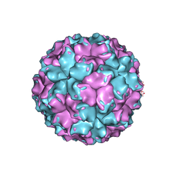 | | X-ray crystallographic structure of the picobirnavirus capsid | | 分子名称: | CAPSID PROTEIN | | 著者 | Duquerroy, S, Da Costa, B, Vigouroux, A, Lepault, J, Navaza, J, Delmas, B, Rey, F.A. | | 登録日 | 2007-10-29 | | 公開日 | 2008-12-16 | | 最終更新日 | 2024-05-01 | | 実験手法 | X-RAY DIFFRACTION (3.4 Å) | | 主引用文献 | The Picobirnavirus Crystal Structure Provides Functional Insights Into Virion Assembly and Cell Entry.
Embo J., 28, 2009
|
|
1WYY
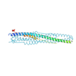 | | Post-fusion hairpin conformation of the sars coronavirus spike glycoprotein | | 分子名称: | CHLORIDE ION, E2 Glycoprotein | | 著者 | Duquerroy, S, Vigouroux, A, Rottier, P.J.M, Rey, F.A, Bosch, B.J. | | 登録日 | 2005-02-18 | | 公開日 | 2005-05-17 | | 最終更新日 | 2023-10-25 | | 実験手法 | X-RAY DIFFRACTION (2.2 Å) | | 主引用文献 | Central ions and lateral asparagine/glutamine zippers stabilize the post-fusion hairpin conformation of the SARS coronavirus spike glycoprotein
Virology, 335, 2005
|
|
6D7G
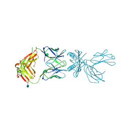 | | Structure of 5F3 TCR in complex with HLA-A2/MART-1 | | 分子名称: | 2-acetamido-2-deoxy-beta-D-glucopyranose, MART1 PEPTIDE-BETA-2-MICROGLOBULIN-HLA-A*02 CHIMERA, T-CELL RECEPTOR GAMMA VARIABLE 8,T-CELL RECEPTOR GAMMA-2 CHAIN C REGION, ... | | 著者 | Roy, S, Adams, E.J. | | 登録日 | 2018-04-24 | | 公開日 | 2019-01-23 | | 最終更新日 | 2020-07-29 | | 実験手法 | X-RAY DIFFRACTION (2.75 Å) | | 主引用文献 | Generation and molecular recognition of melanoma-associated antigen-specific human gamma delta T cells.
Sci Immunol, 3, 2018
|
|
4OUF
 | | Crystal Structure of CBP bromodomain | | 分子名称: | 1,2-ETHANEDIOL, CREB-binding protein, DI(HYDROXYETHYL)ETHER | | 著者 | Roy, S, Das, C, Tyler, J.K, Kutateladze, T.G. | | 登録日 | 2014-02-17 | | 公開日 | 2014-03-12 | | 最終更新日 | 2023-09-20 | | 実験手法 | X-RAY DIFFRACTION (1.4 Å) | | 主引用文献 | Binding of the histone chaperone ASF1 to the CBP bromodomain promotes histone acetylation.
Proc.Natl.Acad.Sci.USA, 111, 2014
|
|
5Z5O
 | | Structure of Pycnonodysostosis disease related I249T mutant of human cathepsin K | | 分子名称: | (4S)-2-METHYL-2,4-PENTANEDIOL, 1,2-ETHANEDIOL, CHLORIDE ION, ... | | 著者 | Biswas, S, Roy, S. | | 登録日 | 2018-01-19 | | 公開日 | 2018-09-26 | | 最終更新日 | 2023-11-22 | | 実験手法 | X-RAY DIFFRACTION (1.92 Å) | | 主引用文献 | Not all pycnodysostosis-related mutants of human cathepsin K are inactive - crystal structure and biochemical studies of an active mutant I249T.
FEBS J., 285, 2018
|
|
7EB1
 | | Solution NMR structure of the RRM domain of RNA binding protein RBM3 from homo sapiens | | 分子名称: | RNA-binding protein 3 | | 著者 | Boral, S, Roy, S, Basak, A.J, Maiti, S, Lee, W, De, S. | | 登録日 | 2021-03-08 | | 公開日 | 2021-12-08 | | 最終更新日 | 2024-05-15 | | 実験手法 | SOLUTION NMR | | 主引用文献 | Structural and dynamic studies of the human RNA binding protein RBM3 reveals the molecular basis of its oligomerization and RNA recognition.
Febs J., 289, 2022
|
|


