Loading
PDBj
メニューPDBj@FacebookPDBj@TwitterPDBj@YouTubewwPDB FoundationwwPDB
RCSB PDBPDBeBMRBAdv. SearchSearch help
Search by PDB author
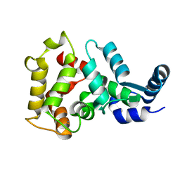
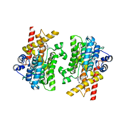
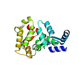
4M2O
DownloadVisualize
BU of 4m2o by Molmil
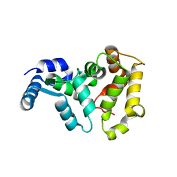
Crystal structure of a non-myristoylated C39A recoverin mutant with one calcium ion bound to EF-hand 3
分子名称: CALCIUM ION, Recoverin
著者Prem Kumar, R, Ranaghan, M.J, Oprian, D.D.
登録日2013-08-05
公開日2013-11-13
最終更新日2023-09-20
実験手法X-RAY DIFFRACTION (1.5 Å)
主引用文献A Highly Conserved Cysteine of Neuronal Calcium-sensing Proteins Controls Cooperative Binding of Ca2+ to Recoverin.
J.Biol.Chem., 288, 2013
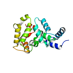
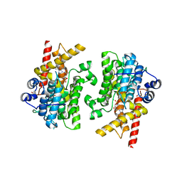
4MLW
DownloadVisualize
BU of 4mlw by Molmil
Crystal structure of non-myristoylated recoverin at 1.45 A resolution with calcium bound to EF-hand 3
分子名称: CALCIUM ION, Recoverin
著者Prem Kumar, R, Ranaghan, M.J, Oprian, D.D.
登録日2013-09-06
公開日2013-11-13
最終更新日2023-09-20
実験手法X-RAY DIFFRACTION (1.45 Å)
主引用文献A highly conserved cysteine of neuronal calcium-sensing proteins controls cooperative binding of Ca2+ to recoverin.
J.Biol.Chem., 288, 2013
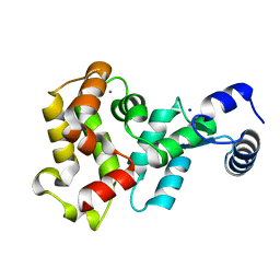
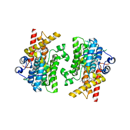
4M2P
DownloadVisualize
BU of 4m2p by Molmil
Crystal structure of a non-myristoylated C39D recoverin mutant with one calcium ion bound to EF-hand 3
分子名称: CALCIUM ION, Recoverin
著者Prem Kumar, R, Ranaghan, M.J, Oprian, D.D.
登録日2013-08-05
公開日2013-11-13
最終更新日2023-09-20
実験手法X-RAY DIFFRACTION (1.45 Å)
主引用文献A Highly Conserved Cysteine of Neuronal Calcium-sensing Proteins Controls Cooperative Binding of Ca2+ to Recoverin.
J.Biol.Chem., 288, 2013
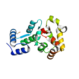
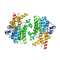
4YI8
DownloadVisualize
BU of 4yi8 by Molmil
Crystal structure of non-myristoylated E153A recoverin at 1.2 A resolution with calcium ions bound to EF-hands 2 and 3
分子名称: CALCIUM ION, Recoverin, SULFATE ION
著者Prem Kumar, R, Ranaghan, M.J, Oprian, D.D.
登録日2015-02-28
公開日2015-12-02
最終更新日2023-09-27
実験手法X-RAY DIFFRACTION (1.2 Å)
主引用文献Crystal Structure of Recoverin with Calcium Ions Bound to Both Functional EF Hands.
Biochemistry, 54, 2015
4YI9
DownloadVisualize
BU of 4yi9 by Molmil
Crystal structure of non-myristoylated E153A recoverin at 1.35 A resolution with a sodium ion bound to EF-hand 2 and calcium ion bound to EF-hand 3
分子名称: CALCIUM ION, Recoverin, SODIUM ION
著者Prem Kumar, R, Ranaghan, M.J, Oprian, D.D.
登録日2015-02-28
公開日2015-12-02
最終更新日2023-09-27
実験手法X-RAY DIFFRACTION (1.35 Å)
主引用文献Crystal Structure of Recoverin with Calcium Ions Bound to Both Functional EF Hands.
Biochemistry, 54, 2015
4M2Q
DownloadVisualize
BU of 4m2q by Molmil
Crystal structure of non-myristoylated recoverin with Cysteine-39 oxidized to sulfenic acid
分子名称: CALCIUM ION, Recoverin
著者Prem Kumar, R, Chakrabarti, K, Kern, D, Oprian, D.D.
登録日2013-08-05
公開日2013-11-13
最終更新日2023-09-20
実験手法X-RAY DIFFRACTION (1.9 Å)
主引用文献A Highly Conserved Cysteine of Neuronal Calcium-sensing Proteins Controls Cooperative Binding of Ca2+ to Recoverin.
J.Biol.Chem., 288, 2013
6V0N
DownloadVisualize
BU of 6v0n by Molmil
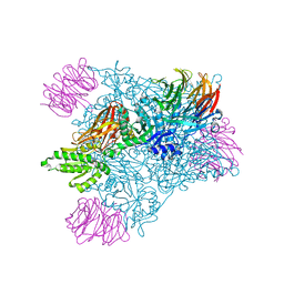
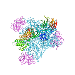
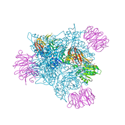
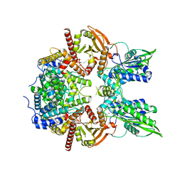
PRMT5 bound to PBM peptide from Riok1
分子名称: Methylosome protein 50, Protein arginine N-methyltransferase 5, Riok1 PBM peptide, ...
著者McMIllan, B.J, Raymond, D.D.
登録日2019-11-19
公開日2020-08-26
最終更新日2023-10-11
実験手法X-RAY DIFFRACTION (2.11 Å)
主引用文献Molecular basis for substrate recruitment to the PRMT5 methylosome.
Mol.Cell, 81, 2021
6V0O
DownloadVisualize
BU of 6v0o by Molmil
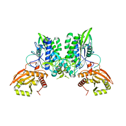
PRMT5 bound to the PBM peptide from pICln
分子名称: ACETYL GROUP, Methylosome protein 50, PBM peptide, ...
著者McMillan, B.J, Raymond, D.D.
登録日2019-11-19
公開日2020-08-26
最終更新日2023-10-11
実験手法X-RAY DIFFRACTION (2.86 Å)
主引用文献Molecular basis for substrate recruitment to the PRMT5 methylosome.
Mol.Cell, 81, 2021
6V0P
DownloadVisualize
BU of 6v0p by Molmil
PRMT5 complex bound to covalent PBM inhibitor BRD6711
分子名称: 2-(5-chloro-6-oxopyridazin-1(6H)-yl)-N-(4-methyl-3-sulfamoylphenyl)acetamide, CHLORIDE ION, GLYCEROL, ...
著者McMillan, B.J, McKinney, D.C.
登録日2019-11-19
公開日2020-11-25
最終更新日2024-10-23
実験手法X-RAY DIFFRACTION (1.88 Å)
主引用文献Discovery of a First-in-Class Inhibitor of the PRMT5-Substrate Adaptor Interaction.
J.Med.Chem., 64, 2021
7M05
DownloadVisualize
BU of 7m05 by Molmil
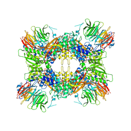
CryoEM structure of PRMT5 bound to covalent PBM-site inhibitor BRD-6988
分子名称: 2-(5-chloro-6-oxopyridazin-1(6H)-yl)-N-(4-methyl-3-{[2-(pyridin-2-yl)ethyl]sulfamoyl}phenyl)acetamide, Methylosome protein 50, Protein arginine N-methyltransferase 5
著者McMillan, B.J, McKinney, D.C, Timm, D.E.
登録日2021-03-10
公開日2021-03-17
最終更新日2021-08-25
実験手法ELECTRON MICROSCOPY (2.39 Å)
主引用文献Discovery of a First-in-Class Inhibitor of the PRMT5-Substrate Adaptor Interaction.
J.Med.Chem., 64, 2021
7L28
DownloadVisualize
BU of 7l28 by Molmil
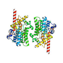
Crystal structure of the catalytic domain of human PDE3A bound to Trequinsin
分子名称: (2E)-9,10-dimethoxy-3-methyl-2-[(2,4,6-trimethylphenyl)imino]-2,3,6,7-tetrahydro-4H-pyrimido[6,1-a]isoquinolin-4-one, ACETATE ION, MAGNESIUM ION, ...
著者Horner, S.W, Garvie, C.
登録日2020-12-16
公開日2021-06-16
最終更新日2023-10-18
実験手法X-RAY DIFFRACTION (2.2 Å)
主引用文献Structure of PDE3A-SLFN12 complex reveals requirements for activation of SLFN12 RNase.
Nat Commun, 12, 2021
7L29
DownloadVisualize
BU of 7l29 by Molmil
Crystal structure of the catalytic domain of human PDE3A bound to AMP
分子名称: ACETATE ION, ADENOSINE MONOPHOSPHATE, CALCIUM ION, ...
著者Horner, S.W, Garvie, C.
登録日2020-12-16
公開日2021-06-16
最終更新日2024-04-03
実験手法X-RAY DIFFRACTION (2.08 Å)
主引用文献Structure of PDE3A-SLFN12 complex reveals requirements for activation of SLFN12 RNase.
Nat Commun, 12, 2021
7KWE
DownloadVisualize
BU of 7kwe by Molmil
Crystal structure of the catalytic domain of human PDE3A bound to DNMDP
分子名称: (4~{R})-3-[4-(diethylamino)-3-[oxidanyl(oxidanylidene)-$l^{4}-azanyl]phenyl]-4-methyl-4,5-dihydro-1~{H}-pyridazin-6-one, ACETATE ION, MAGNESIUM ION, ...
著者Horner, S.W, Garvie, C.
登録日2020-11-30
公開日2021-06-16
最終更新日2023-10-18
実験手法X-RAY DIFFRACTION (2 Å)
主引用文献Structure of PDE3A-SLFN12 complex reveals requirements for activation of SLFN12 RNase.
Nat Commun, 12, 2021
7L27
DownloadVisualize
BU of 7l27 by Molmil
Crystal structure of the catalytic domain of human PDE3A
分子名称: ACETATE ION, CALCIUM ION, MAGNESIUM ION, ...
著者Horner, S.W, Garvie, C.
登録日2020-12-16
公開日2021-06-16
最終更新日2023-10-18
実験手法X-RAY DIFFRACTION (1.7 Å)
主引用文献Structure of PDE3A-SLFN12 complex reveals requirements for activation of SLFN12 RNase.
Nat Commun, 12, 2021
7LRD
DownloadVisualize
BU of 7lrd by Molmil
Cryo-EM of the SLFN12-PDE3A complex: Consensus subset model
分子名称: (4~{R})-3-[4-(diethylamino)-3-[oxidanyl(oxidanylidene)-$l^{4}-azanyl]phenyl]-4-methyl-4,5-dihydro-1~{H}-pyridazin-6-one, MAGNESIUM ION, MANGANESE (II) ION, ...
著者Fuller, J.R, Garvie, C.W, Lemke, C.T.
登録日2021-02-16
公開日2021-06-09
最終更新日2024-03-06
実験手法ELECTRON MICROSCOPY (3.22 Å)
主引用文献Structure of PDE3A-SLFN12 complex reveals requirements for activation of SLFN12 RNase.
Nat Commun, 12, 2021
7LRE
DownloadVisualize
BU of 7lre by Molmil
Cryo-EM of the SLFN12-PDE3A complex: SLFN12 body refinement
分子名称: Schlafen family member 12, ZINC ION
著者Fuller, J.R, Garvie, C.W, Lemke, C.T.
登録日2021-02-16
公開日2021-06-09
最終更新日2024-03-06
実験手法ELECTRON MICROSCOPY (2.76 Å)
主引用文献Structure of PDE3A-SLFN12 complex reveals requirements for activation of SLFN12 RNase.
Nat Commun, 12, 2021
7LRC
DownloadVisualize
BU of 7lrc by Molmil
Cryo-EM of the SLFN12-PDE3A complex: PDE3A body refinement
分子名称: (4~{R})-3-[4-(diethylamino)-3-[oxidanyl(oxidanylidene)-$l^{4}-azanyl]phenyl]-4-methyl-4,5-dihydro-1~{H}-pyridazin-6-one, MAGNESIUM ION, MANGANESE (II) ION, ...
著者Fuller, J.R, Garvie, C.W, Lemke, C.T.
登録日2021-02-16
公開日2021-06-09
最終更新日2024-03-06
実験手法ELECTRON MICROSCOPY (2.97 Å)
主引用文献Structure of PDE3A-SLFN12 complex reveals requirements for activation of SLFN12 RNase.
Nat Commun, 12, 2021

226707

件を2024-10-30に公開中

PDB statisticsPDBj update infoContact PDBjnumon