Loading
PDBj
メニューPDBj@FacebookPDBj@TwitterPDBj@YouTubewwPDB FoundationwwPDB
RCSB PDBPDBeBMRBAdv. SearchSearch help
Search by PDB author
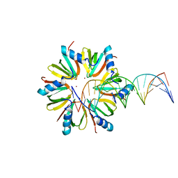
5UK7
DownloadVisualize
BU of 5uk7 by Molmil
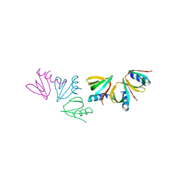
Escherichia coli Hfq bound to dsDNA
分子名称: DNA (5'-D(P*CP*GP*GP*CP*AP*AP*AP*AP*AP*AP*CP*GP*GP*CP*AP*AP*AP*AP*AP*A)-3'), DNA (5'-D(P*TP*TP*TP*TP*TP*TP*GP*CP*CP*GP*TP*TP*TP*TP*TP*TP*GP*CP*CP*G)-3'), RNA-binding protein Hfq, ...
著者Orans, J, Kovach, A.R, Brennan, R.G.
登録日2017-01-20
公開日2018-05-09
最終更新日2023-10-04
実験手法X-RAY DIFFRACTION (3 Å)
主引用文献Crystal structure of Escherichia coli Hfq DNA complex reveals multifunctional nucleic acid binding site
To Be Published
3QE9
DownloadVisualize
BU of 3qe9 by Molmil
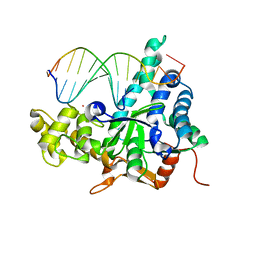
Crystal structure of human exonuclease 1 Exo1 (D173A) in complex with DNA (complex I)
分子名称: CALCIUM ION, DNA (5'-D(*CP*GP*CP*TP*AP*GP*TP*CP*GP*AP*CP*AP*T)-3'), DNA (5'-D(P*TP*CP*GP*AP*CP*TP*AP*GP*CP*G)-3'), ...
著者Orans, J, McSweeney, E.A, Iyer, R.R, Hast, M.A, Hellinga, H.W, Modrich, P, Beese, L.S.
登録日2011-01-20
公開日2011-04-20
最終更新日2024-02-21
実験手法X-RAY DIFFRACTION (2.51 Å)
主引用文献Structures of human exonuclease 1 DNA complexes suggest a unified mechanism for nuclease family.
Cell(Cambridge,Mass.), 145, 2011
3QEA
DownloadVisualize
BU of 3qea by Molmil
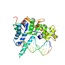
Crystal structure of human exonuclease 1 Exo1 (WT) in complex with DNA (complex II)
分子名称: BARIUM ION, DNA (5'-D(P*CP*GP*CP*TP*AP*GP*TP*CP*GP*AP*CP*AP*T)-3'), DNA (5'-D(P*TP*CP*GP*AP*CP*TP*AP*GP*CP*G)-3'), ...
著者Orans, J, McSweeney, E.A, Iyer, R.R, Hast, M.A, Hellinga, H.W, Modrich, P, Beese, L.S.
登録日2011-01-20
公開日2011-04-20
最終更新日2024-02-21
実験手法X-RAY DIFFRACTION (3.1 Å)
主引用文献Structures of human exonuclease 1 DNA complexes suggest a unified mechanism for nuclease family.
Cell(Cambridge,Mass.), 145, 2011
3QEB
DownloadVisualize
BU of 3qeb by Molmil
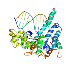
Crystal structure of human exonuclease 1 Exo1 (WT) in complex with DNA and Mn2+ (complex III)
分子名称: DNA (5'-D(*CP*GP*CP*TP*AP*GP*TP*CP*GP*AP*CP*AP*T)-3'), DNA (5'-D(P*TP*CP*GP*AP*CP*TP*AP*GP*CP*G)-3'), Exonuclease 1, ...
著者Orans, J, McSweeney, E.A, Iyer, R.R, Hast, M.A, Hellinga, H.W, Modrich, P, Beese, L.S.
登録日2011-01-20
公開日2011-04-20
最終更新日2024-02-21
実験手法X-RAY DIFFRACTION (3 Å)
主引用文献Structures of human exonuclease 1 DNA complexes suggest a unified mechanism for nuclease family.
Cell(Cambridge,Mass.), 145, 2011
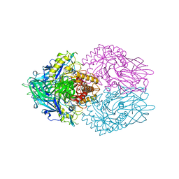
3HX6
DownloadVisualize
BU of 3hx6 by Molmil
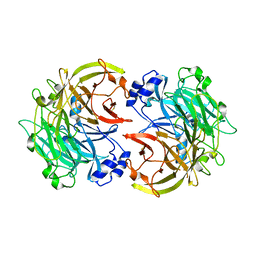
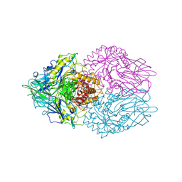
Crystal structure of Pseudomonas aeruginosa PilY1 C-terminal domain
分子名称: CALCIUM ION, Type 4 fimbrial biogenesis protein PilY1
著者Redinbo, M.R, Orans, J.
登録日2009-06-19
公開日2010-01-26
最終更新日2021-10-13
実験手法X-RAY DIFFRACTION (2.1 Å)
主引用文献Crystal structure analysis reveals Pseudomonas PilY1 as an essential calcium-dependent regulator of bacterial surface motility.
Proc.Natl.Acad.Sci.USA, 107, 2010
4JRK
DownloadVisualize
BU of 4jrk by Molmil
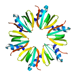
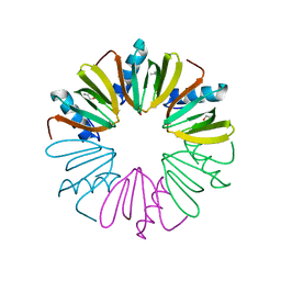
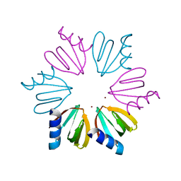
Crystal Structure of Escherichia coli Hfq Surface Mutant
分子名称: Protein hfq
著者Robinson, K.E, Orans, J.
登録日2013-03-21
公開日2013-12-11
最終更新日2024-02-28
実験手法X-RAY DIFFRACTION (1.894 Å)
主引用文献Mapping Hfq-RNA interaction surfaces using tryptophan fluorescence quenching.
Nucleic Acids Res., 42, 2014
4JUV
DownloadVisualize
BU of 4juv by Molmil
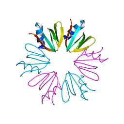
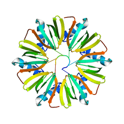
Crystal Structure of Escherichia coli Hfq Distal Face 1 Mutant
分子名称: Protein hfq
著者Robinson, K.E, Orans, J.
登録日2013-03-25
公開日2013-12-11
最終更新日2024-02-28
実験手法X-RAY DIFFRACTION (2.19 Å)
主引用文献Mapping Hfq-RNA interaction surfaces using tryptophan fluorescence quenching.
Nucleic Acids Res., 42, 2014
4JRI
DownloadVisualize
BU of 4jri by Molmil
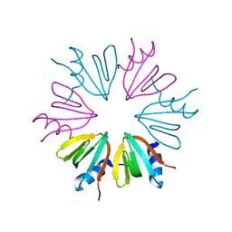
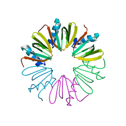
Crystal Structure of Escherichia coli Hfq Proximal Edge Mutant
分子名称: Protein hfq
著者Robinson, K.E, Orans, J.
登録日2013-03-21
公開日2013-12-11
最終更新日2024-02-28
実験手法X-RAY DIFFRACTION (1.829 Å)
主引用文献Mapping Hfq-RNA interaction surfaces using tryptophan fluorescence quenching.
Nucleic Acids Res., 42, 2014
4JLI
DownloadVisualize
BU of 4jli by Molmil
Crystal Structure of Escherichia coli Hfq Proximal Pore Mutant
分子名称: Protein hfq
著者Robinson, K.E, Orans, J.
登録日2013-03-12
公開日2013-12-11
最終更新日2024-02-28
実験手法X-RAY DIFFRACTION (1.79 Å)
主引用文献Mapping Hfq-RNA interaction surfaces using tryptophan fluorescence quenching.
Nucleic Acids Res., 42, 2014
3K4A
DownloadVisualize
BU of 3k4a by Molmil
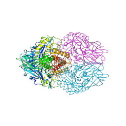
Crystal structure of selenomethionine substituted E. coli beta-glucuronidase
分子名称: Beta-glucuronidase
著者Wallace, B.D, Orans, J, Redinbo, M.R.
登録日2009-10-05
公開日2010-11-17
最終更新日2017-11-01
実験手法X-RAY DIFFRACTION (2.9 Å)
主引用文献Alleviating cancer drug toxicity by inhibiting a bacterial enzyme.
Science, 330, 2010
4NL2
DownloadVisualize
BU of 4nl2 by Molmil
Crystal Structure of Listeria monocytogenes Hfq
分子名称: Protein hfq, S-1,2-PROPANEDIOL
著者Kovach, A.R, Brennan, R.G.
登録日2013-11-13
公開日2014-09-10
最終更新日2024-02-28
実験手法X-RAY DIFFRACTION (2.6 Å)
主引用文献Recognition of U-rich RNA by Hfq from the Gram-positive pathogen Listeria monocytogenes.
Rna, 20, 2014
4NL3
DownloadVisualize
BU of 4nl3 by Molmil
Crystal Structure of Listeria monocytogenes Hfq in complex with U6 RNA
分子名称: 5'-R(*UP*UP*UP*UP*UP*U)-3', Protein hfq
著者Kovach, A.R, Brennan, R.G.
登録日2013-11-13
公開日2014-09-10
最終更新日2023-09-20
実験手法X-RAY DIFFRACTION (3.1 Å)
主引用文献Recognition of U-rich RNA by Hfq from the Gram-positive pathogen Listeria monocytogenes.
Rna, 20, 2014
4NOY
DownloadVisualize
BU of 4noy by Molmil
Crystal structure of Listeria monocytogenes Hfq F43W
分子名称: Protein Hfq, S-1,2-PROPANEDIOL
著者Canty, J.T, Kovach, A.R, Brennan, R.G.
登録日2013-11-20
公開日2014-09-10
最終更新日2023-09-20
実験手法X-RAY DIFFRACTION (2.795 Å)
主引用文献Recognition of U-rich RNA by Hfq from the Gram-positive pathogen Listeria monocytogenes.
Rna, 20, 2014
3LPF
DownloadVisualize
BU of 3lpf by Molmil
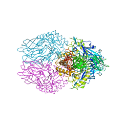
Structure of E. coli beta-Glucuronidase bound with a novel, potent inhibitor 1-((6,7-dimethyl-2-oxo-1,2-dihydroquinolin-3-yl)methyl)-1-(2-hydroxyethyl)-3-(3-methoxyphenyl)thiourea
分子名称: 1-[(6,7-dimethyl-2-oxo-1,2-dihydroquinolin-3-yl)methyl]-1-(2-hydroxyethyl)-3-(3-methoxyphenyl)thiourea, Beta-glucuronidase
著者Wallace, B.D, Redinbo, M.R.
登録日2010-02-05
公開日2010-11-17
最終更新日2023-11-22
実験手法X-RAY DIFFRACTION (2.26 Å)
主引用文献Alleviating cancer drug toxicity by inhibiting a bacterial enzyme.
Science, 330, 2010
3LPG
DownloadVisualize
BU of 3lpg by Molmil
Structure of E. coli beta-Glucuronidase bound with a novel, potent inhibitor 3-(2-fluorophenyl)-1-(2-hydroxyethyl)-1-((6-methyl-2-oxo-1,2-dihydroquinolin-3-yl)methyl)urea
分子名称: 3-(2-fluorophenyl)-1-(2-hydroxyethyl)-1-[(6-methyl-2-oxo-1,2-dihydroquinolin-3-yl)methyl]urea, Beta-glucuronidase
著者Wallace, B.D, Redinbo, M.R.
登録日2010-02-05
公開日2010-11-17
最終更新日2023-11-22
実験手法X-RAY DIFFRACTION (2.425 Å)
主引用文献Alleviating cancer drug toxicity by inhibiting a bacterial enzyme.
Science, 330, 2010
3K46
DownloadVisualize
BU of 3k46 by Molmil
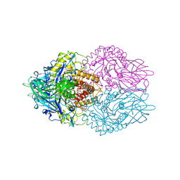
Crystal structure of full-length E. coli beta-glucuronidase
分子名称: Beta-glucuronidase
著者Wallace, B.D, Lane, K.T, Redinbo, M.R.
登録日2009-10-05
公開日2010-11-17
最終更新日2023-09-06
実験手法X-RAY DIFFRACTION (2.5 Å)
主引用文献Alleviating cancer drug toxicity by inhibiting a bacterial enzyme.
Science, 330, 2010
3QSU
DownloadVisualize
BU of 3qsu by Molmil
Structure of Staphylococcus aureus Hfq in complex with A7 RNA
分子名称: RNA (5'-R(P*AP*AP*AP*AP*AP*AP*A)-3'), RNA chaperone Hfq, ZINC ION
著者Brennan, R, Horstmann, N, Link, T.M.
登録日2011-02-21
公開日2012-09-12
最終更新日2024-02-21
実験手法X-RAY DIFFRACTION (2.2 Å)
主引用文献Structural mechanism of Staphylococcus aureus Hfq binding to an RNA A-tract.
Nucleic Acids Res., 40, 2012
3K4D
DownloadVisualize
BU of 3k4d by Molmil
Crystal structure of E. coli beta-glucuronidase with the glucaro-d-lactam inhibitor bound
分子名称: (2S,3R,4S,5R)-3,4,5-trihydroxy-6-oxopiperidine-2-carboxylic acid, Beta-glucuronidase
著者Wallace, B.D, Redinbo, M.R.
登録日2009-10-05
公開日2010-11-17
最終更新日2023-09-06
実験手法X-RAY DIFFRACTION (2.393 Å)
主引用文献Alleviating cancer drug toxicity by inhibiting a bacterial enzyme.
Science, 330, 2010

224931

件を2024-09-11に公開中

PDB statisticsPDBj update infoContact PDBjnumon