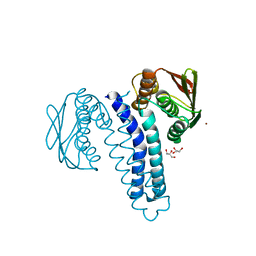
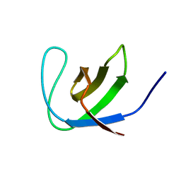
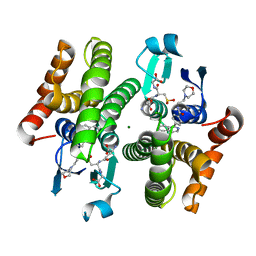
1Y9O
 
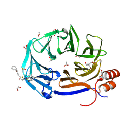
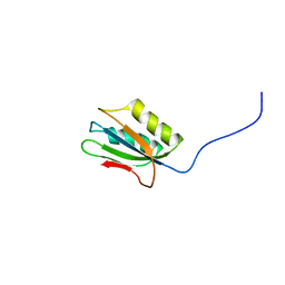 | | 1H NMR Structure of Acylphosphatase from the hyperthermophile Sulfolobus Solfataricus | | 分子名称: | Acylphosphatase | | 著者 | Corazza, A, Rosano, C, Pagano, K, Alverdi, V, Esposito, G, Capanni, C, Bemporad, F, Plakoutsi, G, Stefani, M, Chiti, F, Zuccotti, S, Bolognesi, M, Viglino, P. | | 登録日 | 2004-12-16 | | 公開日 | 2005-11-29 | | 最終更新日 | 2024-05-29 | | 実験手法 | SOLUTION NMR | | 主引用文献 | Structure, conformational stability, and enzymatic properties of acylphosphatase from the hyperthermophile Sulfolobus solfataricus
Proteins, 62, 2006
|
|
8SV0
 | |
8U36
 | |
1G90
 | |
4FPP
 | | Bacterial phosphotransferase | | 分子名称: | GLYCEROL, MAGNESIUM ION, NICKEL (II) ION, ... | | 著者 | Fioravanti, A, Clantin, B, Dewitte, F, Lens, Z, Verger, A, Biondi, E, Villeret, V. | | 登録日 | 2012-06-22 | | 公開日 | 2012-09-12 | | 最終更新日 | 2024-02-28 | | 実験手法 | X-RAY DIFFRACTION (2.2 Å) | | 主引用文献 | Structural insights into ChpT, an essential dimeric histidine phosphotransferase regulating the cell cycle in Caulobacter crescentus.
Acta Crystallogr.,Sect.F, 68, 2012
|
|
7WH4
 | |
6L6O
 | | Crystal structure of stabilized Rab5a GTPase domain from Leishmania donovani | | 分子名称: | ACETATE ION, GLYCEROL, GUANOSINE-5'-DIPHOSPHATE, ... | | 著者 | Arora, A, Zohib, M, Biswal, B.K, Maheshwari, D, Pal, R.K. | | 登録日 | 2019-10-29 | | 公開日 | 2020-11-11 | | 最終更新日 | 2023-11-22 | | 実験手法 | X-RAY DIFFRACTION (1.8 Å) | | 主引用文献 | Crystal structure of the GDP-bound GTPase domain of Rab5a from Leishmania donovani.
Acta Crystallogr.,Sect.F, 76, 2020
|
|
4RP7
 | |
4RP6
 | |
9C0Y
 | | Clathrin terminal domain complexed with Pitstop 2c | | 分子名称: | 1,2-ETHANEDIOL, ACETATE ION, Clathrin heavy chain 1, ... | | 著者 | Bulut, H, Horatscheck, A, Krauss, M, Santos, K.F, McCluskey, A, Wahl, C.W, Nazare, M, Haucke, V. | | 登録日 | 2024-05-28 | | 公開日 | 2024-06-26 | | 実験手法 | X-RAY DIFFRACTION (1.4 Å) | | 主引用文献 | Acute inhibition of clathrin-mediated endocytosis by next-generation small molecule inhibitors of clathrin function
To Be Published
|
|
9C0Z
 | | Clathrin terminal domain complexed with pitstop 2d | | 分子名称: | 1,2-ETHANEDIOL, 2-AMINO-2-HYDROXYMETHYL-PROPANE-1,3-DIOL, ACETATE ION, ... | | 著者 | Bulut, H, Horatscheck, A, Krauss, M, Santos, K.F, McCluskey, A, Wahl, C.W, Nazare, M, Haucke, V. | | 登録日 | 2024-05-28 | | 公開日 | 2024-06-26 | | 実験手法 | X-RAY DIFFRACTION (1.51 Å) | | 主引用文献 | Acute inhibition of clathrin-mediated endocytosis by next-generation small molecule inhibitors of clathrin function
To Be Published
|
|
6HHU
 | |
6QX4
 | |
4QXX
 | |
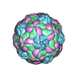
4UDF
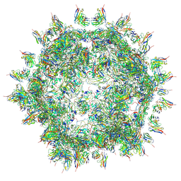 | | STRUCTURAL BASIS OF HUMAN PARECHOVIRUS NEUTRALIZATION BY HUMAN MONOCLONAL ANTIBODIES | | 分子名称: | HUMAN MONOCLONAL ANTIBODY, Protein VP0, Protein VP3 | | 著者 | Shakeel, S, Westerhuis, B.M, Ora, A, Koen, G, Bakker, A.Q, Claassen, Y, Beaumont, T, Wolthers, K.C, Butcher, S.J. | | 登録日 | 2014-12-10 | | 公開日 | 2015-07-22 | | 最終更新日 | 2018-10-03 | | 実験手法 | ELECTRON MICROSCOPY (20 Å) | | 主引用文献 | Structural Basis of Human Parechovirus Neutralization by Human Monoclonal Antibodies.
J. Virol., 89, 2015
|
|
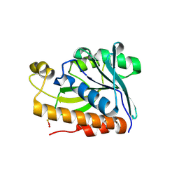
2JTE
 | | Third SH3 domain of CD2AP | | 分子名称: | CD2-associated protein | | 著者 | van Nuland, N.A.J, Ortega, R.J, Romero Romero, M, Ab, E, Ora, A, Lopez, M.O, Azuaga, A.I. | | 登録日 | 2007-07-28 | | 公開日 | 2007-12-11 | | 最終更新日 | 2024-05-29 | | 実験手法 | SOLUTION NMR | | 主引用文献 | The high resolution NMR structure of the third SH3 domain of CD2AP.
J.Biomol.Nmr, 39, 2007
|
|
5MJV
 | | Rebuild and re-refined model for Human Parechovirus 1 | | 分子名称: | CAPSID SUBUNIT VP0, CAPSID SUBUNIT VP1, CAPSID SUBUNIT VP3, ... | | 著者 | Shakeel, S, Dykeman, E.C, White, S.J, Ora, A, Cockburn, J.J.B, Butcher, S.J, Stockley, P.G, Twarock, R. | | 登録日 | 2016-12-01 | | 公開日 | 2017-01-11 | | 最終更新日 | 2024-01-17 | | 実験手法 | X-RAY DIFFRACTION (3.09 Å) | | 主引用文献 | Genomic RNA folding mediates assembly of human parechovirus.
Nat Commun, 8, 2017
|
|
4DHW
 | | Crystal structure of Peptidyl-tRNA hydrolase from Pseudomonas aeruginosa with Adipic acid at 2.4 Angstrom resolution | | 分子名称: | Peptidyl-tRNA hydrolase, hexanedioic acid | | 著者 | Kumar, A, Singh, A, Singh, N, Sinha, M, Sharma, S, Arora, A, Singh, T.P. | | 登録日 | 2012-01-30 | | 公開日 | 2012-02-29 | | 最終更新日 | 2023-11-08 | | 実験手法 | X-RAY DIFFRACTION (2.43 Å) | | 主引用文献 | Crystal structure of Peptidyl-tRNA hydrolase from Pseudomonas aeruginosa with Adipic acid at 2.4 Angstrom resolution
To be Published
|
|
3KXO
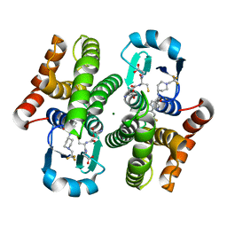 | | An orally active inhibitor bound at the active site of HPGDS | | 分子名称: | 6-(3-fluorophenyl)-N-[1-(2,2,2-trifluoroethyl)piperidin-4-yl]pyridine-3-carboxamide, GLUTATHIONE, Glutathione-requiring prostaglandin D synthase, ... | | 著者 | Kiefer, J.R, Day, J.E, Thorarensen, A. | | 登録日 | 2009-12-03 | | 公開日 | 2010-09-01 | | 最終更新日 | 2024-02-21 | | 実験手法 | X-RAY DIFFRACTION (2.1 Å) | | 主引用文献 | Discovery of an oral potent selective inhibitor of hematopoietic prostaglandin D synthase
TO BE PUBLISHED
|
|
5EKT
 | |
4EE0
 | | Crystal structure of hH-PGDS with water displacing inhibitor | | 分子名称: | 4-(isoquinolin-1-yl)-N-[2-(morpholin-4-yl)ethyl]benzamide, Hematopoietic prostaglandin D synthase, L-GAMMA-GLUTAMYL-3-SULFINO-L-ALANYLGLYCINE, ... | | 著者 | Day, J.E, Thorarensen, A, Trujillo, J.I. | | 登録日 | 2012-03-28 | | 公開日 | 2012-07-18 | | 最終更新日 | 2024-02-28 | | 実験手法 | X-RAY DIFFRACTION (1.75 Å) | | 主引用文献 | Investigation of the binding pocket of human hematopoietic prostaglandin (PG) D2 synthase (hH-PGDS): a tale of two waters.
Bioorg.Med.Chem.Lett., 22, 2012
|
|
4EC0
 | | Crystal structure of hH-PGDS with water displacing inhibitor | | 分子名称: | 4-[2-(aminomethyl)naphthalen-1-yl]-N-[2-(morpholin-4-yl)ethyl]benzamide, GLUTATHIONE, Hematopoietic prostaglandin D synthase, ... | | 著者 | Day, J.E, Thorarensen, A, Trujillo, J.I. | | 登録日 | 2012-03-26 | | 公開日 | 2012-05-16 | | 最終更新日 | 2024-02-28 | | 実験手法 | X-RAY DIFFRACTION (1.85 Å) | | 主引用文献 | Investigation of the binding pocket of human hematopoietic prostaglandin (PG) D2 synthase (hH-PGDS): a tale of two waters.
Bioorg.Med.Chem.Lett., 22, 2012
|
|
7P35
 | | Structure of the SARS-CoV-2 3CL protease in complex with rupintrivir | | 分子名称: | 3C-like proteinase, 4-{2-(4-FLUORO-BENZYL)-6-METHYL-5-[(5-METHYL-ISOXAZOLE-3-CARBONYL)-AMINO]-4-OXO-HEPTANOYLAMINO}-5-(2-OXO-PYRROLIDIN-3-YL)-PENTANOIC ACID ETHYL ESTER | | 著者 | Fabrega-Ferrer, M, Perez-Saavedra, J, Herrera-Morande, A, Coll, M. | | 登録日 | 2021-07-07 | | 公開日 | 2021-07-21 | | 最終更新日 | 2024-05-01 | | 実験手法 | X-RAY DIFFRACTION (2.256 Å) | | 主引用文献 | Structure and inhibition of SARS-CoV-1 and SARS-CoV-2 main proteases by oral antiviral compound AG7404.
Antiviral Res., 208, 2022
|
|
8IOI
 | | Crystal Structure of PadR- family transcriptional regulator Rv1176c from Mycobacterium tuberculosis H37Rv. | | 分子名称: | CHLORIDE ION, GLYCEROL, HEXAETHYLENE GLYCOL, ... | | 著者 | Yadav, V, Zohib, M, Pal, R.K, Biswal, B.K, Arora, A. | | 登録日 | 2023-03-11 | | 公開日 | 2024-03-13 | | 実験手法 | X-RAY DIFFRACTION (2.94 Å) | | 主引用文献 | Structural and biophysical characterization of PadR family protein Rv1176c of Mycobacterium tuberculosis H37Rv.
Int.J.Biol.Macromol., 263, 2024
|
|
5YOZ
 | |


