9Q9E
 
 | |
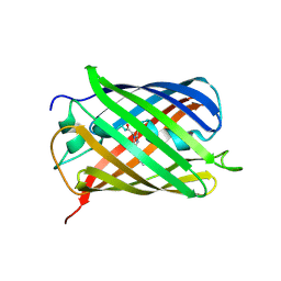
4LQ6
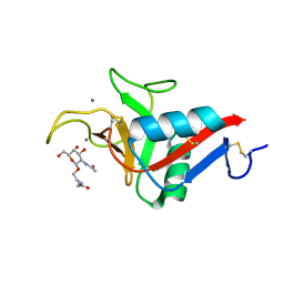
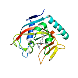
 | | Crystal structure of Rv3717 reveals a novel amidase from M. tuberculosis | | 分子名称: | CHLORIDE ION, N-acetymuramyl-L-alanine amidase-related protein, PLATINUM (II) ION, ... | | 著者 | Kumar, A, Kumar, S, Kumar, D, Mishra, A, Dewangan, R.P, Shrivastava, P, Ramachandran, S, Taneja, B. | | 登録日 | 2013-07-17 | | 公開日 | 2013-12-04 | | 最終更新日 | 2024-11-06 | | 実験手法 | X-RAY DIFFRACTION (1.68 Å) | | 主引用文献 | The structure of Rv3717 reveals a novel amidase from Mycobacterium tuberculosis.
Acta Crystallogr.,Sect.D, 69, 2013
|
|
6IEN
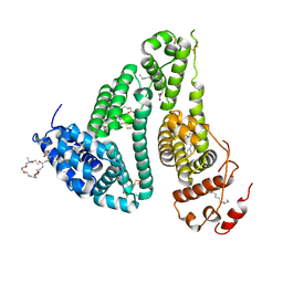
 | | Substrate/product bound Argininosuccinate lyase from Mycobacterium tuberculosis | | 分子名称: | 1,2-ETHANEDIOL, ARGININE, ARGININOSUCCINATE, ... | | 著者 | Paul, A, Mishra, A, Surolia, A, Vijayan, M. | | 登録日 | 2018-09-14 | | 公開日 | 2019-02-20 | | 最終更新日 | 2024-10-02 | | 実験手法 | X-RAY DIFFRACTION (2.7 Å) | | 主引用文献 | Structural studies on M. tuberculosis argininosuccinate lyase and its liganded complex: Insights into catalytic mechanism.
IUBMB Life, 71, 2019
|
|
6IEM
 | | Argininosuccinate lyase from Mycobacterium tuberculosis | | 分子名称: | 1,2-ETHANEDIOL, 3,6,9,12,15-PENTAOXAHEPTADECANE, Argininosuccinate lyase, ... | | 著者 | Paul, A, Mishra, A, Surolia, A, Vijayan, M. | | 登録日 | 2018-09-14 | | 公開日 | 2019-02-20 | | 最終更新日 | 2023-11-22 | | 実験手法 | X-RAY DIFFRACTION (2.2 Å) | | 主引用文献 | Structural studies on M. tuberculosis argininosuccinate lyase and its liganded complex: Insights into catalytic mechanism.
IUBMB Life, 71, 2019
|
|
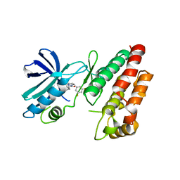
2HV8
 | | Crystal structure of GTP-bound Rab11 in complex with FIP3 | | 分子名称: | 2-(N-MORPHOLINO)-ETHANESULFONIC ACID, GUANOSINE-5'-TRIPHOSPHATE, MAGNESIUM ION, ... | | 著者 | Eathiraj, S, Mishra, A, Prekeris, R, Lambright, D.G. | | 登録日 | 2006-07-27 | | 公開日 | 2006-11-21 | | 最終更新日 | 2023-08-30 | | 実験手法 | X-RAY DIFFRACTION (1.86 Å) | | 主引用文献 | Structural Basis for Rab11-mediated Recruitment of FIP3 to Recycling Endosomes.
J.Mol.Biol., 364, 2006
|
|
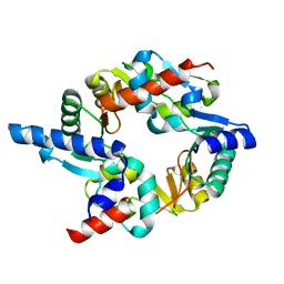
3MJH
 | | Crystal Structure of Human Rab5A in complex with the C2H2 Zinc Finger of EEA1 | | 分子名称: | Early endosome antigen 1, GUANOSINE-5'-TRIPHOSPHATE, MAGNESIUM ION, ... | | 著者 | Mishra, A.K, Eathiraj, S, Lambright, D.G. | | 登録日 | 2010-04-12 | | 公開日 | 2010-05-05 | | 最終更新日 | 2024-02-21 | | 実験手法 | X-RAY DIFFRACTION (2.03 Å) | | 主引用文献 | Structural basis for Rab GTPase recognition and endosome tethering by the C2H2 zinc finger of Early Endosomal Autoantigen 1 (EEA1).
Proc.Natl.Acad.Sci.USA, 107, 2010
|
|
3SNZ
 | |
3SO0
 | |
3QDH
 | |
6YAU
 | | CRYSTAL STRUCTURE OF ASGPR 1 IN COMPLEX WITH GN-A. | | 分子名称: | 5-[(2~{R},3~{R},4~{R},5~{R},6~{R})-3-acetamido-6-(hydroxymethyl)-4,5-bis(oxidanyl)oxan-2-yl]oxy-~{N}-[3-(propanoylamino)propyl]pentanamide, Asialoglycoprotein receptor 1, CALCIUM ION | | 著者 | Schreuder, H.A, Liesum, A. | | 登録日 | 2020-03-13 | | 公開日 | 2021-01-13 | | 最終更新日 | 2024-11-13 | | 実験手法 | X-RAY DIFFRACTION (1.397 Å) | | 主引用文献 | Triantennary GalNAc Molecular Imaging Probes for Monitoring Hepatocyte Function in a Rat Model of Nonalcoholic Steatohepatitis.
Adv Sci, 7, 2020
|
|
6YG9
 | | CRYSTAL STRUCTURE OF HUMAN SERUM ALBUMIN (HSA) IN COMPLEX WITH GN-07. | | 分子名称: | 20-[[(2~{S})-5-[2-[2-[2-[2-[2-[2-(diethylamino)-2-oxidanylidene-ethoxy]ethoxy]ethylamino]-2-oxidanylidene-ethoxy]ethoxy]ethylamino]-1-oxidanyl-1,5-bis(oxidanylidene)pentan-2-yl]amino]-20-oxidanylidene-icosanoic acid, MYRISTIC ACID, Serum albumin | | 著者 | Schreuder, H.A, Liesum, A. | | 登録日 | 2020-03-27 | | 公開日 | 2021-01-13 | | 最終更新日 | 2024-10-09 | | 実験手法 | X-RAY DIFFRACTION (1.89 Å) | | 主引用文献 | Triantennary GalNAc Molecular Imaging Probes for Monitoring Hepatocyte Function in a Rat Model of Nonalcoholic Steatohepatitis.
Adv Sci, 7, 2020
|
|
3SNY
 | |
3SO1
 | |
5XI1
 | | Structural Insight of Flavonoids binding to CAG repeat RNA that causes Huntington's Disease (HD) and Spinocerebellar Ataxia (SCAs) | | 分子名称: | 3,5,7-TRIHYDROXY-2-(3,4,5-TRIHYDROXYPHENYL)-4H-CHROMEN-4-ONE, RNA (5'-R(P*CP*CP*GP*CP*AP*GP*CP*GP*G)-3') | | 著者 | Tawani, A, Mishra, S.K, Khan, E, Kumar, A. | | 登録日 | 2017-04-25 | | 公開日 | 2018-08-08 | | 最終更新日 | 2024-05-15 | | 実験手法 | SOLUTION NMR | | 主引用文献 | Myricetin Reduces Toxic Level of CAG Repeats RNA in Huntington's Disease (HD) and Spino Cerebellar Ataxia (SCAs).
ACS Chem. Biol., 13, 2018
|
|
8RIR
 | |
8RIT
 | |
8P2O
 | | Dimeric mutant S103R of the BTB domain of ZBTB8A from Xenopus laevis | | 分子名称: | 1,2-ETHANEDIOL, Zinc finger and BTB domain-containing protein 8A.1-A | | 著者 | Coste, F, Mance, L, Pukalo, Z, Suskiewicz, M.J. | | 登録日 | 2023-05-16 | | 公開日 | 2024-06-05 | | 最終更新日 | 2024-12-18 | | 実験手法 | X-RAY DIFFRACTION (1.85 Å) | | 主引用文献 | Dynamic BTB-domain filaments promote clustering of ZBTB proteins.
Mol.Cell, 84, 2024
|
|
8P2P
 | |
8P2N
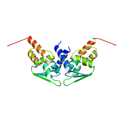 | |
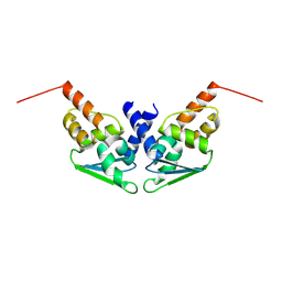
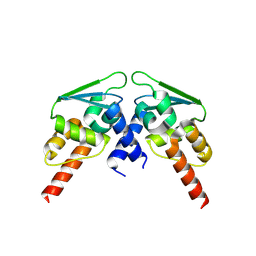
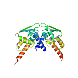
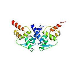
2WD5
 | | SMC hinge heterodimer (Mouse) | | 分子名称: | STRUCTURAL MAINTENANCE OF CHROMOSOMES PROTEIN 1A, STRUCTURAL MAINTENANCE OF CHROMOSOMES PROTEIN 3 | | 著者 | Michie, K.A, Haering, C.H, Nasmyth, K, Lowe, J. | | 登録日 | 2009-03-20 | | 公開日 | 2010-08-11 | | 最終更新日 | 2024-05-08 | | 実験手法 | X-RAY DIFFRACTION (2.7 Å) | | 主引用文献 | A Positively Charged Channel within the Smc1/Smc3 Hinge Required for Sister Chromatid Cohesion.
Embo J., 30, 2011
|
|
6HHO
 | | Crystal structure of RIP1 kinase in complex with GSK547 | | 分子名称: | 6-[4-[(5~{S})-5-[3,5-bis(fluoranyl)phenyl]pyrazolidin-1-yl]carbonylpiperidin-1-yl]pyrimidine-4-carbonitrile, Receptor-interacting serine/threonine-protein kinase 1 | | 著者 | Thorpe, J.H, Harris, P.A. | | 登録日 | 2018-08-28 | | 公開日 | 2018-12-12 | | 最終更新日 | 2024-01-17 | | 実験手法 | X-RAY DIFFRACTION (3.49 Å) | | 主引用文献 | RIP1 Kinase Drives Macrophage-Mediated Adaptive Immune Tolerance in Pancreatic Cancer.
Cancer Cell, 34, 2018
|
|
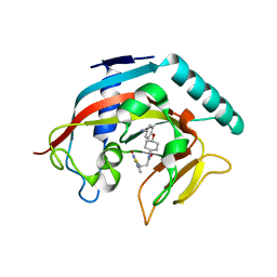
4MSK
 | | Co-crystal structure of tankyrase 1 with compound 34 | | 分子名称: | 3-(4-oxo-3,4-dihydroquinazolin-2-yl)-N-[4-(5-phenyl-1,3,4-oxadiazol-2-yl)phenyl]propanamide, Tankyrase-1, ZINC ION | | 著者 | Huang, X. | | 登録日 | 2013-09-18 | | 公開日 | 2013-12-25 | | 最終更新日 | 2024-10-30 | | 実験手法 | X-RAY DIFFRACTION (2.3 Å) | | 主引用文献 | Development of novel dual binders as potent, selective, and orally bioavailable tankyrase inhibitors.
J.Med.Chem., 56, 2013
|
|
6MZ3
 | |
4MT9
 | | Co-crystal structure of tankyrase 1 with compound 49 | | 分子名称: | N-[trans-4-(4-cyanophenoxy)cyclohexyl]-3-[(4-oxo-3,4-dihydroquinazolin-2-yl)sulfanyl]propanamide, Tankyrase-1, ZINC ION | | 著者 | Huang, X. | | 登録日 | 2013-09-19 | | 公開日 | 2013-12-25 | | 最終更新日 | 2024-02-28 | | 実験手法 | X-RAY DIFFRACTION (2 Å) | | 主引用文献 | Development of novel dual binders as potent, selective, and orally bioavailable tankyrase inhibitors.
J.Med.Chem., 56, 2013
|
|
4MSG
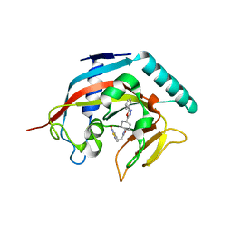 | | Crystal structure of tankyrase 1 with compound 22 | | 分子名称: | 3-[(4-oxo-3,4-dihydroquinazolin-2-yl)sulfanyl]-N-[trans-4-(5-phenyl-1,3,4-oxadiazol-2-yl)cyclohexyl]propanamide, Tankyrase-1, ZINC ION | | 著者 | Huang, X. | | 登録日 | 2013-09-18 | | 公開日 | 2013-12-25 | | 最終更新日 | 2024-02-28 | | 実験手法 | X-RAY DIFFRACTION (1.8 Å) | | 主引用文献 | Development of novel dual binders as potent, selective, and orally bioavailable tankyrase inhibitors.
J.Med.Chem., 56, 2013
|
|


