Loading
PDBj
メニューPDBj@FacebookPDBj@TwitterPDBj@YouTubewwPDB FoundationwwPDB
RCSB PDBPDBeBMRBAdv. SearchSearch help
Search by PDB author
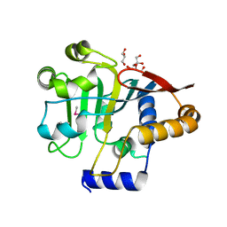
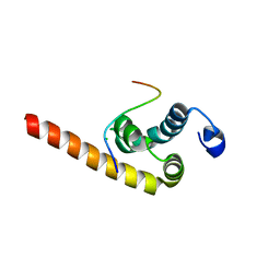
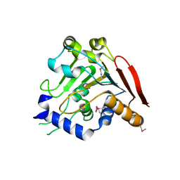
5V45
DownloadVisualize
BU of 5v45 by Molmil
Crystal structure of the F270M, K291M, L318M mutant of SR1 domain of human sacsin
分子名称: Sacsin
著者Menade, M, Kozlov, G, Gehring, K.
登録日2017-03-08
公開日2017-05-24
最終更新日2018-08-29
実験手法X-RAY DIFFRACTION (1.91 Å)
主引用文献Structures of ubiquitin-like (Ubl) and Hsp90-like domains of sacsin provide insight into pathological mutations.
J. Biol. Chem., 293, 2018
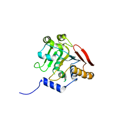
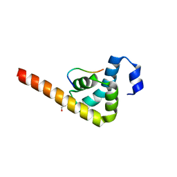
5V46
DownloadVisualize
BU of 5v46 by Molmil
Crystal structure of the I113M, F270M, K291M, L308M mutant of SR1 domain of human sacsin
分子名称: Sacsin
著者Menade, M, Kozlov, G, Gehring, K.
登録日2017-03-08
公開日2017-05-24
最終更新日2023-11-15
実験手法X-RAY DIFFRACTION (1.8 Å)
主引用文献Structures of ubiquitin-like (Ubl) and Hsp90-like domains of sacsin provide insight into pathological mutations.
J. Biol. Chem., 293, 2018
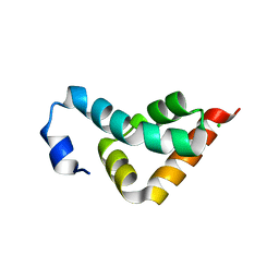
5V44
DownloadVisualize
BU of 5v44 by Molmil
Crystal structure of the SR1 domain of human sacsin
分子名称: GLYCEROL, Sacsin
著者Menade, M, Kozlov, G, Gehring, K.
登録日2017-03-08
公開日2017-05-24
最終更新日2023-11-15
実験手法X-RAY DIFFRACTION (1.56 Å)
主引用文献Structures of ubiquitin-like (Ubl) and Hsp90-like domains of sacsin provide insight into pathological mutations.
J. Biol. Chem., 293, 2018
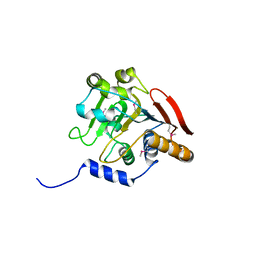
5V47
DownloadVisualize
BU of 5v47 by Molmil
Crystal structure of the SR1 domain of lizard sacsin
分子名称: Lizard sacsin, SULFATE ION
著者Pan, T, Menade, M, Kozlov, G, Gehring, K.
登録日2017-03-08
公開日2017-05-24
最終更新日2023-10-04
実験手法X-RAY DIFFRACTION (1.84 Å)
主引用文献Structures of ubiquitin-like (Ubl) and Hsp90-like domains of sacsin provide insight into pathological mutations.
J. Biol. Chem., 293, 2018
3PT3
DownloadVisualize
BU of 3pt3 by Molmil
Crystal structure of the C-terminal lobe of the human UBR5 HECT domain
分子名称: E3 ubiquitin-protein ligase UBR5
著者Matta-Camacho, E, Kozlov, G, Menade, M, Gehring, K.
登録日2010-12-02
公開日2012-01-25
最終更新日2023-09-06
実験手法X-RAY DIFFRACTION (1.97 Å)
主引用文献Structure of the HECT C-lobe of the UBR5 E3 ubiquitin ligase.
Acta Crystallogr.,Sect.F, 68, 2012
4K95
DownloadVisualize
BU of 4k95 by Molmil
Crystal Structure of Parkin
分子名称: E3 ubiquitin-protein ligase parkin, ZINC ION
著者Seirafi, M, Menade, M, Sauve, V, Kozlov, G, Trempe, J.-F, Nagar, B, Gehring, K.
登録日2013-04-19
公開日2013-05-15
最終更新日2023-09-20
実験手法X-RAY DIFFRACTION (6.499 Å)
主引用文献Structure of parkin reveals mechanisms for ubiquitin ligase activation.
Science, 340, 2013
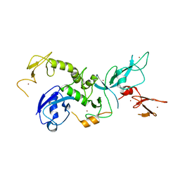
4K7D
DownloadVisualize
BU of 4k7d by Molmil
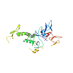
Crystal Structure of Parkin C-terminal RING domains
分子名称: CHLORIDE ION, E3 ubiquitin-protein ligase parkin, MALONATE ION, ...
著者Sauve, V, Trempe, J.-F, Menade, M, Gehring, K.
登録日2013-04-17
公開日2013-05-15
最終更新日2024-02-28
実験手法X-RAY DIFFRACTION (2.8 Å)
主引用文献Structure of parkin reveals mechanisms for ubiquitin ligase activation.
Science, 340, 2013
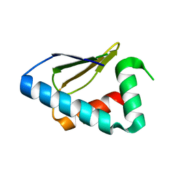
2MJC
DownloadVisualize
BU of 2mjc by Molmil
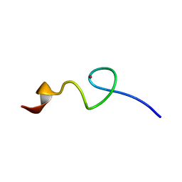
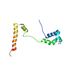
Zn-binding domain of eukaryotic translation initiation factor 3, subunit G
分子名称: Eukaryotic translation initiation factor 3 subunit G, ZINC ION
著者Al-Abdul-Wahid, M, Menade, M, Xie, J, Kozlov, G, Gehring, K.
登録日2014-01-03
公開日2015-01-07
最終更新日2024-05-15
実験手法SOLUTION NMR
主引用文献Solution NMR structure of the Zn-binding domain of eukaryotic translation initiation factor 3, subunit G
To be Published
5VSZ
DownloadVisualize
BU of 5vsz by Molmil
Structure of the Ubl domain of Sacsin mutant L78M
分子名称: Sacsin
著者Trempe, J.-F, Pande, H, Shenker, S, Gehring, K.
登録日2017-05-15
公開日2017-05-31
最終更新日2018-08-29
実験手法X-RAY DIFFRACTION (2.4 Å)
主引用文献Structures of ubiquitin-like (Ubl) and Hsp90-like domains of sacsin provide insight into pathological mutations.
J. Biol. Chem., 293, 2018
5VSX
DownloadVisualize
BU of 5vsx by Molmil
Structure of the Ubl domain of Sacsin
分子名称: Sacsin
著者Trempe, J.-F, Pande, H, Shenker, S, Gehring, K.
登録日2017-05-13
公開日2017-05-31
最終更新日2023-10-04
実験手法X-RAY DIFFRACTION (2.1 Å)
主引用文献Structures of ubiquitin-like (Ubl) and Hsp90-like domains of sacsin provide insight into pathological mutations.
J. Biol. Chem., 293, 2018
5KES
DownloadVisualize
BU of 5kes by Molmil
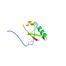
Solution structure of the yeast Ddi1 HDD domain
分子名称: DNA damage-inducible protein 1
著者Trempe, J.-F, Ratcliffe, C, Veverka, V, Saskova, K, Gehring, K.
登録日2016-06-10
公開日2016-10-05
最終更新日2024-05-15
実験手法SOLUTION NMR
主引用文献Structural studies of the yeast DNA damage-inducible protein Ddi1 reveal domain architecture of this eukaryotic protein family.
Sci Rep, 6, 2016
4Z2Z
DownloadVisualize
BU of 4z2z by Molmil
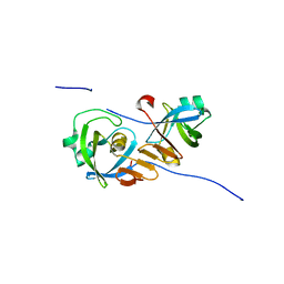
New crystal structure of yeast Ddi1 aspartyl protease reveals substrate engagement mode
分子名称: DNA damage-inducible protein 1
著者Trempe, J.-F, Feng, X, Gehring, K.
登録日2015-03-30
公開日2016-04-13
最終更新日2023-09-27
実験手法X-RAY DIFFRACTION (1.8 Å)
主引用文献Structural studies of the yeast DNA damage-inducible protein Ddi1 reveal domain architecture of this eukaryotic protein family.
Sci Rep, 6, 2016
2N7E
DownloadVisualize
BU of 2n7e by Molmil
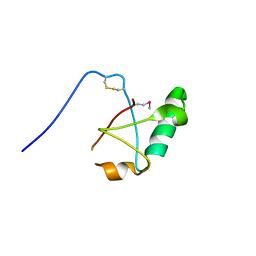
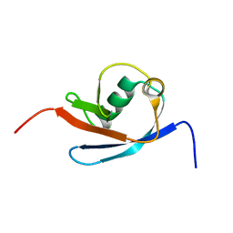
Solution structure of the UBL domain of yeast Ddi1
分子名称: DNA damage-inducible protein 1
著者Siva, M, Grantz Saskova, K, Veverka, V.
登録日2015-09-09
公開日2016-07-20
最終更新日2024-05-15
実験手法SOLUTION NMR
主引用文献Structural studies of the yeast DNA damage-inducible protein Ddi1 reveal domain architecture of this eukaryotic protein family.
Sci Rep, 6, 2016
6B3Y
DownloadVisualize
BU of 6b3y by Molmil
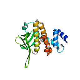
Crystal structure of the PH-like domain from DENND3
分子名称: DENN domain-containing protein 3
著者Kozlov, G, Xu, J, Menade, M, Beaugrand, M, Pan, T, McPherson, P.S, Gehring, K.
登録日2017-09-25
公開日2018-01-24
最終更新日2024-03-13
実験手法X-RAY DIFFRACTION (1.852 Å)
主引用文献A PH-like domain of the Rab12 guanine nucleotide exchange factor DENND3 binds actin and is required for autophagy.
J. Biol. Chem., 293, 2018
3KUT
DownloadVisualize
BU of 3kut by Molmil
Crystal structure of the MLLE domain of poly(A)-binding protein in complex with the binding region of Paip2
分子名称: CHLORIDE ION, PAIP2 protein, Polyadenylate-binding protein 1
著者Kozlov, G, Gehring, K.
登録日2009-11-27
公開日2010-02-09
最終更新日2023-09-06
実験手法X-RAY DIFFRACTION (1.5 Å)
主引用文献Molecular Determinants of PAM2 Recognition by the MLLE Domain of Poly(A)-Binding Protein.
J.Mol.Biol., 397, 2010
3KUS
DownloadVisualize
BU of 3kus by Molmil
Crystal structure of the MLLE domain of poly(A)-binding protein in complex with the binding region of Paip2
分子名称: 4-(2-HYDROXYETHYL)-1-PIPERAZINE ETHANESULFONIC ACID, GLYCEROL, PAIP2 protein, ...
著者Kozlov, G, Gehring, K.
登録日2009-11-27
公開日2010-02-09
最終更新日2023-09-06
実験手法X-RAY DIFFRACTION (1.4 Å)
主引用文献Molecular Determinants of PAM2 Recognition by the MLLE Domain of Poly(A)-Binding Protein.
J.Mol.Biol., 397, 2010
3KUR
DownloadVisualize
BU of 3kur by Molmil
Crystal structure of the MLLE domain of poly(A)-binding protein
分子名称: CHLORIDE ION, Polyadenylate-binding protein 1
著者Kozlov, G, Gehring, K.
登録日2009-11-27
公開日2010-02-09
最終更新日2023-09-06
実験手法X-RAY DIFFRACTION (2.5 Å)
主引用文献Molecular Determinants of PAM2 Recognition by the MLLE Domain of Poly(A)-Binding Protein.
J.Mol.Biol., 397, 2010

222415

件を2024-07-10に公開中

PDB statisticsPDBj update infoContact PDBjnumon