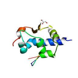
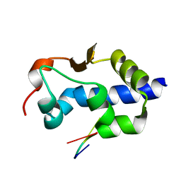
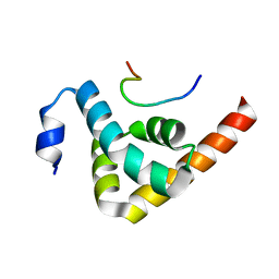
1JH4
 
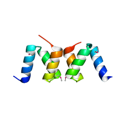
 | | Solution structure of the C-terminal PABC domain of human poly(A)-binding protein in complex with the peptide from Paip1 | | 分子名称: | polyadenylate-binding protein 1, polyadenylate-binding protein-interacting protein-1 | | 著者 | Kozlov, G, Siddiqui, N, Coillet-Matillon, S, Ekiel, I, Gehring, K. | | 登録日 | 2001-06-27 | | 公開日 | 2003-06-24 | | 最終更新日 | 2024-05-22 | | 実験手法 | SOLUTION NMR | | 主引用文献 | Structural basis of ligand recognition by PABC, a highly specific peptide-binding domain found in poly(A)-binding protein and a HECT ubiquitin ligase
EMBO J., 23, 2004
|
|
3EC3
 | |
5V8Z
 | |
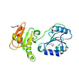
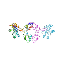
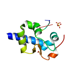
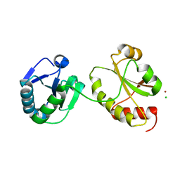
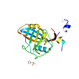
1SSL
 | | Solution structure of the PSI domain from the Met receptor | | 分子名称: | Hepatocyte growth factor receptor | | 著者 | Kozlov, G, Perreault, A, Schrag, J.D, Cygler, M, Gehring, K, Ekiel, I. | | 登録日 | 2004-03-24 | | 公開日 | 2004-10-12 | | 最終更新日 | 2022-03-02 | | 実験手法 | SOLUTION NMR | | 主引用文献 | Insights into function of PSI domains from structure of the Met receptor PSI domain.
Biochem.Biophys.Res.Commun., 321, 2004
|
|
6WUS
 | |
6WUR
 | |
2H8L
 | |
8G91
 | |
8G90
 | |
1D5G
 | |
2QHO
 | |
8EY8
 | |
8EY6
 | |
8EY7
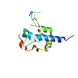 | |
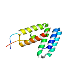
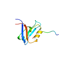
1L1P
 | | Solution Structure of the PPIase Domain from E. coli Trigger Factor | | 分子名称: | trigger factor | | 著者 | Kozlov, G, Trempe, J.-F, Perreault, A, Wong, M, Denisov, A, Ghandi, S, Gehring, K, Ekiel, I, Montreal-Kingston Bacterial Structural Genomics Initiative (BSGI) | | 登録日 | 2002-02-19 | | 公開日 | 2003-06-24 | | 最終更新日 | 2024-05-22 | | 実験手法 | SOLUTION NMR | | 主引用文献 | Solution Structure of the Closed Form of a Peptidyl-Prolyl Isomerase Reveals the Mechanism of Protein Folding
To be Published
|
|
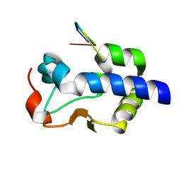
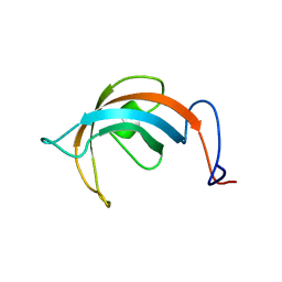
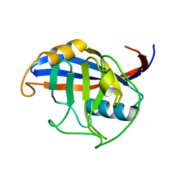
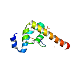
1JGN
 | | Solution structure of the C-terminal PABC domain of human poly(A)-binding protein in complex with the peptide from Paip2 | | 分子名称: | polyadenylate-binding protein 1, polyadenylate-binding protein-interacting protein 2 | | 著者 | Kozlov, G, Siddiqui, N, Coillet-Matillon, S, Ekiel, I, Gehring, K. | | 登録日 | 2001-06-26 | | 公開日 | 2003-06-24 | | 最終更新日 | 2024-05-22 | | 実験手法 | SOLUTION NMR | | 主引用文献 | Structural basis of ligand recognition by PABC, a highly specific peptide-binding domain found in poly(A)-binding protein and a HECT ubiquitin ligase
EMBO J., 23, 2004
|
|
2OO9
 | |
3IDV
 | |
3ICH
 | |
3GZH
 | |
3ICI
 | |
3KTP
 | |
3KUI
 | |
3KUR
 | |
3KTR
 | |


