4DIT
 
 | |
3PG4
 | |
5GMN
 | |
5GMM
 | |
5X6Q
 | |
5X6R
 | |
5X68
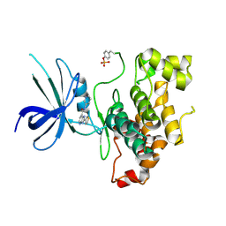
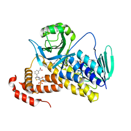
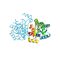
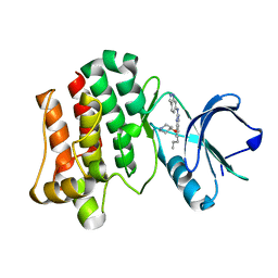
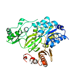
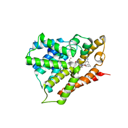
 | | Crystal Structure of Human KMO | | 分子名称: | FLAVIN-ADENINE DINUCLEOTIDE, Kynurenine 3-monooxygenase | | 著者 | Kim, H.T, Hwang, K.Y. | | 登録日 | 2017-02-21 | | 公開日 | 2018-02-21 | | 最終更新日 | 2018-05-02 | | 実験手法 | X-RAY DIFFRACTION (2.1 Å) | | 主引用文献 | Structural Basis for Inhibitor-Induced Hydrogen Peroxide Production by Kynurenine 3-Monooxygenase
Cell Chem Biol, 25, 2018
|
|
5X6P
 | |
5YCX
 | |
5YCS
 | |
5YCR
 | |
5YCV
 | |
5XY1
 | |
3PJF
 | |
3PJD
 | |
3PJE
 | |
2HJW
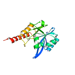
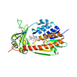
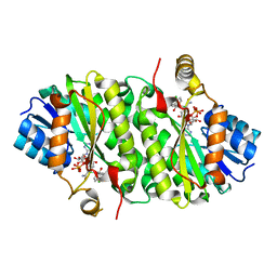
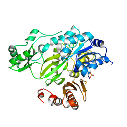
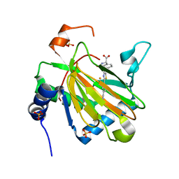
 | | Crystal Structure of the BC domain of ACC2 | | 分子名称: | Acetyl-CoA carboxylase 2 | | 著者 | Cho, Y.S, Lee, J.I, Shin, D, Kim, H.T, Lee, T.G, Heo, Y.S. | | 登録日 | 2006-07-02 | | 公開日 | 2007-07-03 | | 最終更新日 | 2023-10-25 | | 実験手法 | X-RAY DIFFRACTION (2.5 Å) | | 主引用文献 | Crystal structure of the biotin carboxylase domain of human acetyl-CoA carboxylase 2.
Proteins, 70, 2008
|
|
3JRW
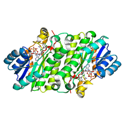
 | | Phosphorylated BC domain of ACC2 | | 分子名称: | Acetyl-CoA carboxylase 2 | | 著者 | Cho, Y.S, Lee, J.I, Shin, D, Kim, H.T, Lee, T.G, Heo, Y.S. | | 登録日 | 2009-09-09 | | 公開日 | 2010-01-12 | | 最終更新日 | 2023-11-01 | | 実験手法 | X-RAY DIFFRACTION (2.6 Å) | | 主引用文献 | Molecular mechanism for the regulation of human ACC2 through phosphorylation by AMPK
Biochem.Biophys.Res.Commun., 391, 2010
|
|
3JRX
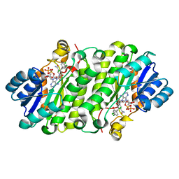
 | | Crystal structure of the BC domain of ACC2 in complex with soraphen A | | 分子名称: | Acetyl-CoA carboxylase 2, SORAPHEN A | | 著者 | Cho, Y.S, Lee, J.I, Shin, D, Kim, H.T, Lee, T.G, Heo, Y.S. | | 登録日 | 2009-09-09 | | 公開日 | 2010-01-12 | | 最終更新日 | 2023-11-01 | | 実験手法 | X-RAY DIFFRACTION (2.5 Å) | | 主引用文献 | Molecular mechanism for the regulation of human ACC2 through phosphorylation by AMPK.
Biochem.Biophys.Res.Commun., 391, 2010
|
|
1ZC1
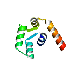
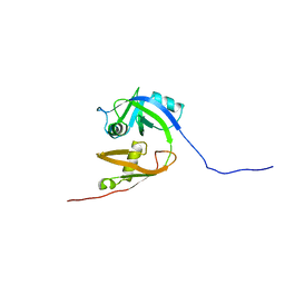 | | Ufd1 exhibits the AAA-ATPase fold with two distinct ubiquitin interaction sites | | 分子名称: | Ubiquitin fusion degradation protein 1 | | 著者 | Park, S, Isaacson, R, Kim, H.T, Silver, P.A, Wagner, G. | | 登録日 | 2005-04-10 | | 公開日 | 2005-07-26 | | 最終更新日 | 2024-05-22 | | 実験手法 | SOLUTION NMR | | 主引用文献 | Ufd1 Exhibits the AAA-ATPase Fold with Two Distinct Ubiquitin Interaction Sites
Structure, 13, 2005
|
|
3V9B
 | |
4KBZ
 | |
3R4Z
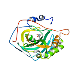
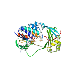
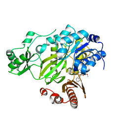
 | | Crystal structure of alpha-neoagarobiose hydrolase (ALPHA-NABH) in complex with alpha-d-galactopyranose from Saccharophagus degradans 2-40 | | 分子名称: | Glycosyl hydrolase family 32, N terminal, alpha-D-galactopyranose | | 著者 | Lee, S, Lee, J.Y, Ha, S.C, Shin, D.H, Kim, K.H, Bang, W.G, Kim, S.H, Choi, I.G. | | 登録日 | 2011-03-18 | | 公開日 | 2012-02-01 | | 最終更新日 | 2024-03-20 | | 実験手法 | X-RAY DIFFRACTION (1.55 Å) | | 主引用文献 | Crystal structure of a key enzyme in the agarolytic pathway, alpha-neoagarobiose hydrolase from Saccharophagus degradans 2-40
Biochem.Biophys.Res.Commun., 412, 2011
|
|
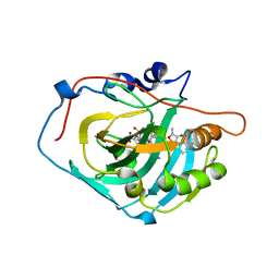
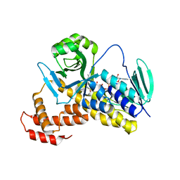
3R4Y
 | | Crystal structure of alpha-neoagarobiose hydrolase (ALPHA-NABH) from Saccharophagus degradans 2-40 | | 分子名称: | Glycosyl hydrolase family 32, N terminal | | 著者 | Lee, S, Lee, J.Y, Ha, S.C, Shin, D.H, Kim, K.H, Bang, W.G, Kim, S.H, Choi, I.G. | | 登録日 | 2011-03-18 | | 公開日 | 2012-02-01 | | 最終更新日 | 2024-03-20 | | 実験手法 | X-RAY DIFFRACTION (2 Å) | | 主引用文献 | Crystal structure of a key enzyme in the agarolytic pathway, alpha-neoagarobiose hydrolase from Saccharophagus degradans 2-40
Biochem.Biophys.Res.Commun., 412, 2011
|
|
5X2E
 | |


