4EGT
 
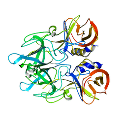 | | Crystal structure of major capsid protein P domain from rabbit hemorrhagic disease virus | | 分子名称: | Major capsid protein VP60 | | 著者 | Wang, X, Xu, F, Zhang, K, Zhai, Y, Sun, F. | | 登録日 | 2012-04-01 | | 公開日 | 2013-01-30 | | 最終更新日 | 2023-09-13 | | 実験手法 | X-RAY DIFFRACTION (2 Å) | | 主引用文献 | Atomic model of rabbit hemorrhagic disease virus by cryo-electron microscopy and crystallography.
Plos Pathog., 9, 2013
|
|
6UUZ
 
 | |
6UV5
 
 | |
6UUW
 
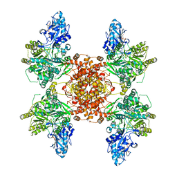 | | Structure of human ATP citrate lyase E599Q mutant in complex with Mg2+, citrate, ATP and CoA | | 分子名称: | (2S)-2-hydroxy-2-[2-oxo-2-(phosphonooxy)ethyl]butanedioic acid, ADENOSINE-5'-DIPHOSPHATE, ATP-citrate synthase, ... | | 著者 | Wei, X, Marmorstein, R. | | 登録日 | 2019-11-01 | | 公開日 | 2019-12-25 | | 最終更新日 | 2024-05-29 | | 実験手法 | ELECTRON MICROSCOPY (2.85 Å) | | 主引用文献 | Molecular basis for acetyl-CoA production by ATP-citrate lyase
Nat.Struct.Mol.Biol., 27, 2020
|
|
7NSH
 
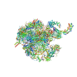 | | 39S mammalian mitochondrial large ribosomal subunit with mtRRF (post) and mtEFG2 | | 分子名称: | 16S rRNA, 39S ribosomal protein L48, mitochondrial, ... | | 著者 | Kummer, E, Schubert, K, Ban, N. | | 登録日 | 2021-03-07 | | 公開日 | 2021-05-05 | | 最終更新日 | 2024-07-10 | | 実験手法 | ELECTRON MICROSCOPY (3.2 Å) | | 主引用文献 | Structural basis of translation termination, rescue, and recycling in mammalian mitochondria.
Mol.Cell, 81, 2021
|
|
7NQL
 
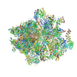 | | 55S mammalian mitochondrial ribosome with ICT1 and P site tRNAMet | | 分子名称: | 12S rRNA, 16S rRNA, 28S ribosomal protein S16, ... | | 著者 | Kummer, E, Schubert, K, Ban, N. | | 登録日 | 2021-03-01 | | 公開日 | 2021-05-05 | | 最終更新日 | 2021-12-15 | | 実験手法 | ELECTRON MICROSCOPY (3.4 Å) | | 主引用文献 | Structural basis of translation termination, rescue, and recycling in mammalian mitochondria.
Mol.Cell, 81, 2021
|
|
7NSI
 
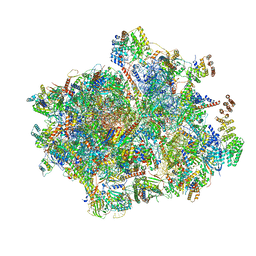 | | 55S mammalian mitochondrial ribosome with mtRRF (pre) and tRNA(P/E) | | 分子名称: | 12S rRNA, 16S rRNA, 28S ribosomal protein S10, ... | | 著者 | Kummer, E, Schubert, K, Ban, N. | | 登録日 | 2021-03-07 | | 公開日 | 2021-06-02 | | 最終更新日 | 2021-12-15 | | 実験手法 | ELECTRON MICROSCOPY (4.6 Å) | | 主引用文献 | Structural basis of translation termination, rescue, and recycling in mammalian mitochondria.
Mol.Cell, 81, 2021
|
|
7NQH
 
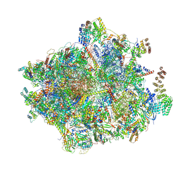 | | 55S mammalian mitochondrial ribosome with mtRF1a and P-site tRNAMet | | 分子名称: | 12S rRNA, 16S rRNA, 28S ribosomal protein S16, ... | | 著者 | Kummer, E, Schubert, K, Ban, N. | | 登録日 | 2021-03-01 | | 公開日 | 2021-05-05 | | 最終更新日 | 2024-10-16 | | 実験手法 | ELECTRON MICROSCOPY (3.5 Å) | | 主引用文献 | Structural basis of translation termination, rescue, and recycling in mammalian mitochondria.
Mol.Cell, 81, 2021
|
|
7NSJ
 
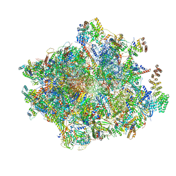 | | 55S mammalian mitochondrial ribosome with tRNA(P/P) and tRNA(E*) | | 分子名称: | 12S rRNA, 16S rRNA, 28S ribosomal protein S16, ... | | 著者 | Kummer, E, Schubert, K, Ban, N. | | 登録日 | 2021-03-07 | | 公開日 | 2021-06-02 | | 最終更新日 | 2024-10-09 | | 実験手法 | ELECTRON MICROSCOPY (3.9 Å) | | 主引用文献 | Structural basis of translation termination, rescue, and recycling in mammalian mitochondria.
Mol.Cell, 81, 2021
|
|
4EJR
 
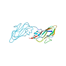 | | Crystal structure of major capsid protein S domain from rabbit hemorrhagic disease virus | | 分子名称: | Major capsid protein VP60 | | 著者 | Xu, F, Ma, J, Zhang, K, Wang, X, Sun, F. | | 登録日 | 2012-04-07 | | 公開日 | 2013-01-30 | | 最終更新日 | 2023-09-13 | | 実験手法 | X-RAY DIFFRACTION (2 Å) | | 主引用文献 | Atomic model of rabbit hemorrhagic disease virus by cryo-electron microscopy and crystallography.
Plos Pathog., 9, 2013
|
|
4EZ5
 
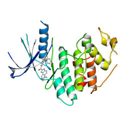 | | CDK6 (monomeric) in complex with inhibitor | | 分子名称: | Cyclin-dependent kinase 6, {5-[4-(dimethylamino)piperidin-1-yl]-1H-imidazo[4,5-b]pyridin-2-yl}[2-(isoquinolin-4-yl)pyridin-4-yl]methanone | | 著者 | Chopra, R, Xu, M. | | 登録日 | 2012-05-02 | | 公開日 | 2013-02-06 | | 最終更新日 | 2023-09-13 | | 実験手法 | X-RAY DIFFRACTION (2.7 Å) | | 主引用文献 | Fragment-Based Discovery of 7-Azabenzimidazoles as Potent, Highly Selective, and Orally Active CDK4/6 Inhibitors.
ACS Med Chem Lett, 3, 2012
|
|
8V0F
 
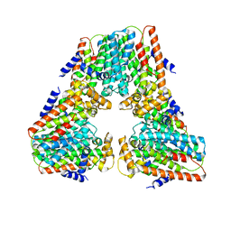 | |
2JMH
 
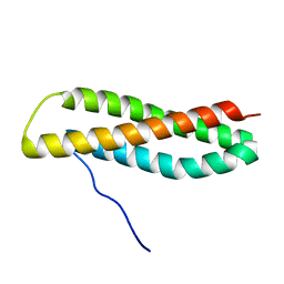 | | NMR solution structure of Blo t 5, a major mite allergen from Blomia tropicalis | | 分子名称: | Mite allergen Blo t 5 | | 著者 | Naik, M.T, Chang, C, Kuo, I, Chua, K, Huang, T. | | 登録日 | 2006-11-12 | | 公開日 | 2007-11-13 | | 最終更新日 | 2023-12-20 | | 実験手法 | SOLUTION NMR | | 主引用文献 | Roles of Structure and Structural Dynamics in the Antibody Recognition of the Allergen Proteins: An NMR Study on Blomia tropicalis Major Allergen
Structure, 16, 2008
|
|
4HND
 
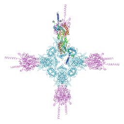 | | Crystal structure of the catalytic domain of Selenomethionine substituted human PI4KIIalpha in complex with ADP | | 分子名称: | ADENOSINE-5'-DIPHOSPHATE, Phosphatidylinositol 4-kinase type 2-alpha | | 著者 | Zhou, Q, Zhai, Y, Zhang, K, Chen, C, Sun, F. | | 登録日 | 2012-10-19 | | 公開日 | 2014-04-09 | | 最終更新日 | 2016-12-28 | | 実験手法 | X-RAY DIFFRACTION (3.2 Å) | | 主引用文献 | Molecular insights into the membrane-associated phosphatidylinositol 4-kinase II alpha.
Nat Commun, 5, 2014
|
|
4HNE
 
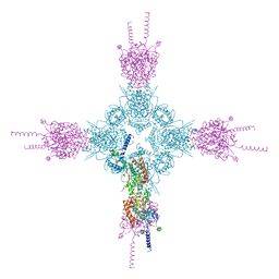 | | Crystal structure of the catalytic domain of human type II alpha Phosphatidylinositol 4-kinase (PI4KIIalpha) in complex with ADP | | 分子名称: | ADENOSINE-5'-DIPHOSPHATE, Phosphatidylinositol 4-kinase type 2-alpha | | 著者 | Zhou, Q, Zhai, Y, Zhang, K, Chen, C, Sun, F. | | 登録日 | 2012-10-19 | | 公開日 | 2014-04-09 | | 最終更新日 | 2023-09-20 | | 実験手法 | X-RAY DIFFRACTION (2.95 Å) | | 主引用文献 | Molecular insights into the membrane-associated phosphatidylinositol 4-kinase II alpha.
Nat Commun, 5, 2014
|
|
8G5C
 
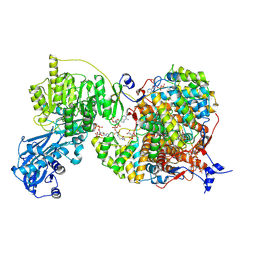 | |
8G1F
 
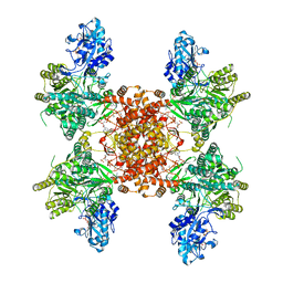 | | Structure of ACLY-D1026A-products | | 分子名称: | (3S)-citryl-Coenzyme A, ACETYL COENZYME *A, ADENOSINE-5'-DIPHOSPHATE, ... | | 著者 | Wei, X, Marmorstein, R. | | 登録日 | 2023-02-02 | | 公開日 | 2023-05-10 | | 最終更新日 | 2024-06-19 | | 実験手法 | ELECTRON MICROSCOPY (2.4 Å) | | 主引用文献 | Allosteric role of the citrate synthase homology domain of ATP citrate lyase.
Nat Commun, 14, 2023
|
|
8G5D
 
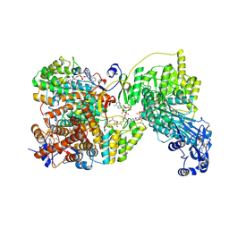 | |
8G1E
 
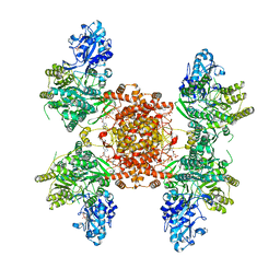 | | Structure of ACLY-D1026A-products-asym | | 分子名称: | (3S)-citryl-Coenzyme A, ACETYL COENZYME *A, ADENOSINE-5'-DIPHOSPHATE, ... | | 著者 | Wei, X, Marmorstein, R. | | 登録日 | 2023-02-02 | | 公開日 | 2023-05-10 | | 最終更新日 | 2024-06-19 | | 実験手法 | ELECTRON MICROSCOPY (2.8 Å) | | 主引用文献 | Allosteric role of the citrate synthase homology domain of ATP citrate lyase.
Nat Commun, 14, 2023
|
|
7PYT
 
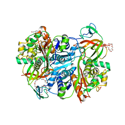 | | Benzoylsuccinyl-CoA thiolase with coenzyme A | | 分子名称: | 3,6,9,12,15,18,21-HEPTAOXATRICOSANE-1,23-DIOL, 4-(2-HYDROXYETHYL)-1-PIPERAZINE ETHANESULFONIC ACID, Benzoylsuccinyl-CoA thiolase subunit, ... | | 著者 | Ermler, U, Heider, J, Weidenweber, S, Demmer, U. | | 登録日 | 2021-10-11 | | 公開日 | 2022-04-06 | | 最終更新日 | 2024-05-01 | | 実験手法 | X-RAY DIFFRACTION (1.7 Å) | | 主引用文献 | Finis tolueni: a new type of thiolase with an integrated Zn-finger subunit catalyzes the final step of anaerobic toluene metabolism.
Febs J., 289, 2022
|
|
7PXP
 
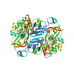 | | Benzoylsuccinyl-CoA thiolase | | 分子名称: | Benzoylsuccinyl-CoA thiolase subunit, ZINC ION | | 著者 | Ermler, U, Heider, H, Weidenweber, S, Demmer, U. | | 登録日 | 2021-10-08 | | 公開日 | 2022-04-06 | | 最終更新日 | 2024-06-19 | | 実験手法 | X-RAY DIFFRACTION (2 Å) | | 主引用文献 | Finis tolueni: a new type of thiolase with an integrated Zn-finger subunit catalyzes the final step of anaerobic toluene metabolism.
Febs J., 289, 2022
|
|
5L4K
 
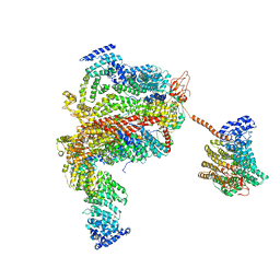 | | The human 26S proteasome lid | | 分子名称: | 26S proteasome complex subunit DSS1, 26S proteasome non-ATPase regulatory subunit 1, 26S proteasome non-ATPase regulatory subunit 11, ... | | 著者 | Schweitzer, A, Aufderheide, A, Rudack, T, Beck, F. | | 登録日 | 2016-05-25 | | 公開日 | 2016-09-07 | | 最終更新日 | 2024-05-08 | | 実験手法 | ELECTRON MICROSCOPY (3.9 Å) | | 主引用文献 | Structure of the human 26S proteasome at a resolution of 3.9 angstrom.
Proc.Natl.Acad.Sci.USA, 113, 2016
|
|
5KTE
 
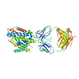 | | Crystal structure of Deinococcus radiodurans MntH, an Nramp-family transition metal transporter | | 分子名称: | Divalent metal cation transporter MntH, Fab Heavy Chain, Fab Light Chain, ... | | 著者 | Bane, L.B, Gaudet, R, Weihofen, W.A, Singharoy, A. | | 登録日 | 2016-07-11 | | 公開日 | 2016-11-23 | | 最終更新日 | 2023-10-04 | | 実験手法 | X-RAY DIFFRACTION (3.941 Å) | | 主引用文献 | Crystal Structure and Conformational Change Mechanism of a Bacterial Nramp-Family Divalent Metal Transporter.
Structure, 24, 2016
|
|
7PCS
 
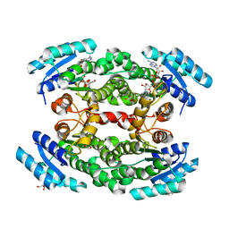 | | Structure of the heterotetrameric SDR family member BbsCD | | 分子名称: | BbsC, BbsD, GLYCEROL, ... | | 著者 | Essen, L.-O, Heider, J, von Horsten, S. | | 登録日 | 2021-08-04 | | 公開日 | 2022-06-15 | | 最終更新日 | 2024-06-19 | | 実験手法 | X-RAY DIFFRACTION (2.25 Å) | | 主引用文献 | Inactive pseudoenzyme subunits in heterotetrameric BbsCD, a novel short-chain alcohol dehydrogenase involved in anaerobic toluene degradation.
Febs J., 289, 2022
|
|
5L4G
 
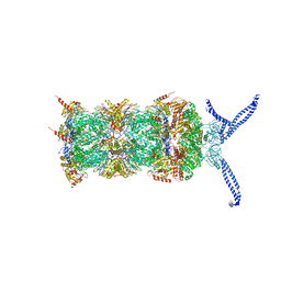 | | The human 26S proteasome at 3.9 A | | 分子名称: | 26S protease regulatory subunit 10B, 26S protease regulatory subunit 4, 26S protease regulatory subunit 6A, ... | | 著者 | Schweitzer, A, Aufderheide, A, Rudack, T, Beck, F. | | 登録日 | 2016-05-25 | | 公開日 | 2016-09-07 | | 最終更新日 | 2024-05-08 | | 実験手法 | ELECTRON MICROSCOPY (3.9 Å) | | 主引用文献 | Structure of the human 26S proteasome at a resolution of 3.9 angstrom.
Proc.Natl.Acad.Sci.USA, 113, 2016
|
|
