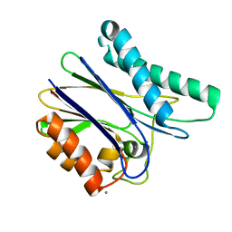
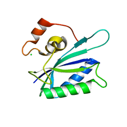
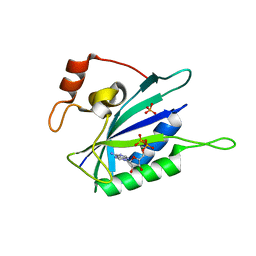
5H1U
 
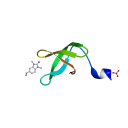
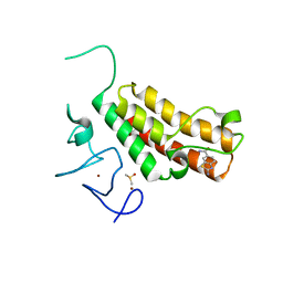 | | Complex structure of TRIM24 PHD-bromodomain and inhibitor 2 | | 分子名称: | 2-amino-1,3-benzothiazole-6-carboxamide, DIMETHYL SULFOXIDE, Transcription intermediary factor 1-alpha, ... | | 著者 | Liu, J. | | 登録日 | 2016-10-11 | | 公開日 | 2017-02-22 | | 最終更新日 | 2023-11-08 | | 実験手法 | X-RAY DIFFRACTION (1.901 Å) | | 主引用文献 | The polar warhead of a TRIM24 bromodomain inhibitor rearranges a water-mediated interaction network
FEBS J., 284, 2017
|
|
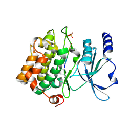
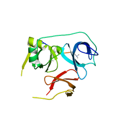
5H1T
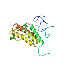 | | Complex structure of TRIM24 PHD-bromodomain and inhibitor 1 | | 分子名称: | DIMETHYL SULFOXIDE, Transcription intermediary factor 1-alpha, ZINC ION, ... | | 著者 | Liu, J. | | 登録日 | 2016-10-11 | | 公開日 | 2017-02-22 | | 最終更新日 | 2023-11-08 | | 実験手法 | X-RAY DIFFRACTION (1.951 Å) | | 主引用文献 | The polar warhead of a TRIM24 bromodomain inhibitor rearranges a water-mediated interaction network
FEBS J., 284, 2017
|
|
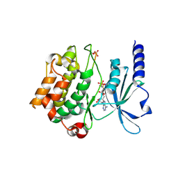
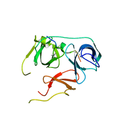
5H1V
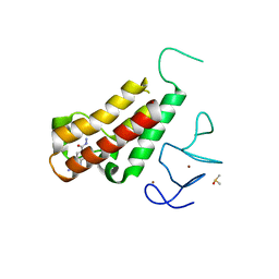 | | Complex structure of TRIM24 PHD-bromodomain and inhibitor 6 | | 分子名称: | 2-Hydrazino-1,3-benzothiazole-6-carbohydrazide, DIMETHYL SULFOXIDE, Transcription intermediary factor 1-alpha, ... | | 著者 | Liu, J. | | 登録日 | 2016-10-11 | | 公開日 | 2017-02-22 | | 最終更新日 | 2023-11-08 | | 実験手法 | X-RAY DIFFRACTION (2.002 Å) | | 主引用文献 | The polar warhead of a TRIM24 bromodomain inhibitor rearranges a water-mediated interaction network
FEBS J., 284, 2017
|
|
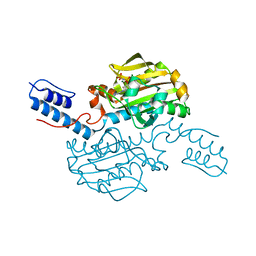
3IMX
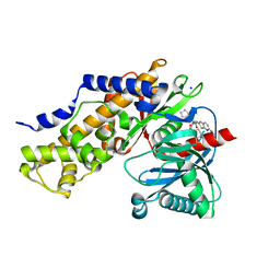 | | Crystal Structure of human glucokinase in complex with a synthetic activator | | 分子名称: | (2R)-3-cyclopentyl-N-(5-methoxy[1,3]thiazolo[5,4-b]pyridin-2-yl)-2-{4-[(4-methylpiperazin-1-yl)sulfonyl]phenyl}propanamide, Glucokinase, SODIUM ION, ... | | 著者 | Stams, T, Vash, B. | | 登録日 | 2009-08-11 | | 公開日 | 2009-10-06 | | 最終更新日 | 2023-09-06 | | 実験手法 | X-RAY DIFFRACTION (2 Å) | | 主引用文献 | Investigation of functionally liver selective glucokinase activators for the treatment of type 2 diabetes.
J.Med.Chem., 52, 2009
|
|
5D2U
 | | Crystal structure of tPphA Variant - H39A | | 分子名称: | CALCIUM ION, Protein serin-threonin phosphatase | | 著者 | Su, J. | | 登録日 | 2015-08-06 | | 公開日 | 2016-05-04 | | 最終更新日 | 2023-11-08 | | 実験手法 | X-RAY DIFFRACTION (1.8 Å) | | 主引用文献 | Structural and Biochemical Characterization of a Cyanobacterial PP2C Phosphatase Reveals Insights into Catalytic Mechanism and Substrate Recognition
Catalysts, 2016
|
|
4FII
 | |
5H1D
 | | Crystal structure of C-terminal of RhoGDI2 | | 分子名称: | Rho GDP-dissociation inhibitor 2 | | 著者 | Liu, J. | | 登録日 | 2016-10-08 | | 公開日 | 2016-10-26 | | 最終更新日 | 2023-11-08 | | 実験手法 | X-RAY DIFFRACTION (1.494 Å) | | 主引用文献 | NMR characterization of weak interactions between RhoGDI2 and fragment screening hits.
Biochim. Biophys. Acta, 1861, 2017
|
|
5HFS
 | | CRYSTAL STRUCTURE OF C-TERMINAL DOMAIN OF CARGO PROTEINS OF TYPE IX SECRETION SYSTEM | | 分子名称: | CALCIUM ION, Gingipain R2, ZINC ION | | 著者 | Golik, P, Szmigielski, B, Ksiazek, M, Nowakowska, Z, Mizgalska, D, Nowak, M, Dubin, G, Potempa, J. | | 登録日 | 2016-01-07 | | 公開日 | 2016-04-06 | | 最終更新日 | 2024-01-10 | | 実験手法 | X-RAY DIFFRACTION (1.97 Å) | | 主引用文献 | The outer-membrane export signal of Porphyromonas gingivalis type IX secretion system (T9SS) is a conserved C-terminal beta-sandwich domain.
Sci Rep, 6, 2016
|
|
4FIF
 | | Catalytic domain of human PAK4 with RPKPLVDP peptide | | 分子名称: | PHOSPHOAMINOPHOSPHONIC ACID-ADENYLATE ESTER, Serine/threonine-protein kinase PAK 4 | | 著者 | Ha, B.H, Boggon, T.J. | | 登録日 | 2012-06-08 | | 公開日 | 2012-09-12 | | 最終更新日 | 2023-09-13 | | 実験手法 | X-RAY DIFFRACTION (2.6 Å) | | 主引用文献 | Type II p21-activated kinases (PAKs) are regulated by an autoinhibitory pseudosubstrate.
Proc.Natl.Acad.Sci.USA, 109, 2012
|
|
5YJ8
 | |
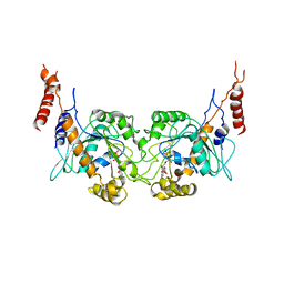
5DG2
 | | Sugar binding protein - human galectin-2 (dimer) | | 分子名称: | Galectin-2, beta-D-galactopyranose-(1-4)-alpha-D-glucopyranose | | 著者 | Su, J.Y, Si, Y.L. | | 登録日 | 2015-08-27 | | 公開日 | 2016-09-14 | | 最終更新日 | 2023-11-08 | | 実験手法 | X-RAY DIFFRACTION (1.612 Å) | | 主引用文献 | Human galectin-2 interacts with carbohydrates and peptides non-classically: new insight from X-ray crystallography and hemagglutination.
Acta Biochim. Biophys. Sin. (Shanghai), 48, 2016
|
|
5DG1
 | | Sugar binding protein - human galectin-2 | | 分子名称: | Galectin-2, beta-D-galactopyranose-(1-4)-beta-D-glucopyranose | | 著者 | Su, J.Y, Si, Y.L. | | 登録日 | 2015-08-27 | | 公開日 | 2016-09-14 | | 最終更新日 | 2023-11-08 | | 実験手法 | X-RAY DIFFRACTION (3.2 Å) | | 主引用文献 | Human galectin-2 interacts with carbohydrates and peptides non-classically: new insight from X-ray crystallography and hemagglutination.
Acta Biochim.Biophys.Sin., 2016
|
|
5EWS
 | | Sugar binding protein - human galectin-2 | | 分子名称: | Galectin-2, beta-D-galactopyranose-(1-4)-beta-D-glucopyranose | | 著者 | Su, J.Y, Si, Y.L. | | 登録日 | 2015-11-21 | | 公開日 | 2016-09-14 | | 最終更新日 | 2024-03-20 | | 実験手法 | X-RAY DIFFRACTION (2 Å) | | 主引用文献 | Human galectin-2 interacts with carbohydrates and peptides non-classically: new insight from X-ray crystallography and hemagglutination.
Acta Biochim. Biophys. Sin. (Shanghai), 48, 2016
|
|
1KBR
 | |
7BU9
 | |
7BQZ
 | |
5K1X
 | |
5K1P
 | |
1EQM
 | | CRYSTAL STRUCTURE OF BINARY COMPLEX OF 6-HYDROXYMETHYL-7,8-DIHYDROPTERIN PYROPHOSPHOKINASE WITH ADENOSINE-5'-DIPHOSPHATE | | 分子名称: | 6-HYDROXYMETHYL-7,8-DIHYDROPTERIN PYROPHOSPHOKINASE, ADENOSINE-5'-DIPHOSPHATE, MAGNESIUM ION, ... | | 著者 | Xiao, B, Blaszczyk, J, Ji, X. | | 登録日 | 2000-04-05 | | 公開日 | 2001-04-05 | | 最終更新日 | 2023-08-30 | | 実験手法 | X-RAY DIFFRACTION (1.5 Å) | | 主引用文献 | Unusual conformational changes in 6-hydroxymethyl-7,8-dihydropterin pyrophosphokinase as revealed by X-ray crystallography and NMR.
J.Biol.Chem., 276, 2001
|
|
6A9F
 | | Crystal structure of a cyclase from Fischerella sp. TAU in complex with 4-(1H-Indol-3-yl)butan-2-one | | 分子名称: | 4-(1~{H}-indol-3-yl)butan-2-one, CALCIUM ION, GLYCEROL, ... | | 著者 | Hu, X.Y, Liu, W.D, Chen, C.C, Guo, R.T. | | 登録日 | 2018-07-13 | | 公開日 | 2018-12-19 | | 最終更新日 | 2023-11-22 | | 実験手法 | X-RAY DIFFRACTION (1.7 Å) | | 主引用文献 | The Crystal Structure of a Class of Cyclases that Catalyze the Cope Rearrangement
Angew. Chem. Int. Ed. Engl., 57, 2018
|
|
3T35
 | | Arabidopsis thaliana dynamin-related protein 1A in postfission state | | 分子名称: | Dynamin-related protein 1A, LINKER, GUANOSINE-5'-DIPHOSPHATE | | 著者 | Yan, L.M, Ma, Y.Y, Sun, Y.N, Lou, Z.Y. | | 登録日 | 2011-07-24 | | 公開日 | 2012-06-06 | | 最終更新日 | 2023-11-01 | | 実験手法 | X-RAY DIFFRACTION (3.592 Å) | | 主引用文献 | Structural basis for mechanochemical role of Arabidopsis thaliana dynamin-related protein in membrane fission
J Mol Cell Biol, 3, 2011
|
|
3T34
 | | Arabidopsis thaliana dynamin-related protein 1A (AtDRP1A) in prefission state | | 分子名称: | Dynamin-related protein 1A, LINKER, GUANOSINE-5'-DIPHOSPHATE, ... | | 著者 | Yan, L.M, Ma, Y.Y, Sun, Y.N, Lou, Z.Y. | | 登録日 | 2011-07-24 | | 公開日 | 2012-06-06 | | 最終更新日 | 2023-11-01 | | 実験手法 | X-RAY DIFFRACTION (2.405 Å) | | 主引用文献 | Structural basis for mechanochemical role of Arabidopsis thaliana dynamin-related protein in membrane fission
J Mol Cell Biol, 3, 2011
|
|
8G3C
 | |
8G3E
 | | Crystal structure of human WDR5 in complex with (1M)-N-[(3,5-difluoro[1,1'-biphenyl]-4-yl)methyl]-6-methyl-4-oxo-1-(pyridin-3-yl)-1,4-dihydropyridazine-3-carboxamide (compound 2, WDR5-MYC inhibitor) | | 分子名称: | (1M)-N-[(3,5-difluoro[1,1'-biphenyl]-4-yl)methyl]-6-methyl-4-oxo-1-(pyridin-3-yl)-1,4-dihydropyridazine-3-carboxamide, WD repeat-containing protein 5 | | 著者 | Zhao, M. | | 登録日 | 2023-02-07 | | 公開日 | 2023-06-28 | | 最終更新日 | 2023-10-25 | | 実験手法 | X-RAY DIFFRACTION (1.33 Å) | | 主引用文献 | Discovery and Structure-Based Design of Inhibitors of the WD Repeat-Containing Protein 5 (WDR5)-MYC Interaction.
J.Med.Chem., 66, 2023
|
|
5GZE
 | |


