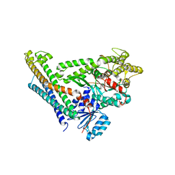
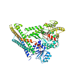
5KAI
 
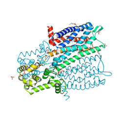
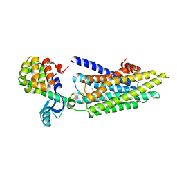
 | | NH3-bound RT XFEL structure of Photosystem II 500 ms after the 2nd illumination (2F) at 2.8 A resolution | | 分子名称: | 1,2-DI-O-ACYL-3-O-[6-DEOXY-6-SULFO-ALPHA-D-GLUCOPYRANOSYL]-SN-GLYCEROL, 1,2-DIPALMITOYL-PHOSPHATIDYL-GLYCEROLE, 1,2-DISTEAROYL-MONOGALACTOSYL-DIGLYCERIDE, ... | | 著者 | Young, I.D, Ibrahim, M, Chatterjee, R, Gul, S, Koroidov, S, Brewster, A.S, Tran, R, Alonso-Mori, R, Fuller, F, Kroll, T, Michels-Clark, T, Laksmono, H, Sierra, R.G, Stan, C.A, Saracini, C, Bean, M.A, Seuffert, I, Sokaras, D, Weng, T.-C, Hunter, M.S, Aquila, A, Koglin, J.E, Robinson, J, Liang, M, Boutet, S, Lyubimov, A.Y, Uervirojnangkoorn, M, Moriarty, N.W, Liebschner, D, Afonine, P.V, Waterman, D.G, Evans, G, Dobbek, H, Weis, W.I, Brunger, A.T, Zwart, P.H, Adams, P.D, Zouni, A, Messinger, J, Bergmann, U, Sauter, N.K, Kern, J, Yachandra, V.K, Yano, J. | | 登録日 | 2016-06-01 | | 公開日 | 2016-11-23 | | 最終更新日 | 2024-10-23 | | 実験手法 | X-RAY DIFFRACTION (2.80000925 Å) | | 主引用文献 | Structure of photosystem II and substrate binding at room temperature.
Nature, 540, 2016
|
|
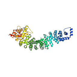
5KTH
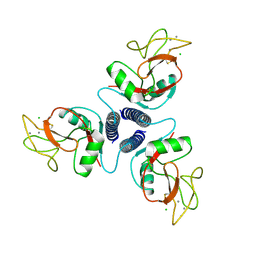
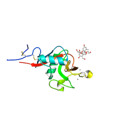 | | Structure of cow mincle complexed with brartemicin | | 分子名称: | 2,4-dihydroxy-6-methyl Benzoic acid, CALCIUM ION, TRIETHYLENE GLYCOL, ... | | 著者 | Feinberg, H, Rambaruth, N.D.S, Jegouzo, S.A.F, Jacobsen, K.M, Djurhuus, R, Poulsen, T.B, Taylor, M.E, Drickamer, K, Weis, W.I. | | 登録日 | 2016-07-11 | | 公開日 | 2016-08-31 | | 最終更新日 | 2024-10-16 | | 実験手法 | X-RAY DIFFRACTION (2.21 Å) | | 主引用文献 | Binding Sites for Acylated Trehalose Analogs of Glycolipid Ligands on an Extended Carbohydrate Recognition Domain of the Macrophage Receptor Mincle.
J.Biol.Chem., 291, 2016
|
|
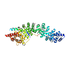
5KJ7
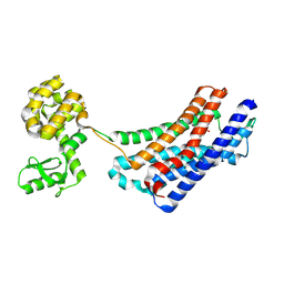
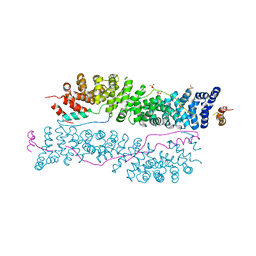
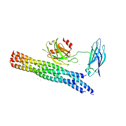 | | Structure of the Ca2+-bound synaptotagmin-1 SNARE complex (long unit cell form) - from XFEL diffraction | | 分子名称: | CALCIUM ION, Synaptosomal-associated protein 25, Synaptotagmin-1, ... | | 著者 | Lyubimov, A.Y, Uervirojnangkoorn, M, Zhou, Q, Zhao, M, Sauter, N.K, Brewster, A.S, Weis, W.I, Brunger, A.T. | | 登録日 | 2016-06-17 | | 公開日 | 2016-10-19 | | 最終更新日 | 2023-09-27 | | 実験手法 | X-RAY DIFFRACTION (3.5 Å) | | 主引用文献 | Advances in X-ray free electron laser (XFEL) diffraction data processing applied to the crystal structure of the synaptotagmin-1 / SNARE complex.
Elife, 5, 2016
|
|
5KJ8
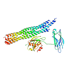 | | Structure of the Ca2+-bound synaptotagmin-1 SNARE complex (long unit cell form) - from synchrotron diffraction | | 分子名称: | CALCIUM ION, Synaptosomal-associated protein 25, Synaptotagmin-1, ... | | 著者 | Lyubimov, A.Y, Uervirojnangkoorn, M, Zhou, Q, Zhao, M, Sauter, N.K, Brewster, A.S, Weis, W.I, Brunger, A.T. | | 登録日 | 2016-06-17 | | 公開日 | 2016-10-19 | | 最終更新日 | 2023-09-27 | | 実験手法 | X-RAY DIFFRACTION (4.1 Å) | | 主引用文献 | Advances in X-ray free electron laser (XFEL) diffraction data processing applied to the crystal structure of the synaptotagmin-1 / SNARE complex.
Elife, 5, 2016
|
|
5KTI
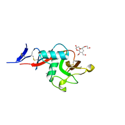 | | Structure of cow mincle complexed with KMJ1 | | 分子名称: | 2,3-dimethoxybenzoic acid, CALCIUM ION, TRIETHYLENE GLYCOL, ... | | 著者 | Feinberg, H, Rambaruth, N.D.S, Jegouzo, S.A.F, Jacobsen, K.M, Djurhuus, R, Poulsen, T.B, Taylor, M.E, Drickamer, K, Weis, W.I. | | 登録日 | 2016-07-11 | | 公開日 | 2016-08-31 | | 最終更新日 | 2024-11-06 | | 実験手法 | X-RAY DIFFRACTION (1.8 Å) | | 主引用文献 | Binding Sites for Acylated Trehalose Analogs of Glycolipid Ligands on an Extended Carbohydrate Recognition Domain of the Macrophage Receptor Mincle.
J.Biol.Chem., 291, 2016
|
|
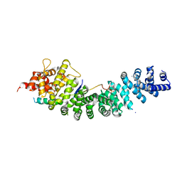
4JEH
 | | Crystal Structure of Munc18a and Syntaxin1 lacking N-peptide complex | | 分子名称: | Syntaxin-1A, Syntaxin-binding protein 1 | | 著者 | Colbert, K.N, Hattendorf, D.A, Weiss, T.M, Burkhardt, P, Fasshauer, D, Weis, W.I. | | 登録日 | 2013-02-27 | | 公開日 | 2013-07-17 | | 最終更新日 | 2023-09-20 | | 実験手法 | X-RAY DIFFRACTION (2.5 Å) | | 主引用文献 | Syntaxin1a variants lacking an N-peptide or bearing the LE mutation bind to Munc18a in a closed conformation.
Proc.Natl.Acad.Sci.USA, 110, 2013
|
|
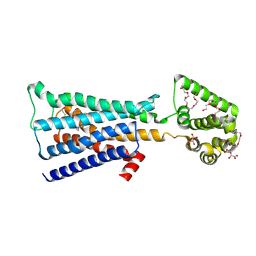
4JEU
 | | Crystal Structure of Munc18a and Syntaxin1 with native N-terminus complex | | 分子名称: | Syntaxin-1A, Syntaxin-binding protein 1 | | 著者 | Colbert, K.N, Hattendorf, D.A, Weiss, T.M, Burkhardt, P, Fasshauer, D, Weis, W.I. | | 登録日 | 2013-02-27 | | 公開日 | 2013-07-17 | | 最終更新日 | 2023-09-20 | | 実験手法 | X-RAY DIFFRACTION (3.2 Å) | | 主引用文献 | Syntaxin1a variants lacking an N-peptide or bearing the LE mutation bind to Munc18a in a closed conformation.
Proc.Natl.Acad.Sci.USA, 110, 2013
|
|
4R0Z
 | | A conserved phosphorylation switch controls the interaction between cadherin and beta-catenin in vitro and in vivo | | 分子名称: | FORMIC ACID, Protein humpback-2 | | 著者 | Choi, H.-J, Loveless, T, Lynch, A, Bang, I, Hardin, J, Weis, W.I. | | 登録日 | 2014-08-03 | | 公開日 | 2015-04-29 | | 最終更新日 | 2023-11-08 | | 実験手法 | X-RAY DIFFRACTION (2.005 Å) | | 主引用文献 | A Conserved Phosphorylation Switch Controls the Interaction between Cadherin and beta-Catenin In Vitro and In Vivo
Dev.Cell, 33, 2015
|
|
4R10
 | | A conserved phosphorylation switch controls the interaction between cadherin and beta-catenin in vitro and in vivo | | 分子名称: | 1,2-ETHANEDIOL, Cadherin-related hmr-1, Protein humpback-2, ... | | 著者 | Choi, H.-J, Loveless, T, Lynch, A, Bang, I, Hardin, J, Weis, W.I. | | 登録日 | 2014-08-03 | | 公開日 | 2015-04-29 | | 最終更新日 | 2024-10-16 | | 実験手法 | X-RAY DIFFRACTION (2.3 Å) | | 主引用文献 | A Conserved Phosphorylation Switch Controls the Interaction between Cadherin and beta-Catenin In Vitro and In Vivo
Dev.Cell, 33, 2015
|
|
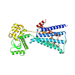
2FO0
 | | Organization of the SH3-SH2 Unit in Active and Inactive Forms of the c-Abl Tyrosine Kinase | | 分子名称: | 6-(2,6-DICHLOROPHENYL)-2-{[3-(HYDROXYMETHYL)PHENYL]AMINO}-8-METHYLPYRIDO[2,3-D]PYRIMIDIN-7(8H)-ONE, GLYCEROL, MYRISTIC ACID, ... | | 著者 | Nagar, B, Hantschel, O, Seeliger, M, Davies, J.M, Weis, W.I, Superti-Furga, G, Kuriyan, J. | | 登録日 | 2006-01-12 | | 公開日 | 2006-03-21 | | 最終更新日 | 2024-10-30 | | 実験手法 | X-RAY DIFFRACTION (2.27 Å) | | 主引用文献 | Organization of the SH3-SH2 unit in active and inactive forms of the c-Abl tyrosine kinase.
Mol.Cell, 21, 2006
|
|
4R11
 | | A conserved phosphorylation switch controls the interaction between cadherin and beta-catenin in vitro and in vivo | | 分子名称: | Cadherin-related hmr-1, IODIDE ION, Protein humpback-2 | | 著者 | Choi, H.-J, Loveless, T, Lynch, A, Bang, I, Hardin, J, Weis, W.I. | | 登録日 | 2014-08-03 | | 公開日 | 2015-04-29 | | 最終更新日 | 2024-11-20 | | 実験手法 | X-RAY DIFFRACTION (2.789 Å) | | 主引用文献 | A Conserved Phosphorylation Switch Controls the Interaction between Cadherin and beta-Catenin In Vitro and In Vivo
Dev.Cell, 33, 2015
|
|
4U15
 | | M3-mT4L receptor bound to tiotropium | | 分子名称: | (1R,2R,4S,5S,7S)-7-{[hydroxy(dithiophen-2-yl)acetyl]oxy}-9,9-dimethyl-3-oxa-9-azoniatricyclo[3.3.1.0~2,4~]nonane, (2R)-2,3-dihydroxypropyl (9Z)-octadec-9-enoate, D(-)-TARTARIC ACID, ... | | 著者 | Thorsen, T.S, Matt, R, Weis, W.I, Kobilka, B. | | 登録日 | 2014-07-15 | | 公開日 | 2014-11-26 | | 最終更新日 | 2024-11-20 | | 実験手法 | X-RAY DIFFRACTION (2.8 Å) | | 主引用文献 | Modified T4 Lysozyme Fusion Proteins Facilitate G Protein-Coupled Receptor Crystallogenesis.
Structure, 22, 2014
|
|
4U14
 | | Structure of the M3 muscarinic acetylcholine receptor bound to the antagonist tiotropium crystallized with disulfide-stabilized T4 lysozyme (dsT4L) | | 分子名称: | (1R,2R,4S,5S,7S)-7-{[hydroxy(dithiophen-2-yl)acetyl]oxy}-9,9-dimethyl-3-oxa-9-azoniatricyclo[3.3.1.0~2,4~]nonane, Muscarinic acetylcholine receptor M3,Endolysin,Muscarinic acetylcholine receptor M3 | | 著者 | Thorsen, T.S, Matt, R.A, Weis, W.I, Kobilka, B.K. | | 登録日 | 2014-07-15 | | 公開日 | 2014-11-26 | | 最終更新日 | 2024-11-06 | | 実験手法 | X-RAY DIFFRACTION (3.57 Å) | | 主引用文献 | Modified T4 Lysozyme Fusion Proteins Facilitate G Protein-Coupled Receptor Crystallogenesis.
Structure, 22, 2014
|
|
4U16
 | | M3-mT4L receptor bound to NMS | | 分子名称: | D(-)-TARTARIC ACID, Muscarinic acetylcholine receptor M3,Lysozyme,Muscarinic acetylcholine receptor M3, N-methyl scopolamine | | 著者 | Thorsen, T.S, Matt, R, Weis, W.I, Kobilka, B. | | 登録日 | 2014-07-15 | | 公開日 | 2014-11-26 | | 最終更新日 | 2024-11-20 | | 実験手法 | X-RAY DIFFRACTION (3.7 Å) | | 主引用文献 | Modified T4 Lysozyme Fusion Proteins Facilitate G Protein-Coupled Receptor Crystallogenesis.
Structure, 22, 2014
|
|
4EJ4
 | | Structure of the delta opioid receptor bound to naltrindole | | 分子名称: | (4bS,8R,8aS,14bR)-7-(cyclopropylmethyl)-5,6,7,8,14,14b-hexahydro-4,8-methano[1]benzofuro[2,3-a]pyrido[4,3-b]carbazole-1,8a(9H)-diol, Delta-type opioid receptor, Lysozyme chimera | | 著者 | Granier, S, Manglik, A, Kruse, A.C, Kobilka, T.S, Thian, F.S, Weis, W.I, Kobilka, B.K. | | 登録日 | 2012-04-06 | | 公開日 | 2012-05-16 | | 最終更新日 | 2024-10-30 | | 実験手法 | X-RAY DIFFRACTION (3.4 Å) | | 主引用文献 | Structure of the delta opioid receptor bound to naltrindole
Nature, 485, 2012
|
|
4E17
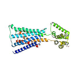
 | | Alpha-E-catenin is an autoinhibited molecule that co-activates vinculin | | 分子名称: | Catenin alpha-1, Vinculin | | 著者 | Choi, H.-J, Pokutta, S, Cadwell, G.W, Bankston, L.A, Liddington, R.C, Weis, W.I. | | 登録日 | 2012-03-05 | | 公開日 | 2012-05-16 | | 最終更新日 | 2023-09-13 | | 実験手法 | X-RAY DIFFRACTION (2.304 Å) | | 主引用文献 | Conformational plasticity of alpha-catenin revealed by binding interactions with vinculin
To be Published
|
|
4E18
 | | Alpha-E-catenin is an autoinhibited molecule that co-activates vinculin | | 分子名称: | Catenin alpha-1, Vinculin | | 著者 | Choi, H.-J, Pokutta, S, Cadwell, G.W, Bankston, L.A, Liddington, R.C, Weis, W.I. | | 登録日 | 2012-03-05 | | 公開日 | 2012-05-16 | | 最終更新日 | 2023-09-13 | | 実験手法 | X-RAY DIFFRACTION (2.403 Å) | | 主引用文献 | Conformational plasticity of alpha-catenin revealed by binding interactions with vinculin
To be Published
|
|
4DKL
 | | Crystal structure of the mu-opioid receptor bound to a morphinan antagonist | | 分子名称: | CHLORIDE ION, CHOLESTEROL, Mu-type opioid receptor, ... | | 著者 | Manglik, A, Kruse, A.C, Kobilka, T.S, Thian, F.S, Mathiesen, J.M, Sunahara, R.K, Pardo, L, Weis, W.I, Kobilka, B.K, Granier, S. | | 登録日 | 2012-02-03 | | 公開日 | 2012-03-21 | | 最終更新日 | 2024-11-27 | | 実験手法 | X-RAY DIFFRACTION (2.8 Å) | | 主引用文献 | Crystal structure of the {mu}-opioid receptor bound to a morphinan antagonist.
Nature, 485, 2012
|
|
4DAJ
 | | Structure of the M3 Muscarinic Acetylcholine Receptor | | 分子名称: | (1R,2R,4S,5S,7S)-7-{[hydroxy(dithiophen-2-yl)acetyl]oxy}-9,9-dimethyl-3-oxa-9-azoniatricyclo[3.3.1.0~2,4~]nonane, Muscarinic acetylcholine receptor M3, Lysozyme, ... | | 著者 | Kruse, A.C, Hu, J, Pan, A.C, Arlow, D.H, Rosenbaum, D.M, Rosemond, E, Green, H.F, Liu, T, Chae, P.S, Dror, R.O, Shaw, D.E, Weis, W.I, Wess, J, Kobilka, B. | | 登録日 | 2012-01-12 | | 公開日 | 2012-02-22 | | 最終更新日 | 2024-10-30 | | 実験手法 | X-RAY DIFFRACTION (3.4 Å) | | 主引用文献 | Structure and dynamics of the M3 muscarinic acetylcholine receptor.
Nature, 482, 2012
|
|
4GBR
 | |
1KMB
 | | SELECTIN-LIKE MUTANT OF MANNOSE-BINDING PROTEIN A | | 分子名称: | CALCIUM ION, CHLORIDE ION, MANNOSE-BINDING PROTEIN-A | | 著者 | Ng, K.K.-S, Weis, W.I. | | 登録日 | 1996-11-07 | | 公開日 | 1997-02-12 | | 最終更新日 | 2024-11-20 | | 実験手法 | X-RAY DIFFRACTION (2.1 Å) | | 主引用文献 | Structure of a selectin-like mutant of mannose-binding protein complexed with sialylated and sulfated Lewis(x) oligosaccharides.
Biochemistry, 36, 1997
|
|
3IFQ
 | | Interction of plakoglobin and beta-catenin with desmosomal cadherins | | 分子名称: | E-cadherin, SULFATE ION, plakoglobin | | 著者 | Choi, H.-J, Gross, J.C, Pokutta, S, Weis, W.I. | | 登録日 | 2009-07-24 | | 公開日 | 2009-09-15 | | 最終更新日 | 2024-11-27 | | 実験手法 | X-RAY DIFFRACTION (2.8 Å) | | 主引用文献 | Interactions of plakoglobin and beta-catenin with desmosomal cadherins: basis of selective exclusion of alpha- and beta-catenin from desmosomes.
J.Biol.Chem., 284, 2009
|
|
3JQH
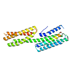
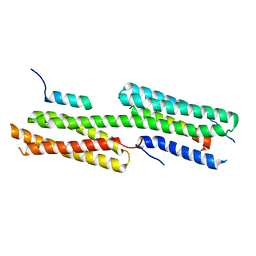
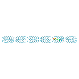 | | Structure of the neck region of the glycan-binding receptor DC-SIGNR | | 分子名称: | C-type lectin domain family 4 member M | | 著者 | Feinberg, H, Tso, C.K.W, Taylor, M.E, Drickamer, K, Weis, W.I. | | 登録日 | 2009-09-06 | | 公開日 | 2009-11-10 | | 最終更新日 | 2024-02-21 | | 実験手法 | X-RAY DIFFRACTION (2.201 Å) | | 主引用文献 | Segmented helical structure of the neck region of the glycan-binding receptor DC-SIGNR.
J.Mol.Biol., 394, 2009
|
|
2M4N
 | | Solution structure of the putative Ras interaction domain of AFD-1, isoform a from Caenorhabditis elegans | | 分子名称: | Protein AFD-1, isoform a | | 著者 | Harris, R, Hillerich, B, Ahmed, M, Bonanno, J.B, Chamala, S, Evans, B, Lafleur, J, Hammonds, J, Washington, E, Stead, M, Love, J, Attonito, J, Seidel, R.D, Liddington, R.C, Weis, W.I, Nelson, W.J, Girvin, M.E, Almo, S.C, New York Structural Genomics Research Consortium (NYSGRC), Assembly, Dynamics and Evolution of Cell-Cell and Cell-Matrix Adhesions (CELLMAT) | | 登録日 | 2013-02-07 | | 公開日 | 2013-03-20 | | 最終更新日 | 2024-05-15 | | 実験手法 | SOLUTION NMR | | 主引用文献 | Solution structure of the putative Ras interaction domain of AFD-1, isoform a from Caenorhabditis elegans
To be Published
|
|
4ZRW
 | | Structure of cow mincle complexed with trehalose | | 分子名称: | CALCIUM ION, alpha-D-glucopyranose-(1-1)-alpha-D-glucopyranose, mincle protein | | 著者 | Feinberg, H, Rambaruth, N.D.S, Taylor, M.E, Drickamer, K, Weis, W.I. | | 登録日 | 2015-05-12 | | 公開日 | 2016-05-25 | | 最終更新日 | 2024-11-06 | | 実験手法 | X-RAY DIFFRACTION (2.6 Å) | | 主引用文献 | Binding Sites for Acylated Trehalose Analogs of Glycolipid Ligands on an Extended Carbohydrate Recognition Domain of the Macrophage Receptor Mincle.
J.Biol.Chem., 291, 2016
|
|


