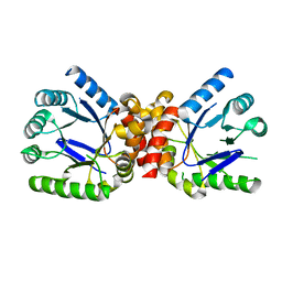
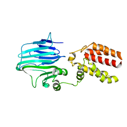
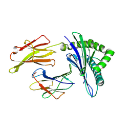
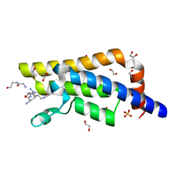
5KCC
 
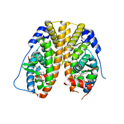 | | Crystal Structure of the ER-alpha Ligand-binding Domain (Y537S) in Complex with Oxabicyclic Heptene Sulfonamide (OBHS-N) | | 分子名称: | (1S,2R,4S)-5,6-bis(4-hydroxyphenyl)-N-phenyl-7-oxabicyclo[2.2.1]hept-5-ene-2-sulfonamide, Estrogen receptor, NCOA2 | | 著者 | Nwachukwu, J.C, Srinivasan, S, Zheng, Y, Wang, S, Min, J, Dong, C, Liao, Z, Cavett, V, Nowak, J, Houtman, R, Carlson, K.E, Josan, J.S, Elemento, O, Katzenellenbogen, J.A, Zhou, H.B, Nettles, K.W. | | 登録日 | 2016-06-06 | | 公開日 | 2016-11-16 | | 最終更新日 | 2024-03-06 | | 実験手法 | X-RAY DIFFRACTION (2.386 Å) | | 主引用文献 | Full antagonism of the estrogen receptor without a prototypical ligand side chain.
Nat. Chem. Biol., 13, 2017
|
|
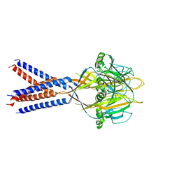
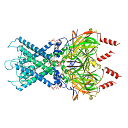
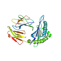
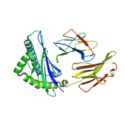
2EXX
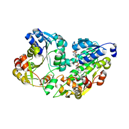 | | Crystal structure of HSCARG from Homo sapiens in complex with NADP | | 分子名称: | GLYCEROL, HSCARG protein, NADP NICOTINAMIDE-ADENINE-DINUCLEOTIDE PHOSPHATE | | 著者 | Dai, X, Chen, Q, Yao, D, Liang, Y, Dong, Y, Gu, X, Zheng, X, Luo, M. | | 登録日 | 2005-11-09 | | 公開日 | 2006-11-21 | | 最終更新日 | 2017-10-18 | | 実験手法 | X-RAY DIFFRACTION (2.4 Å) | | 主引用文献 | Restructuring of the dinucleotide-binding fold in an NADP(H) sensor protein.
Proc.Natl.Acad.Sci.USA, 104, 2007
|
|
3TYZ
 | |
3TZF
 | |
3TZN
 | |
7JI3
 | |
4Q2M
 | |
4Q2L
 | |
5Y83
 | |
6XEU
 | | CryoEM structure of GIRK2PIP2* - G protein-gated inwardly rectifying potassium channel GIRK2 with PIP2 | | 分子名称: | G protein-activated inward rectifier potassium channel 2, POTASSIUM ION, SODIUM ION, ... | | 著者 | Mathiharan, Y.K, Glaaser, I.W, Skiniotis, G, Slesinger, P.A. | | 登録日 | 2020-06-13 | | 公開日 | 2021-09-01 | | 実験手法 | ELECTRON MICROSCOPY (3.2 Å) | | 主引用文献 | Structural insights into GIRK2 channel modulation by cholesterol and PIP2
Cell Rep, 36, 2021
|
|
6XEV
 | | CryoEM structure of GIRK2-PIP2/CHS - G protein-gated inwardly rectifying potassium channel GIRK2 with modulators cholesteryl hemisuccinate and PIP2 | | 分子名称: | CHOLESTEROL HEMISUCCINATE, G protein-activated inward rectifier potassium channel 2, POTASSIUM ION, ... | | 著者 | Mathiharan, Y.K, Glaaser, I.W, Skiniotis, G, Slesinger, P.A. | | 登録日 | 2020-06-14 | | 公開日 | 2021-09-01 | | 実験手法 | ELECTRON MICROSCOPY (3.5 Å) | | 主引用文献 | Structural insights into GIRK2 channel modulation by cholesterol and PIP2
Cell Rep, 36, 2021
|
|
6J2A
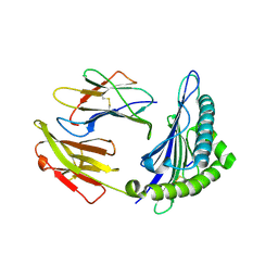 | | The structure of HLA-A*3003/NP44 | | 分子名称: | Beta-2-microglobulin, HLA-A*3003, NP44 | | 著者 | Zhu, S.Y, Liu, K.F, Chai, Y, Ding, C.M, Lv, J.X, Gao, F.G, Lou, Y.L, Liu, W.J. | | 登録日 | 2018-12-31 | | 公開日 | 2019-09-25 | | 実験手法 | X-RAY DIFFRACTION (1.4 Å) | | 主引用文献 | Divergent Peptide Presentations of HLA-A*30 Alleles Revealed by Structures With Pathogen Peptides.
Front Immunol, 10, 2019
|
|
6J1W
 | | The structure of HLA-A*3001/RT313 | | 分子名称: | ALA-ILE-PHE-GLN-SER-SER-MET-THR-LYS, Beta-2-microglobulin, HLA-A*3001 | | 著者 | Zhu, S.Y, Liu, K.F, Chai, Y, Ding, C.M, Lv, J.X, Gao, G.F, Lou, Y.L, Liu, W.J. | | 登録日 | 2018-12-29 | | 公開日 | 2019-09-25 | | 実験手法 | X-RAY DIFFRACTION (1.501 Å) | | 主引用文献 | Divergent Peptide Presentations of HLA-A*30 Alleles Revealed by Structures With Pathogen Peptides.
Front Immunol, 10, 2019
|
|
6J29
 | | The structure of HLA-A*3003/MTB | | 分子名称: | Beta-2-microglobulin, HLA-A*3003, MTB | | 著者 | Zhu, S.Y, Liu, K.F, Chai, Y, Ding, C.M, Lv, J.X, Gao, F.G, Lou, Y.L, Liu, W.J. | | 登録日 | 2018-12-31 | | 公開日 | 2019-09-25 | | 実験手法 | X-RAY DIFFRACTION (1.6 Å) | | 主引用文献 | Divergent Peptide Presentations of HLA-A*30 Alleles Revealed by Structures With Pathogen Peptides.
Front Immunol, 10, 2019
|
|
4QUT
 | | Structure of the bromodomain of human ATPase family AAA domain-containing protein 2 (ATAD2) complexed with Histone H4-K(ac)12 | | 分子名称: | 1,2-ETHANEDIOL, ATPase family AAA domain-containing protein 2, Histone H4, ... | | 著者 | Chaikuad, A, Felletar, I, von Delft, F, Arrowsmith, C.H, Edwards, A.M, Bountra, C, Knapp, S, Structural Genomics Consortium (SGC) | | 登録日 | 2014-07-12 | | 公開日 | 2014-07-30 | | 最終更新日 | 2023-12-06 | | 実験手法 | X-RAY DIFFRACTION (1.7 Å) | | 主引用文献 | Atad2 is a generalist facilitator of chromatin dynamics in embryonic stem cells.
J Mol Cell Biol, 8, 2016
|
|
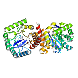
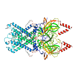
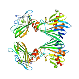
4R99
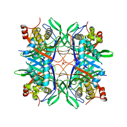 | | Crystal structure of a uricase from Bacillus fastidious | | 分子名称: | SULFATE ION, Uricase | | 著者 | Feng, J, Wang, L, Liu, H.B, Liu, L, Liao, F. | | 登録日 | 2014-09-03 | | 公開日 | 2015-05-27 | | 最終更新日 | 2023-11-08 | | 実験手法 | X-RAY DIFFRACTION (1.8 Å) | | 主引用文献 | Crystal structure of Bacillus fastidious uricase reveals an unexpected folding of the C-terminus residues crucial for thermostability under physiological conditions.
Appl.Microbiol.Biotechnol., 99, 2015
|
|
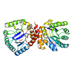
4R8X
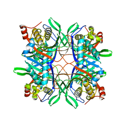 | | Crystal structure of a uricase from Bacillus fastidious | | 分子名称: | Uricase | | 著者 | Feng, J, Wang, L, Liu, H.B, Liu, L, Liao, F. | | 登録日 | 2014-09-03 | | 公開日 | 2015-05-27 | | 最終更新日 | 2023-11-08 | | 実験手法 | X-RAY DIFFRACTION (1.401 Å) | | 主引用文献 | Crystal structure of Bacillus fastidious uricase reveals an unexpected folding of the C-terminus residues crucial for thermostability under physiological conditions.
Appl.Microbiol.Biotechnol., 99, 2015
|
|
4QUU
 | | Structure of the bromodomain of human ATPase family AAA domain-containing protein 2 (ATAD2) complexed with Histone H4-K(ac)5 | | 分子名称: | 1,2-ETHANEDIOL, ATPase family AAA domain-containing protein 2, Histone H4, ... | | 著者 | Chaikuad, A, Felletar, I, von Delft, F, Arrowsmith, C.H, Edwards, A.M, Bountra, C, Knapp, S, Structural Genomics Consortium (SGC) | | 登録日 | 2014-07-12 | | 公開日 | 2014-07-30 | | 最終更新日 | 2023-12-06 | | 実験手法 | X-RAY DIFFRACTION (1.8 Å) | | 主引用文献 | Atad2 is a generalist facilitator of chromatin dynamics in embryonic stem cells.
J Mol Cell Biol, 8, 2016
|
|
6J1V
 | | The structure of HLA-A*3003/RT313 | | 分子名称: | Beta-2-microglobulin, HLA-A*3003, RT313 | | 著者 | Zhu, S.Y, Liu, K.F, Chai, Y, Ding, C.M, Lv, J.X, Gao, G.F, Lou, Y.L, Liu, W.J. | | 登録日 | 2018-12-29 | | 公開日 | 2019-09-25 | | 実験手法 | X-RAY DIFFRACTION (2 Å) | | 主引用文献 | Divergent Peptide Presentations of HLA-A*30 Alleles Revealed by Structures With Pathogen Peptides.
Front Immunol, 10, 2019
|
|
5Y82
 | |
6JMD
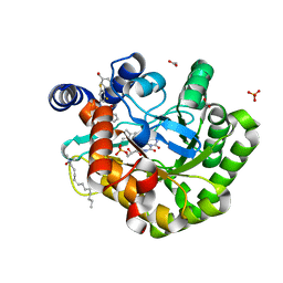 | | Crystal structure of human DHODH in complex with inhibitor 1223 | | 分子名称: | 3-[3,5-bis(fluoranyl)-4-[3-(hydroxymethyl)phenyl]phenyl]benzo[f]benzotriazole-4,9-dione, ACETATE ION, Dihydroorotate dehydrogenase (quinone), ... | | 著者 | Yu, Y, Chen, Q. | | 登録日 | 2019-03-08 | | 公開日 | 2020-03-18 | | 最終更新日 | 2024-03-27 | | 実験手法 | X-RAY DIFFRACTION (1.781 Å) | | 主引用文献 | Bifunctional Naphtho[2,3- d ][1,2,3]triazole-4,9-dione Compounds Exhibit Antitumor Effects In Vitro and In Vivo by Inhibiting Dihydroorotate Dehydrogenase and Inducing Reactive Oxygen Species Production.
J.Med.Chem., 63, 2020
|
|
6XJY
 | |
6XJQ
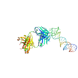
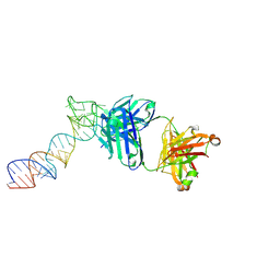
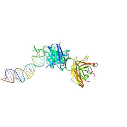
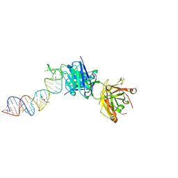 | | Crystal structure of a self-alkylating ribozyme - alkylated form with biotinylated epoxide substrate | | 分子名称: | 2-{[(4R)-4-hydroxyhexyl]oxy}ethyl 5-[(3aS,4S,6aR)-2-oxohexahydro-1H-thieno[3,4-d]imidazol-4-yl]pentanoate, Fab HAVx Heavy Chain, Fab HAVx Light Chain, ... | | 著者 | Koirala, D, Piccirilli, J.A. | | 登録日 | 2020-06-24 | | 公開日 | 2022-01-19 | | 最終更新日 | 2023-10-18 | | 実験手法 | X-RAY DIFFRACTION (1.708 Å) | | 主引用文献 | Structural basis for substrate binding and catalysis by a self-alkylating ribozyme.
Nat.Chem.Biol., 18, 2022
|
|
6XJZ
 | |
6XJW
 | |


