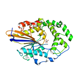
1AQL
 
 | |
1AKN
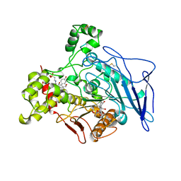
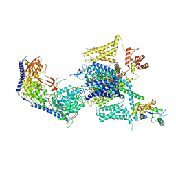
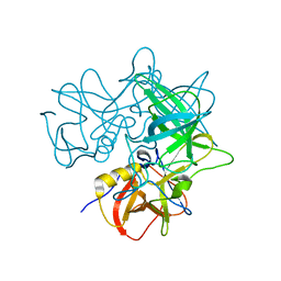
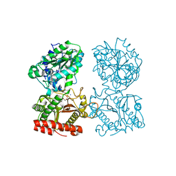
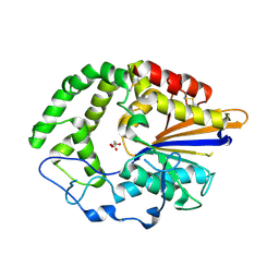
 | | STRUCTURE OF BILE-SALT ACTIVATED LIPASE | | 分子名称: | 2-acetamido-2-deoxy-beta-D-glucopyranose, BILE-SALT ACTIVATED LIPASE | | 著者 | Wang, X, Zhang, X. | | 登録日 | 1997-05-23 | | 公開日 | 1998-05-27 | | 最終更新日 | 2023-08-02 | | 実験手法 | X-RAY DIFFRACTION (2.8 Å) | | 主引用文献 | The crystal structure of bovine bile salt activated lipase: insights into the bile salt activation mechanism.
Structure, 5, 1997
|
|
8HMB
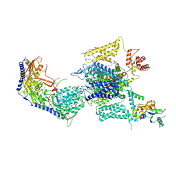 | | Cryo-EM structure of human high-voltage activated L-type calcium channel CaV1.2 in complex with benidipine (BEN) | | 分子名称: | (3R)-1-benzylpiperidin-3-yl methyl (2R,3R,4R,5R,6S)-2,6-dimethyl-4-(3-nitrophenyl)piperidine-3,5-dicarboxylate, 1,2-Distearoyl-sn-glycerophosphoethanolamine, 2-acetamido-2-deoxy-beta-D-glucopyranose, ... | | 著者 | Wei, Y, Yu, Z, Zhao, Y. | | 登録日 | 2022-12-02 | | 公開日 | 2024-04-24 | | 実験手法 | ELECTRON MICROSCOPY (3.3 Å) | | 主引用文献 | Structural bases of inhibitory mechanism of Ca V 1.2 channel inhibitors.
Nat Commun, 15, 2024
|
|
8HMA
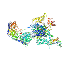 | | Cryo-EM structure of human high-voltage activated L-type calcium channel CaV1.2 in complex with tetrandrine (TET) | | 分子名称: | 1,2-Distearoyl-sn-glycerophosphoethanolamine, 2-acetamido-2-deoxy-beta-D-glucopyranose, 6,6',7,12-tetramethoxy-2,2'-dimethyl-1beta-3,4-didehydroberbaman, ... | | 著者 | Wei, Y, Yu, Z, Zhao, Y. | | 登録日 | 2022-12-02 | | 公開日 | 2024-04-24 | | 実験手法 | ELECTRON MICROSCOPY (3.4 Å) | | 主引用文献 | Structural bases of inhibitory mechanism of Ca V 1.2 channel inhibitors.
Nat Commun, 15, 2024
|
|
8HLP
 | | Cryo-EM structure of human high-voltage activated L-type calcium channel CaV1.2 (apo) | | 分子名称: | 1,2-Distearoyl-sn-glycerophosphoethanolamine, 2-acetamido-2-deoxy-beta-D-glucopyranose, CALCIUM ION, ... | | 著者 | Wei, Y, Yu, Z, Zhao, Y. | | 登録日 | 2022-11-30 | | 公開日 | 2024-04-24 | | 実験手法 | ELECTRON MICROSCOPY (3.5 Å) | | 主引用文献 | Structural bases of inhibitory mechanism of Ca V 1.2 channel inhibitors.
Nat Commun, 15, 2024
|
|
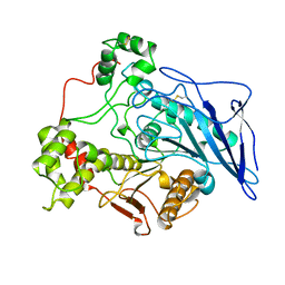
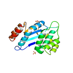
3N2N
 | | The Crystal Structure of Tumor Endothelial Marker 8 (TEM8) extracellular domain | | 分子名称: | ACETATE ION, Anthrax toxin receptor 1, MAGNESIUM ION | | 著者 | Fu, S, Tong, X.H, Wu, Y, Li, Y.Y, Rao, Z.H. | | 登録日 | 2010-05-18 | | 公開日 | 2011-01-05 | | 最終更新日 | 2023-11-01 | | 実験手法 | X-RAY DIFFRACTION (1.8 Å) | | 主引用文献 | The structure of tumor endothelial marker 8 (TEM8) extracellular domain and implications for its receptor function for recognizing anthrax toxin.
Plos One, 5, 2010
|
|
5D1I
 | |
2GTH
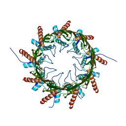
 | | crystal structure of the wildtype MHV coronavirus non-structural protein nsp15 | | 分子名称: | Replicase polyprotein 1ab | | 著者 | Xu, X, Zhai, Y, Sun, F, Lou, Z, Su, D, Rao, Z. | | 登録日 | 2006-04-28 | | 公開日 | 2006-08-15 | | 最終更新日 | 2023-10-25 | | 実験手法 | X-RAY DIFFRACTION (2.7 Å) | | 主引用文献 | New Antiviral Target Revealed by the Hexameric Structure of Mouse Hepatitis Virus Nonstructural Protein nsp15
J.Virol., 80, 2006
|
|
2OBT
 | |
2G9T
 | | Crystal structure of the SARS coronavirus nsp10 at 2.1A | | 分子名称: | ZINC ION, orf1a polyprotein | | 著者 | Su, D, Lou, Z, Yang, H, Sun, F, Rao, Z. | | 登録日 | 2006-03-07 | | 公開日 | 2006-08-15 | | 最終更新日 | 2024-03-13 | | 実験手法 | X-RAY DIFFRACTION (2.1 Å) | | 主引用文献 | Dodecamer Structure of Severe Acute Respiratory Syndrome Coronavirus Nonstructural Protein nsp10
J.Virol., 80, 2006
|
|
2GTI
 | | mutation of MHV coronavirus non-structural protein nsp15 (F307L) | | 分子名称: | GLYCEROL, Replicase polyprotein 1ab, SULFATE ION | | 著者 | Xu, X, Zhai, Y, Sun, F, Lou, Z, Su, D, Rao, Z. | | 登録日 | 2006-04-28 | | 公開日 | 2006-08-15 | | 最終更新日 | 2021-11-10 | | 実験手法 | X-RAY DIFFRACTION (2.15 Å) | | 主引用文献 | New Antiviral Target Revealed by the Hexameric Structure of Mouse Hepatitis Virus Nonstructural Protein nsp15
J.Virol., 80, 2006
|
|
2OBS
 | |
2GA6
 | | The crystal structure of SARS nsp10 without zinc ion as additive | | 分子名称: | ZINC ION, orf1a polyprotein | | 著者 | Su, D, Lou, Z, Sun, F, Zhai, Y, Yang, H, Rao, Z. | | 登録日 | 2006-03-08 | | 公開日 | 2006-08-15 | | 最終更新日 | 2023-10-25 | | 実験手法 | X-RAY DIFFRACTION (2.7 Å) | | 主引用文献 | Dodecamer Structure of Severe Acute Respiratory Syndrome Coronavirus Nonstructural Protein nsp10
J.Virol., 80, 2006
|
|
4Q65
 | | Structure of the E. coli Peptide Transporter YbgH | | 分子名称: | Dipeptide permease D | | 著者 | Zhang, C, Zhao, Y, Mao, G, Liu, M, Wang, X. | | 登録日 | 2014-04-21 | | 公開日 | 2014-08-13 | | 最終更新日 | 2024-05-29 | | 実験手法 | X-RAY DIFFRACTION (3.4 Å) | | 主引用文献 | Crystal structure of the E. coli peptide transporter YbgH.
Structure, 22, 2014
|
|
3CTZ
 | | Structure of human cytosolic X-prolyl aminopeptidase | | 分子名称: | CALCIUM ION, HEXAETHYLENE GLYCOL, MANGANESE (II) ION, ... | | 著者 | Li, X, Lou, Z, Rao, Z. | | 登録日 | 2008-04-15 | | 公開日 | 2008-05-27 | | 最終更新日 | 2024-03-20 | | 実験手法 | X-RAY DIFFRACTION (1.6 Å) | | 主引用文献 | Structure of human cytosolic X-prolyl aminopeptidase: a double Mn(II)-dependent dimeric enzyme with a novel three-domain subunit
J.Biol.Chem., 283, 2008
|
|
3WDO
 | | Structure of E. coli YajR transporter | | 分子名称: | MFS Transporter | | 著者 | Jiang, D. | | 登録日 | 2013-06-19 | | 公開日 | 2013-08-07 | | 最終更新日 | 2024-03-20 | | 実験手法 | X-RAY DIFFRACTION (3.15 Å) | | 主引用文献 | Structure of the YajR transporter suggests a transport mechanism based on the conserved motif A
Proc.Natl.Acad.Sci.USA, 110, 2013
|
|
4Q79
 | | Structure of a HG-derivative CsgG | | 分子名称: | CsgG, MERCURY (II) ION | | 著者 | Huang, Y, Zhang, C.X, Cao, B, Zhao, Y, Kou, Y, Ni, D. | | 登録日 | 2014-04-24 | | 公開日 | 2014-12-17 | | 最終更新日 | 2024-03-20 | | 実験手法 | X-RAY DIFFRACTION (3.1 Å) | | 主引用文献 | Structure of the nonameric bacterial amyloid secretion channel
Proc.Natl.Acad.Sci.USA, 111, 2014
|
|
3HVN
 | | Crystal structure of cytotoxin protein suilysin from Streptococcus suis | | 分子名称: | 1,1,1,3,3,3-hexafluoropropan-2-ol, HEPTANE-1,2,3-TRIOL, Hemolysin | | 著者 | Xu, L, Huang, B, Du, H, Zhang, C.X, Xu, J, Li, X, Rao, Z. | | 登録日 | 2009-06-16 | | 公開日 | 2010-03-02 | | 最終更新日 | 2024-05-29 | | 実験手法 | X-RAY DIFFRACTION (2.852 Å) | | 主引用文献 | Crystal structure of cytotoxin protein suilysin from Streptococcus suis.
Protein Cell, 1, 2010
|
|
4JOD
 | |
4JOB
 | |
4JOC
 | |
4JR6
 | | Crystal structure of DsbA from Mycobacterium tuberculosis (reduced) | | 分子名称: | Possible conserved membrane or secreted protein, SULFATE ION | | 著者 | Wang, L. | | 登録日 | 2013-03-21 | | 公開日 | 2013-07-17 | | 最終更新日 | 2017-11-15 | | 実験手法 | X-RAY DIFFRACTION (1.902 Å) | | 主引用文献 | Structure analysis of the extracellular domain reveals disulfide bond forming-protein properties of Mycobacterium tuberculosis Rv2969c.
Protein Cell, 4, 2013
|
|
4JR4
 | | Crystal structure of Mtb DsbA (Oxidized) | | 分子名称: | Possible conserved membrane or secreted protein, SULFATE ION | | 著者 | Wang, L. | | 登録日 | 2013-03-21 | | 公開日 | 2013-07-17 | | 最終更新日 | 2017-11-15 | | 実験手法 | X-RAY DIFFRACTION (2.498 Å) | | 主引用文献 | Structure analysis of the extracellular domain reveals disulfide bond forming-protein properties of Mycobacterium tuberculosis Rv2969c.
Protein Cell, 4, 2013
|
|
4MG3
 | | Crystal Structural Analysis of 2A Protease from Coxsackievirus A16 | | 分子名称: | 2-AMINO-2-HYDROXYMETHYL-PROPANE-1,3-DIOL, PENTAETHYLENE GLYCOL, Protease 2A, ... | | 著者 | Sun, Y, Wang, X, Dang, M, Yuan, S. | | 登録日 | 2013-08-28 | | 公開日 | 2014-03-26 | | 最終更新日 | 2017-11-15 | | 実験手法 | X-RAY DIFFRACTION (1.798 Å) | | 主引用文献 | An open conformation determined by a structural switch for 2A protease from coxsackievirus A16.
Protein Cell, 4, 2013
|
|
5ZOR
 | |


