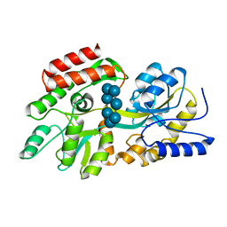
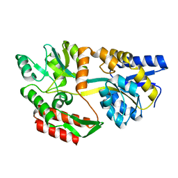
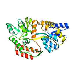
1QUL
 
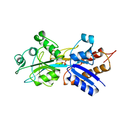 | | PHOSPHATE-BINDING PROTEIN MUTANT WITH ASP 137 REPLACED BY THR COMPLEX WITH CHLORINE AND PHOSPHATE | | 分子名称: | CHLORIDE ION, PHOSPHATE ION, PHOSPHATE-BINDING PROTEIN | | 著者 | Yao, N, Choudhary, A, Ledvina, P.S, Quiocho, F.A. | | 登録日 | 1995-11-11 | | 公開日 | 1996-07-11 | | 最終更新日 | 2024-02-14 | | 実験手法 | X-RAY DIFFRACTION (1.7 Å) | | 主引用文献 | Modulation of a salt link does not affect binding of phosphate to its specific active transport receptor.
Biochemistry, 35, 1996
|
|
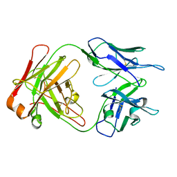
1M7D
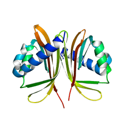
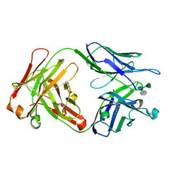 | | Crystal structure of a Monoclonal Fab Specific for Shigella flexneri Y Lipopolysaccharide complexed with a trisaccharide | | 分子名称: | alpha-L-rhamnopyranose-(1-3)-alpha-L-Olivopyranose-(1-3)-methyl 2-acetamido-2-deoxy-beta-D-glucopyranoside, heavy chain of the monoclonal antibody Fab SYA/J6, light chain of the monoclonal antibody Fab SYA/J6 | | 著者 | Vyas, N.K, Vyas, M.N, Chervenak, M.C, Johnson, M.A, Pinto, B.M, Bundle, D.R, Quiocho, F.A. | | 登録日 | 2002-07-19 | | 公開日 | 2003-07-22 | | 最終更新日 | 2020-07-29 | | 実験手法 | X-RAY DIFFRACTION (2.3 Å) | | 主引用文献 | Molecular Recognition of Oligosaccharide Epitopes by a Monoclonal Fab Specific for Shigella flexneri
Y Lipopolysaccharide: X-ray Structures and Thermodynamics
Biochemistry, 41, 2002
|
|
1LWS
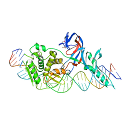 | | Crystal structure of the intein homing endonuclease PI-SceI bound to its recognition sequence | | 分子名称: | CALCIUM ION, ENDONUCLEASE PI-SCEI, PI-SceI DNA recognition region bottom strand, ... | | 著者 | Moure, C.M, Gimble, F.S, Quiocho, F.A. | | 登録日 | 2002-06-03 | | 公開日 | 2002-09-27 | | 最終更新日 | 2011-07-13 | | 実験手法 | X-RAY DIFFRACTION (3.5 Å) | | 主引用文献 | Crystal structure of the intein homing endonuclease PI-SceI bound to its recognition sequence.
Nat.Struct.Biol., 9, 2002
|
|
1LWT
 | |
1M71
 | | Crystal structure of a Monoclonal Fab Specific for Shigella Flexneri Y lipopolysaccharide | | 分子名称: | heavy chain of the monoclonal antibody Fab SYA/J6, light chain of the monoclonal antibody Fab SYA/J6 | | 著者 | Vyas, N.K, Vyas, M.N, Chervenak, M.C, Johnson, M.A, Pinto, B.M, Bundle, D.R, Quiocho, F.A. | | 登録日 | 2002-07-18 | | 公開日 | 2003-07-22 | | 最終更新日 | 2011-07-13 | | 実験手法 | X-RAY DIFFRACTION (2.8 Å) | | 主引用文献 | Molecular Recognition of Oligosaccharide Epitopes by a Monoclonal Fab Specific for
Shigella flexneri Y Lipopolysaccharide: X-ray Structures and Thermodyanamics
Biochemistry, 41, 2002
|
|
1OMP
 | |
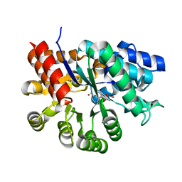
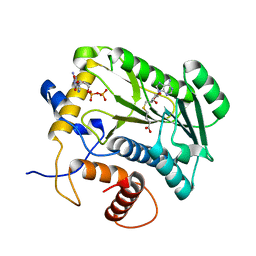
1A40
 | | PHOSPHATE-BINDING PROTEIN WITH ALA 197 REPLACED WITH TRP | | 分子名称: | PHOSPHATE ION, PHOSPHATE-BINDING PERIPLASMIC PROTEIN PRECURSOR | | 著者 | Ledivina, P.S, Wang, Z, Tsai, A, Koehl, E, Quiocho, F.A. | | 登録日 | 1998-02-10 | | 公開日 | 1999-03-23 | | 最終更新日 | 2024-05-22 | | 実験手法 | X-RAY DIFFRACTION (1.7 Å) | | 主引用文献 | Dominant role of local dipolar interactions in phosphate binding to a receptor cleft with an electronegative charge surface: equilibrium, kinetic, and crystallographic studies.
Protein Sci., 7, 1998
|
|
1A4L
 | |
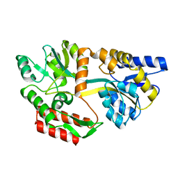
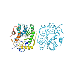
1A4M
 | | ADA STRUCTURE COMPLEXED WITH PURINE RIBOSIDE AT PH 7.0 | | 分子名称: | 6-HYDROXY-1,6-DIHYDRO PURINE NUCLEOSIDE, ADENOSINE DEAMINASE, ZINC ION | | 著者 | Wang, Z, Quiocho, F.A. | | 登録日 | 1998-01-31 | | 公開日 | 1998-10-14 | | 最終更新日 | 2024-05-22 | | 実験手法 | X-RAY DIFFRACTION (1.95 Å) | | 主引用文献 | Complexes of adenosine deaminase with two potent inhibitors: X-ray structures in four independent molecules at pH of maximum activity.
Biochemistry, 37, 1998
|
|
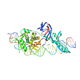
1R7M
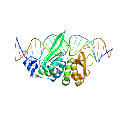 | | The homing endonuclease I-SceI bound to its DNA recognition region | | 分子名称: | 5'-D(*CP*AP*CP*GP*CP*TP*AP*GP*GP*GP*AP*TP*AP*AP*CP*AP*GP*GP*GP*TP*AP*AP*TP*AP*C)-3', 5'-D(*GP*GP*TP*AP*TP*TP*AP*CP*CP*CP*TP*GP*TP*TP*AP*TP*CP*CP*CP*TP*AP*GP*CP*GP*T)-3', CALCIUM ION, ... | | 著者 | Moure, C.M, Gimble, F.S, Quiocho, F.A. | | 登録日 | 2003-10-21 | | 公開日 | 2004-10-26 | | 最終更新日 | 2024-02-14 | | 実験手法 | X-RAY DIFFRACTION (2.25 Å) | | 主引用文献 | The crystal structure of the gene targeting homing endonuclease I-SceI reveals the origins of its target site specificity
J.Mol.Biol., 334, 2003
|
|
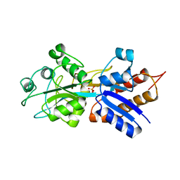
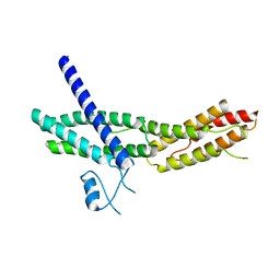
1U5O
 | | Structure of the D23A mutant of the nuclear transport carrier NTF2 | | 分子名称: | Nuclear transport factor 2 | | 著者 | Cushman, I, Bowman, B.R, Sowa, M.E, Lichtarge, O, Quiocho, F.A, Moore, M.S. | | 登録日 | 2004-07-28 | | 公開日 | 2004-12-07 | | 最終更新日 | 2024-02-14 | | 実験手法 | X-RAY DIFFRACTION (2.5 Å) | | 主引用文献 | Computational and biochemical identification of a nuclear pore complex binding site on the nuclear transport carrier NTF2.
J.Mol.Biol., 344, 2004
|
|
1TT9
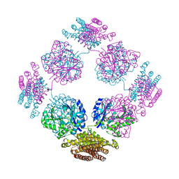 | | Structure of the bifunctional and Golgi associated formiminotransferase cyclodeaminase octamer | | 分子名称: | Formimidoyltransferase-cyclodeaminase (Formiminotransferase- cyclodeaminase) (FTCD) (58 kDa microtubule-binding protein) | | 著者 | Mao, Y, Vyas, N.K, Vyas, M.N, Chen, D.H, Ludtke, S.J, Chiu, W, Quiocho, F.A. | | 登録日 | 2004-06-22 | | 公開日 | 2005-06-28 | | 最終更新日 | 2024-02-14 | | 実験手法 | X-RAY DIFFRACTION (3.42 Å) | | 主引用文献 | Structure of the bifunctional and Golgi-associated formiminotransferase cyclodeaminase octamer
Embo J., 23, 2004
|
|
1VDE
 | |
1VPT
 | | AS11 VARIANT OF VACCINIA VIRUS PROTEIN VP39 IN COMPLEX WITH S-ADENOSYL-L-METHIONINE | | 分子名称: | S-ADENOSYLMETHIONINE, VP39 | | 著者 | Hodel, A.E, Gershon, P.D, Shi, X, Quiocho, F.A. | | 登録日 | 1996-03-20 | | 公開日 | 1996-08-17 | | 最終更新日 | 2024-02-14 | | 実験手法 | X-RAY DIFFRACTION (1.8 Å) | | 主引用文献 | The 1.85 A structure of vaccinia protein VP39: a bifunctional enzyme that participates in the modification of both mRNA ends.
Cell(Cambridge,Mass.), 85, 1996
|
|
1DMB
 | |
1ADD
 | |
1P39
 | |
2A2F
 | | Crystal Structure of Sec15 C-terminal domain | | 分子名称: | Exocyst complex component Sec15 | | 著者 | Wu, S, Mehta, S.Q, Pichaud, F, Bellen, H.J, Quiocho, F.A. | | 登録日 | 2005-06-22 | | 公開日 | 2005-09-13 | | 最終更新日 | 2024-02-14 | | 実験手法 | X-RAY DIFFRACTION (2.5 Å) | | 主引用文献 | Sec15 interacts with Rab11 via a novel domain and affects Rab11 localization in vivo.
Nat.Struct.Mol.Biol., 12, 2005
|
|
1XKT
 | | Human fatty acid synthase: Structure and substrate selectivity of the thioesterase domain | | 分子名称: | fatty acid synthase | | 著者 | Chakravarty, B, Gu, Z, Chirala, S.S, Wakil, S.J, Quiocho, F.A. | | 登録日 | 2004-09-29 | | 公開日 | 2004-10-26 | | 最終更新日 | 2024-02-14 | | 実験手法 | X-RAY DIFFRACTION (2.6 Å) | | 主引用文献 | Human fatty acid synthase: structure and substrate selectivity of the thioesterase domain.
Proc.Natl.Acad.Sci.Usa, 101, 2004
|
|
1JW5
 | |
1JTF
 | | Crystal Structure Analysis of VP39-F180W mutant and m7GpppG complex | | 分子名称: | 7N-METHYL-8-HYDROGUANOSINE-5'-DIPHOSPHATE, S-ADENOSYL-L-HOMOCYSTEINE, VP39 | | 著者 | Hu, G, Oguro, A, Gershon, P.D, Quiocho, F.A. | | 登録日 | 2001-08-20 | | 公開日 | 2002-07-10 | | 最終更新日 | 2024-03-06 | | 実験手法 | X-RAY DIFFRACTION (2.6 Å) | | 主引用文献 | The "cap-binding slot" of an mRNA cap-binding protein: quantitative effects of aromatic side chain choice in the double-stacking sandwich with cap.
Biochemistry, 41, 2002
|
|
1JSZ
 | | Crystal Structure Analysis of N7,9-dimethylguanine-VP39 complex | | 分子名称: | 7,9-DIMETHYLGUANINE, S-ADENOSYL-L-HOMOCYSTEINE, VP39 | | 著者 | Hu, G, Oguro, A, Gershon, P.D, Quiocho, F.A. | | 登録日 | 2001-08-19 | | 公開日 | 2002-07-10 | | 最終更新日 | 2024-03-13 | | 実験手法 | X-RAY DIFFRACTION (1.93 Å) | | 主引用文献 | The "cap-binding slot" of an mRNA cap-binding protein: quantitative effects of aromatic side chain choice in the double-stacking sandwich with cap.
Biochemistry, 41, 2002
|
|
1JTE
 | | Crystal Structure Analysis of VP39 F180W mutant | | 分子名称: | S-ADENOSYL-L-HOMOCYSTEINE, VP39 | | 著者 | Hu, G, Oguro, A, Gershon, P.D, Quiocho, F.A. | | 登録日 | 2001-08-20 | | 公開日 | 2002-07-10 | | 最終更新日 | 2024-05-29 | | 実験手法 | X-RAY DIFFRACTION (2 Å) | | 主引用文献 | The "cap-binding slot" of an mRNA cap-binding protein: quantitative effects of aromatic side chain choice in the double-stacking sandwich with cap.
Biochemistry, 41, 2002
|
|
1JW4
 | |
1CLL
 | |


