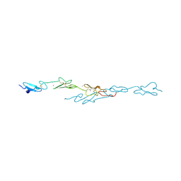
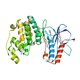
2I1N
 
 | | Crystal structure of the 1st PDZ domain of Human DLG3 | | 分子名称: | Discs, large homolog 3, SODIUM ION | | 著者 | Turnbull, A.P, Phillips, C, Bunkoczi, G, Debreczeni, J, Ugochukwu, E, Pike, A.C.W, Gorrec, F, Umeano, C, Elkins, J, Berridge, G, Savitsky, P, Gileadi, O, von Delft, F, Weigelt, J, Edwards, A, Arrowsmith, C, Sundstrom, M, Doyle, D, Structural Genomics Consortium (SGC) | | 登録日 | 2006-08-14 | | 公開日 | 2006-09-05 | | 最終更新日 | 2023-08-30 | | 実験手法 | X-RAY DIFFRACTION (1.85 Å) | | 主引用文献 | Structure of PICK1 and other PDZ domains obtained with the help of self-binding C-terminal extensions.
Protein Sci., 16, 2007
|
|
2P0A
 | | The crystal structure of human synapsin III (SYN3) in complex with AMPPNP | | 分子名称: | 1,2-ETHANEDIOL, CHLORIDE ION, PHOSPHOAMINOPHOSPHONIC ACID-ADENYLATE ESTER, ... | | 著者 | Turnbull, A.P, Phillips, C, Pike, A.C.W, Elkins, J.M, Gileadi, C, Salah, E, Niesen, F.H, Burgess, N, Gileadi, O, Gorrec, F, Umeano, C, von Delft, F, Weigelt, J, Edwards, A, Arrowsmith, C.H, Sundstrom, M, Doyle, D, Structural Genomics Consortium (SGC) | | 登録日 | 2007-02-28 | | 公開日 | 2007-03-27 | | 最終更新日 | 2023-08-30 | | 実験手法 | X-RAY DIFFRACTION (1.9 Å) | | 主引用文献 | The crystal structure of human synapsin III (SYN3) in complex with AMPPNP
To be Published
|
|
2Q3H
 | | The crystal structure of RhouA in the GDP-bound state. | | 分子名称: | GUANOSINE-5'-DIPHOSPHATE, MAGNESIUM ION, Ras homolog gene family, ... | | 著者 | Gileadi, C, Yang, X, Papagrigoriou, E, Elkins, J, Zhao, Y, Bray, J, Gileadi, O, Umeano, C, Ugochukwu, E, Uppenberg, J, Bunkoczi, G, von Delft, F, Pike, A.C.W, Phillips, C, Savitsky, P, Fedorov, O, Edwards, A, Weigelt, J, Arrowsmith, C.H, Sundstrom, M, Doyle, D.A, Structural Genomics Consortium (SGC) | | 登録日 | 2007-05-30 | | 公開日 | 2007-06-19 | | 最終更新日 | 2024-04-03 | | 実験手法 | X-RAY DIFFRACTION (1.73 Å) | | 主引用文献 | The crystal structure of RhouA in the GDP-bound state.
To be Published
|
|
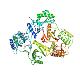
5NP0
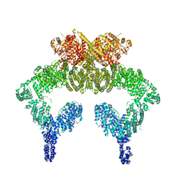 | | Closed dimer of human ATM (Ataxia telangiectasia mutated) | | 分子名称: | Serine-protein kinase ATM | | 著者 | Baretic, D, Pollard, H.K, Fisher, D.I, Johnson, C.M, Santhanam, B, Truman, C.M, Kouba, T, Fersht, A.R, Phillips, C, Williams, R.L. | | 登録日 | 2017-04-13 | | 公開日 | 2017-05-17 | | 最終更新日 | 2024-05-15 | | 実験手法 | ELECTRON MICROSCOPY (5.7 Å) | | 主引用文献 | Structures of closed and open conformations of dimeric human ATM.
Sci Adv, 3, 2017
|
|
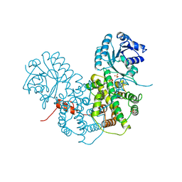
5NP1
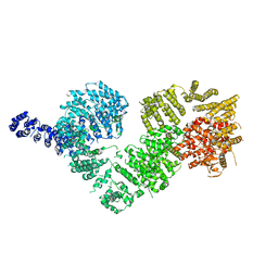 | | Open protomer of human ATM (Ataxia telangiectasia mutated) | | 分子名称: | Serine-protein kinase ATM | | 著者 | Baretic, D, Pollard, H.K, Fisher, D.I, Johnson, C.M, Santhanam, B, Truman, C.M, Kouba, T, Fersht, A.R, Phillips, C, Williams, R.L. | | 登録日 | 2017-04-13 | | 公開日 | 2017-05-17 | | 最終更新日 | 2024-05-15 | | 実験手法 | ELECTRON MICROSCOPY (5.7 Å) | | 主引用文献 | Structures of closed and open conformations of dimeric human ATM.
Sci Adv, 3, 2017
|
|
5O2S
 | | Human KRAS in complex with darpin K27 | | 分子名称: | GTPase KRas, GUANOSINE-5'-DIPHOSPHATE, MAGNESIUM ION, ... | | 著者 | Debreczeni, J.E, Guillard, S, Kolasinska-Zwierz, P, Breed, J, Zhang, J, Bery, N, Marwood, R, Tart, J, Stocki, P, Mistry, B, Phillips, C, Rabbitts, T, Jackson, R, Minter, R. | | 登録日 | 2017-05-22 | | 公開日 | 2017-07-26 | | 最終更新日 | 2024-05-08 | | 実験手法 | X-RAY DIFFRACTION (3.22 Å) | | 主引用文献 | Structural and functional characterization of a DARPin which inhibits Ras nucleotide exchange.
Nat Commun, 8, 2017
|
|
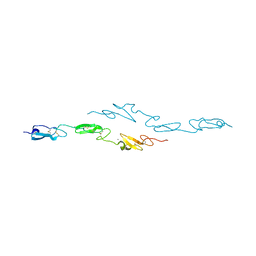
2JIK
 | | Crystal structure of PDZ domain of Synaptojanin-2 binding protein | | 分子名称: | SYNAPTOJANIN-2 BINDING PROTEIN | | 著者 | Tickle, J, Phillips, C, Pike, A.C.W, Cooper, C, Salah, E, Elkins, J, Turnbull, A.P, Edwards, A, Arrowsmith, C.H, Weigelt, J, Sundstrom, M, Doyle, D. | | 登録日 | 2007-06-28 | | 公開日 | 2007-07-10 | | 最終更新日 | 2023-12-13 | | 実験手法 | X-RAY DIFFRACTION (1.35 Å) | | 主引用文献 | Crystal Structure of Pdz Domain of Synaptojanin-2 Binding Protein
To be Published
|
|
2JIN
 | | Crystal structure of PDZ domain of Synaptojanin-2 binding protein | | 分子名称: | SODIUM ION, SULFATE ION, SYNAPTOJANIN-2 BINDING PROTEIN | | 著者 | Tickle, J, Phillips, C, Pike, A.C.W, Cooper, C, Salah, E, Elkins, J, Turnbull, A.P, Edwards, A, Arrowsmith, C.H, Weigelt, J, Sundstrom, M, Doyle, D. | | 登録日 | 2007-06-28 | | 公開日 | 2007-07-10 | | 最終更新日 | 2023-12-13 | | 実験手法 | X-RAY DIFFRACTION (1.5 Å) | | 主引用文献 | Crystal Structure of Pdz Domain of Synaptojanin-2 Binding Protein
To be Published
|
|
5O2T
 | | Human KRAS in complex with darpin K27 | | 分子名称: | 5'-GUANOSINE-DIPHOSPHATE-MONOTHIOPHOSPHATE, GTPase KRas, MAGNESIUM ION, ... | | 著者 | Debreczeni, J.E, Guillard, S, Kolasinska-Zwierz, P, Breed, J, Zhang, J, Bery, N, Marwood, R, Tart, J, Stocki, P, Mistry, B, Phillips, C, Rabbitts, T, Jackson, R, Minter, R. | | 登録日 | 2017-05-22 | | 公開日 | 2017-07-26 | | 最終更新日 | 2024-05-08 | | 実験手法 | X-RAY DIFFRACTION (2.19 Å) | | 主引用文献 | Structural and functional characterization of a DARPin which inhibits Ras nucleotide exchange.
Nat Commun, 8, 2017
|
|
2JLE
 | | Novel indazole nnrtis created using molecular template hybridization based on crystallographic overlays | | 分子名称: | 5-[(5-fluoro-3-methyl-1H-indazol-4-yl)oxy]benzene-1,3-dicarbonitrile, REVERSE TRANSCRIPTASE/RNASEH | | 著者 | Jones, L.H, Allan, G, Barba, O, Burt, C, Corbau, R, Dupont, T, Irving, S, Mowbray, C.E, Phillips, C, Swain, N.A, Webster, R, Westby, M. | | 登録日 | 2008-09-08 | | 公開日 | 2009-08-04 | | 最終更新日 | 2024-05-08 | | 実験手法 | X-RAY DIFFRACTION (2.9 Å) | | 主引用文献 | Novel Indazole Non-Nucleoside Reverse Transcriptase Inhibitors Using Molecular Hybridization Based on Crystallographic Overlays.
J.Med.Chem., 52, 2009
|
|
2V90
 | | Crystal structure of the 3rd PDZ domain of intestine- and kidney- enriched PDZ domain IKEPP (PDZD3) | | 分子名称: | PDZ DOMAIN-CONTAINING PROTEIN 3, SULFATE ION | | 著者 | Uppenberg, J, Gileadi, C, Phillips, C, Elkins, J, Bunkoczi, G, Cooper, C, Pike, A.C.W, Salah, E, Ugochukwu, E, Arrowsmith, C.H, Edwards, A, Sundstrom, M, Weigelt, J, Doyle, D.A. | | 登録日 | 2007-08-16 | | 公開日 | 2007-08-28 | | 最終更新日 | 2023-12-13 | | 実験手法 | X-RAY DIFFRACTION (2 Å) | | 主引用文献 | Crystal Structure of the 3Rd Pdz Domain of Intestine- and Kidney-Enriched Pdz Domain Ikepp (Pdzd3)
To be Published
|
|
2PGD
 | |
5MLB
 | | Crystal structure of human RAS in complex with darpin K27 | | 分子名称: | DARPin K27, GTPase KRas, GUANOSINE-5'-DIPHOSPHATE, ... | | 著者 | Debreczeni, J.E, Guillard, S, Kolasinska-Zwierz, P, Breed, J, Zhang, J, Bery, N, Marwood, R, Tart, J, Overman, R, Stocki, P, Mistry, B, Phillips, C, Rabbitts, T, Jackson, R, Minter, R. | | 登録日 | 2016-12-06 | | 公開日 | 2017-12-20 | | 最終更新日 | 2024-05-08 | | 実験手法 | X-RAY DIFFRACTION (3.22 Å) | | 主引用文献 | INhibition of RAS nucleotide exchange by a DARPin: structural characterisation and effects on downstream signalling by active RAS
To Be Published
|
|
5MLA
 | | Crystal structure of human RAS in complex with darpin K55 | | 分子名称: | 5'-GUANOSINE-DIPHOSPHATE-MONOTHIOPHOSPHATE, GTPase KRas, MAGNESIUM ION, ... | | 著者 | Debreczeni, J.E, Guillard, S, Kolasinska-Zwierz, P, Breed, J, Zhang, J, Bery, N, Marwood, R, Tart, J, Overman, R, Stocki, P, Mistry, B, Phillips, C, Rabbitts, T, Jackson, R, Minter, R. | | 登録日 | 2016-12-06 | | 公開日 | 2017-12-20 | | 最終更新日 | 2024-05-08 | | 実験手法 | X-RAY DIFFRACTION (2.19 Å) | | 主引用文献 | Inhibition of RAS nucleotide exchange by a DARPin: structural characterisation and effects on downstream signalling by active RAS
To Be Published
|
|
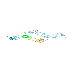
2HE2
 | | Crystal structure of the 3rd PDZ domain of human discs large homologue 2, DLG2 | | 分子名称: | Discs large homolog 2 | | 著者 | Turnbull, A.P, Phillips, C, Berridge, G, Savitsky, P, Smee, C.E.A, Papagrigoriou, E, Debreczeni, J, Gorrec, F, Elkins, J.M, von Delft, F, Weigelt, J, Edwards, A, Arrowsmith, C, Sundstrom, M, Doyle, D.A, Structural Genomics Consortium (SGC) | | 登録日 | 2006-06-21 | | 公開日 | 2006-07-04 | | 最終更新日 | 2023-08-30 | | 実験手法 | X-RAY DIFFRACTION (1.5 Å) | | 主引用文献 | Structure of PICK1 and other PDZ domains obtained with the help of self-binding C-terminal extensions.
Protein Sci., 16, 2007
|
|
2QG1
 | | Crystal structure of the 11th PDZ domain of MPDZ (MUPP1) | | 分子名称: | 1,2-ETHANEDIOL, Multiple PDZ domain protein | | 著者 | Papagrigoriou, E, Salah, E, Phillips, C, Savitsky, P, Boisguerin, P, Oschkinat, H, Gileadi, C, Yang, X, Elkins, J.M, Ugochukwu, E, Bunkoczi, G, Uppenberg, J, Sundstrom, M, Arrowsmith, C.H, Weigelt, J, Edwards, A, von Delft, F, Doyle, D, Structural Genomics Consortium (SGC) | | 登録日 | 2007-06-28 | | 公開日 | 2007-07-24 | | 最終更新日 | 2023-08-30 | | 実験手法 | X-RAY DIFFRACTION (1.4 Å) | | 主引用文献 | Crystal structure of the 11th PDZ domain of MPDZ (MUPP1).
To be Published
|
|
4CUF
 | | Human Notch1 EGF domains 11-13 mutant T466S | | 分子名称: | 1,2-ETHANEDIOL, CALCIUM ION, NEUROGENIC LOCUS NOTCH HOMOLOG PROTEIN 1 | | 著者 | Taylor, P, Takeuchi, H, Sheppard, D, Chillakuri, C, Lea, S.M, Haltiwanger, R.S, Handford, P.A. | | 登録日 | 2014-03-18 | | 公開日 | 2014-05-14 | | 最終更新日 | 2024-11-06 | | 実験手法 | X-RAY DIFFRACTION (2.29 Å) | | 主引用文献 | Fringe-Mediated Extension of O-Linked Fucose in the Ligand-Binding Region of Notch1 Increases Binding to Mammalian Notch Ligands.
Proc.Natl.Acad.Sci.USA, 111, 2014
|
|
4D0E
 | | Human Notch1 EGF domains 11-13 mutant GlcNAc-fucose disaccharide modified at T466 | | 分子名称: | 2-acetamido-2-deoxy-beta-D-glucopyranose-(1-3)-alpha-L-fucopyranose, CALCIUM ION, NEUROGENIC LOCUS NOTCH HOMOLOG PROTEIN 1 | | 著者 | Taylor, P, Takeuchi, H, Sheppard, D, Chillakuri, C, Lea, S.M, Haltiwanger, R.S, Handford, P.A. | | 登録日 | 2014-04-25 | | 公開日 | 2014-05-21 | | 最終更新日 | 2023-12-20 | | 実験手法 | X-RAY DIFFRACTION (1.61 Å) | | 主引用文献 | Fringe-Mediated Extension of O-Linked Fucose in the Ligand-Binding Region of Notch1 Increases Binding to Mammalian Notch Ligands.
Proc.Natl.Acad.Sci.USA, 111, 2014
|
|
4CUD
 | | Human Notch1 EGF domains 11-13 mutant fucosylated at T466 | | 分子名称: | CALCIUM ION, NEUROGENIC LOCUS NOTCH HOMOLOG PROTEIN 1, alpha-L-fucopyranose | | 著者 | Taylor, P, Takeuchi, H, Sheppard, D, Chillakuri, C, Lea, S.M, Haltiwanger, R.S, Handford, P.A. | | 登録日 | 2014-03-18 | | 公開日 | 2014-05-21 | | 最終更新日 | 2023-12-20 | | 実験手法 | X-RAY DIFFRACTION (1.85 Å) | | 主引用文献 | Fringe-Mediated Extension of O-Linked Fucose in the Ligand-Binding Region of Notch1 Increases Binding to Mammalian Notch Ligands.
Proc.Natl.Acad.Sci.USA, 111, 2014
|
|
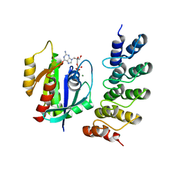
2W4O
 | | Crystal structure of Human CAMK4 in complex with 4-Amino(sulfamoyl- phenylamino)-triazole-carbothioic acid (2,6-difluoro-phenyl)-amide) | | 分子名称: | 5-AMINO-3-{[4-(AMINOSULFONYL)PHENYL]AMINO}-N-(2,6-DIFLUOROPHENYL)-1H-1,2,4-TRIAZOLE-1-CARBOTHIOAMIDE, CALCIUM/CALMODULIN-DEPENDENT PROTEIN KINASE TYPE IV | | 著者 | Muniz, J.R.C, Rellos, P, Gileadi, O, Fedorov, O, Filippakopoulos, P, Salah, E, Pike, A, Phillips, C, Niesen, F, Shrestha, L, Burgess-Brown, N, Bullock, A, Berridge, G, von Delft, F, Edwards, A.M, Bountra, C, Arrowsmith, C.H, Weigelt, J, Knapp, S. | | 登録日 | 2008-11-28 | | 公開日 | 2009-01-20 | | 最終更新日 | 2024-02-07 | | 実験手法 | X-RAY DIFFRACTION (2.17 Å) | | 主引用文献 | Crystal Structure of Human Camk4 in Complex with 4-Amino(Sulfamoyl-Phenylamino)-Triazole- Carbothioic Acid (2,6-Difluoro-Phenyl)-Amide)
To be Published
|
|
4D0F
 | | Human Notch1 EGF domains 11-13 mutant T466A | | 分子名称: | 1,2-ETHANEDIOL, CALCIUM ION, NEUROGENIC LOCUS NOTCH HOMOLOG PROTEIN 1 | | 著者 | Taylor, P, Takeuchi, H, Sheppard, D, Chillakuri, C, Lea, S.M, Haltiwanger, R.S, Handford, P.A. | | 登録日 | 2014-04-25 | | 公開日 | 2014-05-21 | | 最終更新日 | 2024-10-16 | | 実験手法 | X-RAY DIFFRACTION (2.8 Å) | | 主引用文献 | Fringe-Mediated Extension of O-Linked Fucose in the Ligand-Binding Region of Notch1 Increases Binding to Mammalian Notch Ligands.
Proc.Natl.Acad.Sci.USA, 111, 2014
|
|
4CUE
 | | Human Notch1 EGF domains 11-13 mutant T466V | | 分子名称: | CALCIUM ION, NEUROGENIC LOCUS NOTCH HOMOLOG PROTEIN 1 | | 著者 | Taylor, P, Takeuchi, H, Sheppard, D, Chillakuri, C, Lea, S.M, Haltiwanger, R.S, Handford, P.A. | | 登録日 | 2014-03-18 | | 公開日 | 2014-05-21 | | 最終更新日 | 2023-12-20 | | 実験手法 | X-RAY DIFFRACTION (3 Å) | | 主引用文献 | Fringe-Mediated Extension of O-Linked Fucose in the Ligand-Binding Region of Notch1 Increases Binding to Mammalian Notch Ligands.
Proc.Natl.Acad.Sci.USA, 111, 2014
|
|
2YB9
 | | Crystal Structure of Human Neutral Endopeptidase complexed with a heteroarylalanine diacid. | | 分子名称: | HETEROARYLALANINE 5-PHENYL OXAZOLE, NEPRILYSIN, ZINC ION | | 著者 | Glossop, M.S, Bazin, R.J, Dack, K.N, Done, S, Fox, D.N.A, MacDonald, G.A, Mills, M, Owen, D.R, Phillips, C, Reeves, K.A, Ringer, T.J, Strang, R.S, Watson, C.A.L. | | 登録日 | 2011-03-02 | | 公開日 | 2011-05-25 | | 最終更新日 | 2011-11-02 | | 実験手法 | X-RAY DIFFRACTION (2.4 Å) | | 主引用文献 | Synthesis and Evaluation of Heteroarylalanine Diacids as Potent and Selective Neutral Endopeptidase Inhibitors.
Bioorg.Med.Chem.Lett., 21, 2011
|
|
2YIW
 | | triazolopyridine inhibitors of p38 kinase | | 分子名称: | 1-(3-tert-butyl-1-phenyl-1H-pyrazol-5-yl)-3-(2-{[3-(1-methylethyl)[1,2,4]triazolo[4,3-a]pyridin-6-yl]sulfanyl}benzyl)urea, 2-fluoro-4-[4-(4-fluorophenyl)-1H-pyrazol-3-yl]pyridine, MITOGEN-ACTIVATED PROTEIN KINASE 14 | | 著者 | Millan, D.S, Anderson, M, Bunnage, M.E, Burrows, J.L, Butcher, K.J, Dodd, P.G, Evans, T.J, Fairman, D.A, Hughes, S.J, Irving, S.L, Kilty, I.C, Lemaitre, A, Lewthwaite, R.A, Mahnke, A, Mathais, J.P, Philip, J, Phillips, C, Smith, R.T, Stefamiak, M.H, Yeadon, M. | | 登録日 | 2011-05-17 | | 公開日 | 2011-11-30 | | 最終更新日 | 2024-05-08 | | 実験手法 | X-RAY DIFFRACTION (2 Å) | | 主引用文献 | Design and Synthesis of Inhaled P38 Inhibitors for the Treatment of Chronic Obstructive Pulmonary Disease.
J.Med.Chem., 54, 2011
|
|
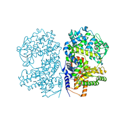
2BU6
 | | crystal structures of human pyruvate dehydrogenase kinase 2 containing physiological and synthetic ligands | | 分子名称: | (N-{4-[(ETHYLANILINO)SULFONYL]-2-METHYLPHENYL}-3,3,3-TRIFLUORO-2-HYDROXY-2-METHYLPROPANAMIDE, PYRUVATE DEHYDROGENASE KINASE ISOENZYME 2 | | 著者 | Knoechel, T.R, Tucker, A.D, Robinson, C.M, Phillips, C, Taylor, W, Bungay, P.J, Kasten, S.A, Roche, T.E, Brown, D.G. | | 登録日 | 2005-06-08 | | 公開日 | 2006-02-02 | | 最終更新日 | 2024-05-08 | | 実験手法 | X-RAY DIFFRACTION (2.4 Å) | | 主引用文献 | Regulatory Roles of the N-Terminal Domain Based on Crystal Structures of Human Pyruvate Dehydrogenase Kinase 2 Containing Physiological and Synthetic Ligands.
Biochemistry, 45, 2006
|
|


