2Q3L
 
 | |
2HUJ
 | |
2P55
 | |
2GVI
 | |
2HAG
 | |
6NDL
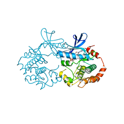
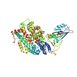
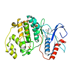
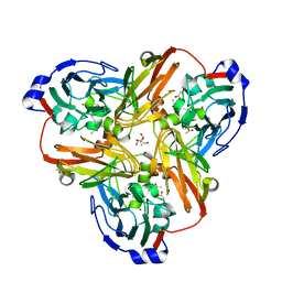
 | | Crystal structure of Staphylococcus aureus biotin protein ligase in complex with a sulfonamide inhibitor | | 分子名称: | 1-[4-(6-aminopurin-9-yl)butylsulfamoyl]-3-[4-[(4~{S})-2-oxidanylidene-1,3,3~{a},4,6,6~{a}-hexahydrothieno[3,4-d]imidazol-4-yl]butyl]urea, Biotin Protein Ligase, GLYCEROL | | 著者 | Marshall, A.C, Polyak, S.W, Bruning, J.B, Lee, K. | | 登録日 | 2018-12-13 | | 公開日 | 2019-12-18 | | 最終更新日 | 2023-10-11 | | 実験手法 | X-RAY DIFFRACTION (2 Å) | | 主引用文献 | Sulfonamide-Based Inhibitors of Biotin Protein Ligase as New Antibiotic Leads.
Acs Chem.Biol., 14, 2019
|
|
2Q8U
 | |
7EFR
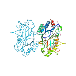
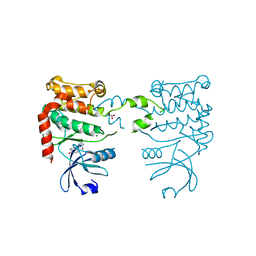
 | | Structure of SARS-CoV-2 spike receptor-binding domain in complex with high affinity ACE2 mutant (T27W,N330Y) | | 分子名称: | 2-acetamido-2-deoxy-beta-D-glucopyranose, Processed angiotensin-converting enzyme 2, Spike glycoprotein | | 著者 | Lu, G.W, Ye, F, Lin, X. | | 登録日 | 2021-03-23 | | 公開日 | 2021-11-24 | | 最終更新日 | 2024-10-16 | | 実験手法 | X-RAY DIFFRACTION (2.494 Å) | | 主引用文献 | S19W, T27W, and N330Y mutations in ACE2 enhance SARS-CoV-2 S-RBD binding toward both wild-type and antibody-resistant viruses and its molecular basis.
Signal Transduct Target Ther, 6, 2021
|
|
7EFP
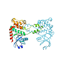
 | | Structure of SARS-CoV-2 spike receptor-binding domain in complex with high affinity ACE2 mutant (S19W,N330Y) | | 分子名称: | 2-acetamido-2-deoxy-beta-D-glucopyranose, Processed angiotensin-converting enzyme 2, Spike glycoprotein | | 著者 | Lu, G.W, Ye, F, Lin, X. | | 登録日 | 2021-03-22 | | 公開日 | 2021-11-24 | | 最終更新日 | 2023-11-29 | | 実験手法 | X-RAY DIFFRACTION (2.698 Å) | | 主引用文献 | S19W, T27W, and N330Y mutations in ACE2 enhance SARS-CoV-2 S-RBD binding toward both wild-type and antibody-resistant viruses and its molecular basis.
Signal Transduct Target Ther, 6, 2021
|
|
6NLR
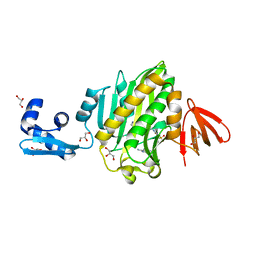
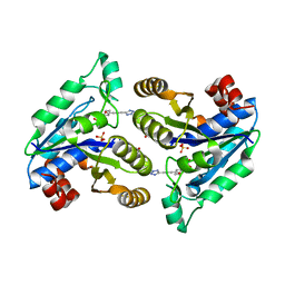
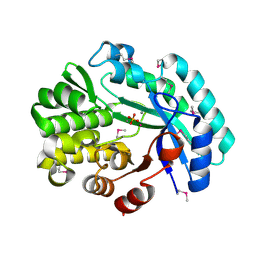 | | Crystal structure of the putative histidinol phosphatase hisK from Listeria monocytogenes with trinuclear metals determined by PIXE revealing sulphate ion in active site. Based on PIXE analysis and original date from 3DCP | | 分子名称: | CALCIUM ION, COBALT (II) ION, FE (III) ION, ... | | 著者 | Snell, E.H, Garman, E.F, Lowe, E.D. | | 登録日 | 2019-01-09 | | 公開日 | 2019-12-25 | | 最終更新日 | 2020-01-22 | | 実験手法 | X-RAY DIFFRACTION (2.1 Å) | | 主引用文献 | High-Throughput PIXE as an Essential Quantitative Assay for Accurate Metalloprotein Structural Analysis: Development and Application.
J.Am.Chem.Soc., 142, 2020
|
|
2OJI
 | |
2OK1
 | |
2OJJ
 | |
2QTP
 | |
2RA9
 | |
2OOC
 | |
7L1J
 | |
2CN8
 | |
2CN5
 | |
2FJS
 | |
2FNA
 | |
2G36
 | |
7LX0
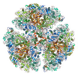 | | Quantitative assessment of chlorophyll types in cryo-EM maps of photosystem I acclimated to far-red light | | 分子名称: | 1,2-DIPALMITOYL-PHOSPHATIDYL-GLYCEROLE, 1,2-DISTEAROYL-MONOGALACTOSYL-DIGLYCERIDE, BETA-CAROTENE, ... | | 著者 | Gisriel, C.J, Wang, J. | | 登録日 | 2021-03-02 | | 公開日 | 2021-07-28 | | 実験手法 | ELECTRON MICROSCOPY (2.96 Å) | | 主引用文献 | Quantitative assessment of chlorophyll types in cryo-EM maps of photosystem I acclimated to far-red light
BBA Adv, 1, 2021
|
|
2FHW
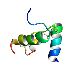 | | Solution structure of human relaxin-3 | | 分子名称: | Relaxin 3 (Prorelaxin H3) (Insulin-like peptide INSL7) (Insulin-like peptide 7) | | 著者 | Rosengren, K.J, Craik, D.J. | | 登録日 | 2005-12-27 | | 公開日 | 2006-01-24 | | 最終更新日 | 2022-03-09 | | 実験手法 | SOLUTION NMR | | 主引用文献 | Solution structure and novel insights into the determinants of the receptor specificity of human relaxin-3.
J.Biol.Chem., 281, 2006
|
|
2FNO
 | |


