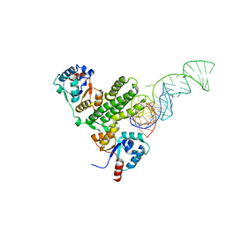
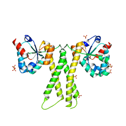
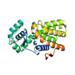
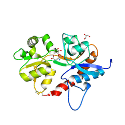
5Z6Z
 
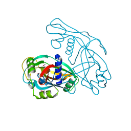
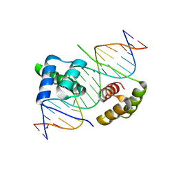 | | Crystal structure of human DUX4 homeodomains bound to DNA | | 分子名称: | DNA (5'-D(*CP*CP*AP*CP*TP*AP*AP*CP*CP*TP*AP*AP*TP*CP*AP*CP*AP*CP*C)-3'), DNA (5'-D(*GP*GP*TP*GP*TP*GP*AP*TP*TP*AP*GP*GP*TP*TP*AP*GP*TP*GP*G)-3'), Double homeobox protein 4 | | 著者 | Li, Y.Y, Wu, B.X, Gan, J.H. | | 登録日 | 2018-01-26 | | 公開日 | 2018-10-31 | | 最終更新日 | 2024-03-27 | | 実験手法 | X-RAY DIFFRACTION (2.301 Å) | | 主引用文献 | Structural basis for multiple gene regulation by human DUX4.
Biochem. Biophys. Res. Commun., 505, 2018
|
|
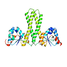
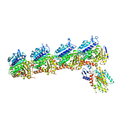
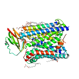
8HM0
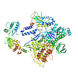 | | F8-A22-E4 complex of MPXV in trimeric form | | 分子名称: | DNA polymerase, DNA polymerase processivity factor component A20, E4R | | 著者 | Li, Y.N, Shen, Y.P, Hu, Z.W, Yan, R.H. | | 登録日 | 2022-12-02 | | 公開日 | 2023-05-31 | | 最終更新日 | 2023-12-13 | | 実験手法 | ELECTRON MICROSCOPY (3.1 Å) | | 主引用文献 | Structural basis for the assembly of the DNA polymerase holoenzyme from a monkeypox virus variant.
Sci Adv, 9, 2023
|
|
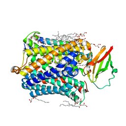
8HLZ
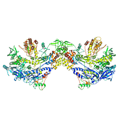 | | F8-A22-E4 complex of MPXV in hexameric form | | 分子名称: | DNA polymerase, DNA polymerase processivity factor component A20, E4R | | 著者 | Li, Y.N, Shen, Y.P, Hu, Z.W, Yan, R.H. | | 登録日 | 2022-12-02 | | 公開日 | 2023-05-31 | | 最終更新日 | 2023-12-13 | | 実験手法 | ELECTRON MICROSCOPY (3.5 Å) | | 主引用文献 | Structural basis for the assembly of the DNA polymerase holoenzyme from a monkeypox virus variant.
Sci Adv, 9, 2023
|
|
7E8O
 | |
7E8K
 | |
7E8J
 | |
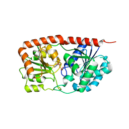
1BM6
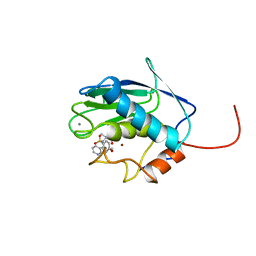 | | SOLUTION STRUCTURE OF THE CATALYTIC DOMAIN OF HUMAN STROMELYSIN-1 COMPLEXED TO A POTENT NON-PEPTIDIC INHIBITOR, NMR, 20 STRUCTURES | | 分子名称: | 1-METHYLOXY-4-SULFONE-BENZENE, 3-METHYLPYRIDINE, CALCIUM ION, ... | | 著者 | Li, Y, Zhang, X, Melton, R, Ganu, V, Gonnella, N.C. | | 登録日 | 1998-07-29 | | 公開日 | 1999-07-29 | | 最終更新日 | 2024-05-22 | | 実験手法 | SOLUTION NMR | | 主引用文献 | Solution structure of the catalytic domain of human stromelysin-1 complexed to a potent, nonpeptidic inhibitor.
Biochemistry, 37, 1998
|
|
5ICT
 | |
4HRO
 | |
4HRS
 | |
7EXC
 | | Crystal structure of T2R-TTL-1129A2 complex | | 分子名称: | 2-(N-MORPHOLINO)-ETHANESULFONIC ACID, CALCIUM ION, GLYCEROL, ... | | 著者 | Yang, J.H, Yan, W. | | 登録日 | 2021-05-26 | | 公開日 | 2022-06-01 | | 最終更新日 | 2023-11-29 | | 実験手法 | X-RAY DIFFRACTION (2.39 Å) | | 主引用文献 | Structure-Based Design and Synthesis of N-Substituted 3-Amino-beta-Carboline Derivatives as Potent alpha beta-Tubulin Degradation Agents
J.Med.Chem., 65, 2022
|
|
6UUQ
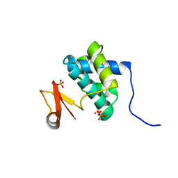
 | | Structure of Calcineurin bound to RCAN1 | | 分子名称: | Calcipressin-1, FE (III) ION, PHOSPHATE ION, ... | | 著者 | Sheftic, S, Page, R, Peti, W. | | 登録日 | 2019-10-31 | | 公開日 | 2020-09-09 | | 最終更新日 | 2023-10-11 | | 実験手法 | X-RAY DIFFRACTION (1.849 Å) | | 主引用文献 | The structure of the RCAN1:CN complex explains the inhibition of and substrate recruitment by calcineurin.
Sci Adv, 6, 2020
|
|
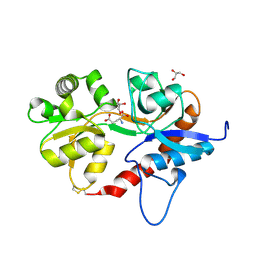
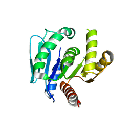
7C6C
 | | Crystal structure of native chitosanase from Bacillus subtilis MY002 | | 分子名称: | (2S)-2-hydroxybutanedioic acid, Chitosanase | | 著者 | Gou, Y, Liu, Z.C, Xie, T, Wang, G.G. | | 登録日 | 2020-05-21 | | 公開日 | 2021-03-31 | | 最終更新日 | 2023-11-29 | | 実験手法 | X-RAY DIFFRACTION (1.258 Å) | | 主引用文献 | Structure-based rational design of chitosanase CsnMY002 for high yields of chitobiose.
Colloids Surf B Biointerfaces, 202, 2021
|
|
7C6D
 | | Crystal structure of E19A mutant chitosanase from Bacillus subtilis MY002 complexed with 6 GlcN. | | 分子名称: | 2-amino-2-deoxy-beta-D-glucopyranose-(1-4)-2-amino-2-deoxy-beta-D-glucopyranose-(1-4)-2-amino-2-deoxy-beta-D-glucopyranose-(1-4)-2-amino-2-deoxy-beta-D-glucopyranose-(1-4)-2-amino-2-deoxy-beta-D-glucopyranose-(1-4)-2-amino-2-deoxy-beta-D-glucopyranose, Chitosanase | | 著者 | Gou, Y, Liu, Z.C, Xie, T, Wang, G.G. | | 登録日 | 2020-05-21 | | 公開日 | 2021-03-31 | | 最終更新日 | 2023-11-29 | | 実験手法 | X-RAY DIFFRACTION (1.451 Å) | | 主引用文献 | Structure-based rational design of chitosanase CsnMY002 for high yields of chitobiose.
Colloids Surf B Biointerfaces, 202, 2021
|
|
6K9V
 | | Crystal structure of tubulin in complex with inhibitor D64 | | 分子名称: | (5-methoxy-1H-indol-2-yl)-phenyl-methanone, 2-(N-MORPHOLINO)-ETHANESULFONIC ACID, CALCIUM ION, ... | | 著者 | Yu, Y, Chen, Q. | | 登録日 | 2019-06-18 | | 公開日 | 2019-08-28 | | 最終更新日 | 2023-11-22 | | 実験手法 | X-RAY DIFFRACTION (2.543 Å) | | 主引用文献 | Structural insights into the design of indole derivatives as tubulin polymerization inhibitors.
Febs Lett., 594, 2020
|
|
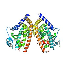
6KIH
 | | Sucrose-phosphate synthase (tll1590) from Thermosynechococcus elongatus | | 分子名称: | 6-O-phosphono-beta-D-fructofuranose-(2-1)-alpha-D-glucopyranose, Tll1590 protein, URIDINE-5'-DIPHOSPHATE | | 著者 | Su, J. | | 登録日 | 2019-07-18 | | 公開日 | 2020-05-27 | | 最終更新日 | 2023-11-22 | | 実験手法 | X-RAY DIFFRACTION (3 Å) | | 主引用文献 | Co-crystal Structure ofThermosynechococcus elongatusSucrose Phosphate Synthase With UDP and Sucrose-6-Phosphate Provides Insight Into Its Mechanism of Action Involving an Oxocarbenium Ion and the Glycosidic Bond.
Front Microbiol, 11, 2020
|
|
6LDQ
 | |
8JYV
 | |
8JYW
 | |
4F55
 | |
3K8S
 | | Crystal Structure of PPARg in complex with T2384 | | 分子名称: | 2-chloro-N-{3-chloro-4-[(5-chloro-1,3-benzothiazol-2-yl)sulfanyl]phenyl}-4-(trifluoromethyl)benzenesulfonamide, Peroxisome proliferator-activated receptor gamma | | 著者 | Wang, Z. | | 登録日 | 2009-10-14 | | 公開日 | 2009-11-03 | | 最終更新日 | 2023-09-06 | | 実験手法 | X-RAY DIFFRACTION (2.55 Å) | | 主引用文献 | T2384, a novel antidiabetic agent with unique peroxisome proliferator-activated receptor gamma binding properties
J.Biol.Chem., 283, 2008
|
|
5DT6
 | |
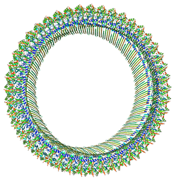
6A6B
 | | cryo-em structure of alpha-synuclein fiber | | 分子名称: | Alpha-synuclein | | 著者 | Li, Y.W, Zhao, C.Y, Luo, F, Liu, Z, Gui, X, Luo, Z, Zhang, X, Li, D, Liu, C, Li, X. | | 登録日 | 2018-06-27 | | 公開日 | 2018-07-11 | | 最終更新日 | 2024-03-27 | | 実験手法 | ELECTRON MICROSCOPY (3.07 Å) | | 主引用文献 | Amyloid fibril structure of alpha-synuclein determined by cryo-electron microscopy
Cell Res., 28, 2018
|
|
4FAA
 | |
4FA7
 | |


