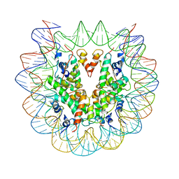
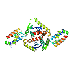
6TS0
 
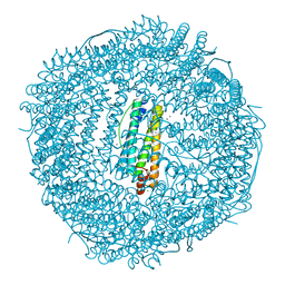
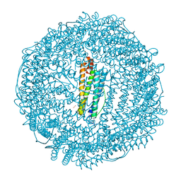 | | Crystal structure of human L ferritin (HuLf) triple variant E60A-E61A-E64A Fe(III)-loaded for 30 minutes | | 分子名称: | CADMIUM ION, FE (III) ION, Ferritin light chain, ... | | 著者 | Pozzi, C, Ciambellotti, S, Turano, P, Mangani, S. | | 登録日 | 2019-12-19 | | 公開日 | 2020-02-19 | | 最終更新日 | 2024-01-24 | | 実験手法 | X-RAY DIFFRACTION (2.2 Å) | | 主引用文献 | Iron Biomineral Growth from the Initial Nucleation Seed in L-Ferritin.
Chemistry, 26, 2020
|
|
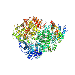
6TSX
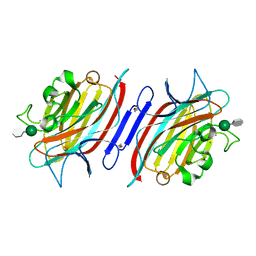
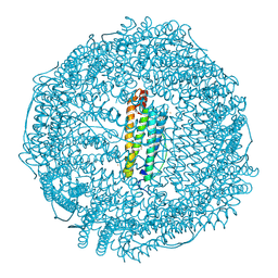 | | Crystal structure of horse L ferritin (HoLf) Fe(III)-loaded for 30 minutes | | 分子名称: | CADMIUM ION, CHLORIDE ION, FE (III) ION, ... | | 著者 | Pozzi, C, Ciambellotti, S, Turano, P, Mangani, S. | | 登録日 | 2019-12-21 | | 公開日 | 2020-02-19 | | 最終更新日 | 2024-01-24 | | 実験手法 | X-RAY DIFFRACTION (2.021 Å) | | 主引用文献 | Iron Biomineral Growth from the Initial Nucleation Seed in L-Ferritin.
Chemistry, 26, 2020
|
|
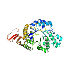
1P8L
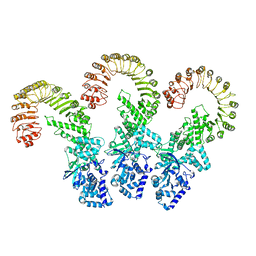
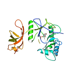 | | New Crystal Structure of Chlorella Virus DNA Ligase-Adenylate | | 分子名称: | ADENOSINE MONOPHOSPHATE, PBCV-1 DNA ligase | | 著者 | Odell, M, Malinina, L, Teplova, M, Shuman, S. | | 登録日 | 2003-05-07 | | 公開日 | 2003-08-26 | | 最終更新日 | 2023-08-16 | | 実験手法 | X-RAY DIFFRACTION (2.95 Å) | | 主引用文献 | Analysis of the DNA Joining Repertoire of Chlorella Virus DNA ligase and a New Crystal Structure of the Ligase-Adenylate Intermediate
Nucleic Acids Res., 31, 2003
|
|
3GHF
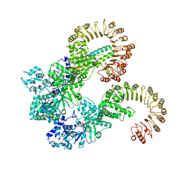
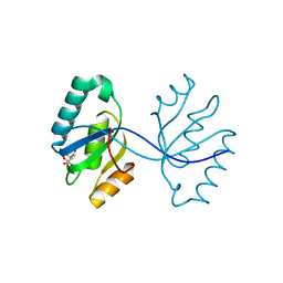 | | Crystal structure of the septum site-determining protein minC from Salmonella typhimurium | | 分子名称: | CITRIC ACID, Septum site-determining protein minC | | 著者 | Bonanno, J.B, Gilmore, M, Bain, K.T, Chang, S, Romero, R, Wasserman, S, Sauder, J.M, Burley, S.K, Almo, S.C, New York SGX Research Center for Structural Genomics (NYSGXRC) | | 登録日 | 2009-03-03 | | 公開日 | 2009-03-24 | | 最終更新日 | 2024-02-21 | | 実験手法 | X-RAY DIFFRACTION (2.2 Å) | | 主引用文献 | Crystal structure of the septum site-determining protein minC from Salmonella typhimurium
To be Published
|
|
6PWE
 | |
3G9A
 | | Green fluorescent protein bound to minimizer nanobody | | 分子名称: | Green fluorescent protein, Minimizer | | 著者 | Kirchhofer, A, Helma, J, Schmidthals, K, Frauer, C, Cui, S, Karcher, A, Pellis, M, Muyldermans, S, Delucci, C.C, Cardoso, M.C, Leonhardt, H, Hopfner, K.-P, Rothbauer, U. | | 登録日 | 2009-02-13 | | 公開日 | 2009-12-08 | | 最終更新日 | 2023-11-15 | | 実験手法 | X-RAY DIFFRACTION (1.614 Å) | | 主引用文献 | Modulation of protein properties in living cells using nanobodies
Nat.Struct.Mol.Biol., 17, 2010
|
|
6U5O
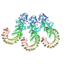
 | | Structure of the Human Metapneumovirus Polymerase bound to the phosphoprotein tetramer | | 分子名称: | Phosphoprotein, RNA-directed RNA polymerase L | | 著者 | Pan, J, Qian, X, Lattmann, S, Sahili, A.E, Yeo, T.H, Kalocsay, M, Fearns, R, Lescar, J. | | 登録日 | 2019-08-28 | | 公開日 | 2019-09-25 | | 最終更新日 | 2024-03-20 | | 実験手法 | ELECTRON MICROSCOPY (3.7 Å) | | 主引用文献 | Structure of the human metapneumovirus polymerase phosphoprotein complex.
Nature, 577, 2020
|
|
6IAF
 | | Fifteen minutes iron loaded Rana Catesbeiana H' ferritin variant H54N | | 分子名称: | CHLORIDE ION, FE (II) ION, Ferritin, ... | | 著者 | Pozzi, C, Di Pisa, F, Mangani, S. | | 登録日 | 2018-11-26 | | 公開日 | 2019-05-22 | | 最終更新日 | 2024-01-24 | | 実験手法 | X-RAY DIFFRACTION (1.35 Å) | | 主引用文献 | Effect of the point mutation H54N on the ferroxidase process of Rana catesbeiana H' ferritin.
J.Inorg.Biochem., 197, 2019
|
|
1NM4
 | | Solution structure of Cu(I)-CopC from Pseudomonas syringae | | 分子名称: | Copper resistance protein C | | 著者 | Arnesano, F, Banci, L, Bertini, I, Mangani, S, Thompsett, A.R, Structural Proteomics in Europe (SPINE) | | 登録日 | 2003-01-09 | | 公開日 | 2003-04-08 | | 最終更新日 | 2024-05-22 | | 実験手法 | SOLUTION NMR | | 主引用文献 | A redox switch in CopC: An intriguing copper trafficking protein that binds copper(I) and copper(II)
at different sites
Proc.Natl.Acad.Sci.USA, 100, 2003
|
|
6I9T
 | | Two minutes iron loaded Rana Catesbeiana H' ferritin variant H54N | | 分子名称: | CHLORIDE ION, FE (II) ION, Ferritin, ... | | 著者 | Pozzi, C, Di Pisa, F, Mangani, S. | | 登録日 | 2018-11-25 | | 公開日 | 2019-05-22 | | 最終更新日 | 2024-01-24 | | 実験手法 | X-RAY DIFFRACTION (1.2 Å) | | 主引用文献 | Effect of the point mutation H54N on the ferroxidase process of Rana catesbeiana H' ferritin.
J.Inorg.Biochem., 197, 2019
|
|
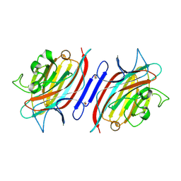
1Q8P
 | | Pterocarpus angolensis lectin PAL in complex with the dimannoside Man(alpha1-3)Man | | 分子名称: | CALCIUM ION, MANGANESE (II) ION, alpha-D-mannopyranose-(1-3)-methyl alpha-D-mannopyranoside, ... | | 著者 | Loris, R, Van Walle, I, De Greve, H, Beeckmans, S, Deboeck, F, Wyns, L, Bouckaert, J. | | 登録日 | 2003-08-22 | | 公開日 | 2004-02-10 | | 最終更新日 | 2020-07-29 | | 実験手法 | X-RAY DIFFRACTION (1.75 Å) | | 主引用文献 | Structural Basis of Oligomannose Recognition by the Pterocarpus angolensis Seed Lectin
J.Mol.Biol., 335, 2004
|
|
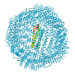
8FW2
 | | Cryo-EM structure of full-length human NLRC4 inflammasome with C11 symmetry | | 分子名称: | NLR family CARD domain-containing protein 4 | | 著者 | Matico, R.E, Yu, X, Miller, R, Somani, S, Ricketts, M.D, Kumar, N, Steele, R.A, Medley, Q, Berger, S, Faustin, B, Sharma, S. | | 登録日 | 2023-01-20 | | 公開日 | 2024-01-03 | | 最終更新日 | 2024-01-31 | | 実験手法 | ELECTRON MICROSCOPY (3.8 Å) | | 主引用文献 | Structural basis of the human NAIP/NLRC4 inflammasome assembly and pathogen sensing.
Nat.Struct.Mol.Biol., 31, 2024
|
|
8FVU
 | | Cryo-EM structure of human Needle/NAIP/NLRC4 (R288A) | | 分子名称: | ADENOSINE-5'-TRIPHOSPHATE, Baculoviral IAP repeat-containing protein 1, Lethal factor,Type III secretion system protein, ... | | 著者 | Matico, R.E, Yu, X, Miller, R, Somani, S, Ricketts, M.D, Kumar, N, Steele, R.A, Medley, Q, Berger, S, Faustin, B, Sharma, S. | | 登録日 | 2023-01-19 | | 公開日 | 2024-01-03 | | 最終更新日 | 2024-01-31 | | 実験手法 | ELECTRON MICROSCOPY (3.6 Å) | | 主引用文献 | Structural basis of the human NAIP/NLRC4 inflammasome assembly and pathogen sensing.
Nat.Struct.Mol.Biol., 31, 2024
|
|
8FW9
 | | Cryo-EM structure of full-length human NLRC4 inflammasome with C12 symmetry | | 分子名称: | NLR family CARD domain-containing protein 4 | | 著者 | Matico, R.E, Yu, X, Miller, R, Somani, S, Ricketts, M.D, Kumar, N, Steele, R.A, Medley, Q, Berger, S, Faustin, B, Sharma, S. | | 登録日 | 2023-01-20 | | 公開日 | 2024-01-03 | | 最終更新日 | 2024-01-31 | | 実験手法 | ELECTRON MICROSCOPY (4.46 Å) | | 主引用文献 | Structural basis of the human NAIP/NLRC4 inflammasome assembly and pathogen sensing.
Nat.Struct.Mol.Biol., 31, 2024
|
|
6SUI
 | | AMICOUMACIN KINASE AMIN | | 分子名称: | PENTAETHYLENE GLYCOL, Phosphotransferase enzyme family protein | | 著者 | Bourenkov, G.P, Mokrushina, Y.A, Terekhov, S.S, Smirnov, I.V, Gabibov, A.G, Altman, S. | | 登録日 | 2019-09-14 | | 公開日 | 2020-07-22 | | 最終更新日 | 2024-05-15 | | 実験手法 | X-RAY DIFFRACTION (1.6 Å) | | 主引用文献 | A kinase bioscavenger provides antibiotic resistance by extremely tight substrate binding.
Sci Adv, 6, 2020
|
|
3EBA
 | | CAbHul6 FGLW mutant (humanized) in complex with human lysozyme | | 分子名称: | CAbHul6, Lysozyme C, SULFATE ION | | 著者 | Loris, R, Vincke, C, Saerens, D, Martinez-Rodriguez, S, Muyldermans, S, Conrath, K. | | 登録日 | 2008-08-27 | | 公開日 | 2008-12-02 | | 最終更新日 | 2023-11-01 | | 実験手法 | X-RAY DIFFRACTION (1.85 Å) | | 主引用文献 | General Strategy to Humanize a Camelid Single-domain Antibody and Identification of a Universal Humanized Nanobody Scaffold
J.Biol.Chem., 284, 2009
|
|
3E2V
 | | Crystal structure of an uncharacterized amidohydrolase from Saccharomyces cerevisiae | | 分子名称: | 3'-5'-exonuclease, GLYCEROL, MAGNESIUM ION | | 著者 | Bonanno, J.B, Dickey, M, Bain, K.T, Hu, S, Romero, R, Smith, D, Wasserman, S, Sauder, J.M, Burley, S.K, Almo, S.C, New York SGX Research Center for Structural Genomics (NYSGXRC) | | 登録日 | 2008-08-06 | | 公開日 | 2008-08-26 | | 最終更新日 | 2021-02-10 | | 実験手法 | X-RAY DIFFRACTION (1.5 Å) | | 主引用文献 | Crystal structure of an uncharacterized amidohydrolase from Saccharomyces cerevisiae
To be Published
|
|
2B4J
 | | Structural basis for the recognition between HIV-1 integrase and LEDGF/p75 | | 分子名称: | GLYCEROL, Integrase (IN), PC4 and SFRS1 interacting protein, ... | | 著者 | Cherepanov, P, Ambrosio, A.L, Rahman, S, Ellenberger, T, Engelman, A. | | 登録日 | 2005-09-24 | | 公開日 | 2005-10-25 | | 最終更新日 | 2023-08-23 | | 実験手法 | X-RAY DIFFRACTION (2.02 Å) | | 主引用文献 | Structural basis for the recognition between HIV-1 integrase and transcriptional coactivator p75
Proc.Natl.Acad.Sci.Usa, 102, 2005
|
|
3DHU
 | | Crystal structure of an alpha-amylase from Lactobacillus plantarum | | 分子名称: | Alpha-amylase | | 著者 | Bonanno, J.B, Dickey, M, Bain, K.T, Iizuka, M, Ozyurt, S, Smith, D, Wasserman, S, Sauder, J.M, Burley, S.K, Almo, S.C, New York SGX Research Center for Structural Genomics (NYSGXRC) | | 登録日 | 2008-06-18 | | 公開日 | 2008-08-12 | | 最終更新日 | 2024-02-21 | | 実験手法 | X-RAY DIFFRACTION (2 Å) | | 主引用文献 | Crystal structure of an alpha-amylase from Lactobacillus plantarum
To be Published
|
|
5DO2
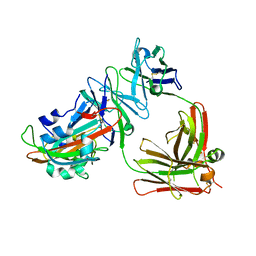 | | Complex structure of MERS-RBD bound with 4C2 antibody | | 分子名称: | 2-acetamido-2-deoxy-beta-D-glucopyranose, 4C2 heavy chain, 4C2 light chain, ... | | 著者 | Li, Y, Wan, Y, Liu, P, Zhao, J, Lu, G, Qi, J, Wang, Q, Lu, X, Wu, Y, Liu, W, Yuen, K.Y, Perlman, S, Gao, G.F, Yan, J. | | 登録日 | 2015-09-10 | | 公開日 | 2015-10-14 | | 最終更新日 | 2023-11-08 | | 実験手法 | X-RAY DIFFRACTION (2.409 Å) | | 主引用文献 | A humanized neutralizing antibody against MERS-CoV targeting the receptor-binding domain of the spike protein.
Cell Res., 25, 2015
|
|
1Q8Q
 | | Pterocarpus angolensis lectin (PAL) in complex with the dimannoside Man(alpha1-4)Man | | 分子名称: | CALCIUM ION, MANGANESE (II) ION, alpha-D-mannopyranose-(1-4)-methyl alpha-D-mannopyranoside, ... | | 著者 | Loris, R, Van Walle, I, De Greve, H, Beeckmans, S, Deboeck, F, Wyns, L, Bouckaert, J. | | 登録日 | 2003-08-22 | | 公開日 | 2004-02-10 | | 最終更新日 | 2020-07-29 | | 実験手法 | X-RAY DIFFRACTION (2.05 Å) | | 主引用文献 | Structural Basis of Oligomannose Recognition by the Pterocarpus angolensis Seed Lectin
J.Mol.Biol., 335, 2004
|
|
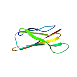
1JTO
 | | Degenerate interfaces in antigen-antibody complexes | | 分子名称: | Lysozyme, Vh Single-Domain Antibody | | 著者 | Decanniere, K, Transue, T.R, Desmyter, A, Maes, D, Muyldermans, S, Wyns, L. | | 登録日 | 2001-08-21 | | 公開日 | 2001-10-31 | | 最終更新日 | 2023-08-16 | | 実験手法 | X-RAY DIFFRACTION (2.5 Å) | | 主引用文献 | Degenerate interfaces in antigen-antibody complexes.
J.Mol.Biol., 313, 2001
|
|
1BXU
 | | OXIDIZED PLASTOCYANIN FROM SYNECHOCOCCUS SP. | | 分子名称: | COPPER (II) ION, PLASTOCYANIN | | 著者 | Inoue, T, Sugawara, H, Hamanaka, S, Tsukui, H, Suzuki, E, Kohzuma, T, Kai, Y. | | 登録日 | 1998-10-09 | | 公開日 | 1999-06-15 | | 最終更新日 | 2024-05-22 | | 実験手法 | X-RAY DIFFRACTION (1.9 Å) | | 主引用文献 | Crystal structure determinations of oxidized and reduced plastocyanin from the cyanobacterium Synechococcus sp. PCC 7942.
Biochemistry, 38, 1999
|
|
3E8V
 | | Crystal structure of a possible transglutaminase-family protein proteolytic fragment from Bacteroides fragilis | | 分子名称: | Possible transglutaminase-family protein, UNKNOWN LIGAND | | 著者 | Bonanno, J.B, Rutter, M, Bain, K.T, Hu, S, Romero, R, Smith, D, Wasserman, S, Sauder, J.M, Burley, S.K, Almo, S.C, New York SGX Research Center for Structural Genomics (NYSGXRC) | | 登録日 | 2008-08-20 | | 公開日 | 2008-09-02 | | 最終更新日 | 2024-02-21 | | 実験手法 | X-RAY DIFFRACTION (2.4 Å) | | 主引用文献 | Crystal structure of a possible transglutaminase-family protein proteolytic fragment from Bacteroides fragilis
To be Published
|
|
4J7Z
 | | Thermus thermophilus DNAJ J- and G/F-DOMAINS | | 分子名称: | Chaperone protein DnaJ 2, GLYCEROL | | 著者 | Barends, T.R.M, Brosi, R.W, Steinmetz, A, Scherer, A, Hartmann, E, Eschenbach, J, Lorenz, T, Seidel, R, Shoeman, R, Zimmermann, S, Bittl, R, Schlichting, I, Reinstein, J. | | 登録日 | 2013-02-14 | | 公開日 | 2013-07-31 | | 最終更新日 | 2024-02-28 | | 実験手法 | EPR (1.64 Å), X-RAY DIFFRACTION | | 主引用文献 | Combining crystallography and EPR: crystal and solution structures of the multidomain cochaperone DnaJ.
Acta Crystallogr.,Sect.D, 69, 2013
|
|


