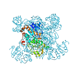
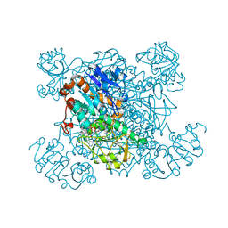
6M04
 
 | | Structure of the human homo-hexameric LRRC8D channel at 4.36 Angstroms | | 分子名称: | Volume-regulated anion channel subunit LRRC8D | | 著者 | Nakamura, R, Kasuya, G, Yokoyama, T, Shirouzu, M, Ishitani, R, Nureki, O. | | 登録日 | 2020-02-20 | | 公開日 | 2020-06-17 | | 実験手法 | ELECTRON MICROSCOPY (4.36 Å) | | 主引用文献 | Cryo-EM structure of the volume-regulated anion channel LRRC8D isoform identifies features important for substrate permeation.
Commun Biol, 3, 2020
|
|
4WLM
 | |
4WLG
 | |
4WMB
 | |
4WMK
 | |
4WLZ
 | |
4WMI
 | |
4WM0
 | |
4WN2
 | |
6LVM
 | | Crystal structure of FGFR3 in complex with pyrimidine derivative | | 分子名称: | 2-[[5-[2-(3,5-dimethoxyphenyl)ethyl]-2-[[3-methoxy-4-[4-(4-methylpiperazin-1-yl)piperidin-1-yl]phenyl]amino]pyrimidin-4-yl]amino]-N-ethyl-benzenesulfonamide, Fibroblast growth factor receptor 3 | | 著者 | Echizen, Y, Tateishi, Y, Amano, Y. | | 登録日 | 2020-02-04 | | 公開日 | 2020-04-08 | | 最終更新日 | 2023-11-29 | | 実験手法 | X-RAY DIFFRACTION (2.53 Å) | | 主引用文献 | Structure-based drug design of 1,3,5-triazine and pyrimidine derivatives as novel FGFR3 inhibitors with high selectivity over VEGFR2.
Bioorg.Med.Chem., 28, 2020
|
|
6LVK
 | | Crystal structure of FGFR2 in complex with 1,3,5-triazine derivative | | 分子名称: | Fibroblast growth factor receptor 2, N-ethyl-2-[[4-[[4-(4-methylpiperazin-1-yl)-3-(2-morpholin-4-ylethoxy)phenyl]amino]-1,3,5-triazin-2-yl]amino]benzenesulfonamide, SULFATE ION | | 著者 | Echizen, Y, Amano, Y, Tateishi, Y. | | 登録日 | 2020-02-04 | | 公開日 | 2020-04-08 | | 最終更新日 | 2023-11-29 | | 実験手法 | X-RAY DIFFRACTION (2.29 Å) | | 主引用文献 | Structure-based drug design of 1,3,5-triazine and pyrimidine derivatives as novel FGFR3 inhibitors with high selectivity over VEGFR2.
Bioorg.Med.Chem., 28, 2020
|
|
4YUU
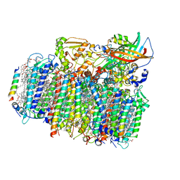 | | Crystal structure of oxygen-evolving photosystem II from a red alga | | 分子名称: | 1,2-DI-O-ACYL-3-O-[6-DEOXY-6-SULFO-ALPHA-D-GLUCOPYRANOSYL]-SN-GLYCEROL, 1,2-DIPALMITOYL-PHOSPHATIDYL-GLYCEROLE, 1,2-DISTEAROYL-MONOGALACTOSYL-DIGLYCERIDE, ... | | 著者 | Ago, H, Shen, J.-R. | | 登録日 | 2015-03-19 | | 公開日 | 2016-01-20 | | 最終更新日 | 2020-02-05 | | 実験手法 | X-RAY DIFFRACTION (2.7700038 Å) | | 主引用文献 | Novel Features of Eukaryotic Photosystem II Revealed by Its Crystal Structure Analysis from a Red Alga
J.Biol.Chem., 291, 2016
|
|
1QTO
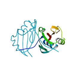 | | 1.5 A CRYSTAL STRUCTURE OF A BLEOMYCIN RESISTANCE DETERMINANT FROM BLEOMYCIN-PRODUCING STREPTOMYCES VERTICILLUS | | 分子名称: | BLEOMYCIN-BINDING PROTEIN | | 著者 | Kawano, Y, Kumagai, T, Muta, K, Matoba, Y, Davies, J, Sugiyama, M. | | 登録日 | 1999-06-28 | | 公開日 | 2000-06-28 | | 最終更新日 | 2024-02-14 | | 実験手法 | X-RAY DIFFRACTION (1.5 Å) | | 主引用文献 | The 1.5 A crystal structure of a bleomycin resistance determinant from bleomycin-producing Streptomyces verticillus.
J.Mol.Biol., 295, 2000
|
|
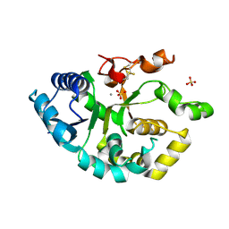
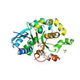
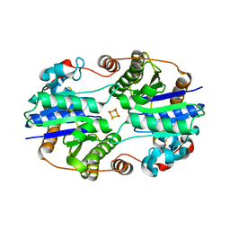
1PJB
 | | L-ALANINE DEHYDROGENASE | | 分子名称: | L-ALANINE DEHYDROGENASE | | 著者 | Baker, P.J, Sawa, Y, Shibata, H, Sedelnikova, S.E, Rice, D.W. | | 登録日 | 1998-06-05 | | 公開日 | 1999-06-08 | | 最終更新日 | 2024-02-14 | | 実験手法 | X-RAY DIFFRACTION (2.1 Å) | | 主引用文献 | Analysis of the structure and substrate binding of Phormidium lapideum alanine dehydrogenase.
Nat.Struct.Biol., 5, 1998
|
|
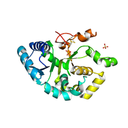
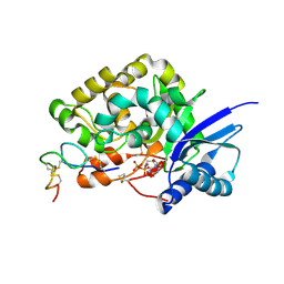
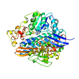
1PJC
 | | L-ALANINE DEHYDROGENASE COMPLEXED WITH NAD | | 分子名称: | NICOTINAMIDE-ADENINE-DINUCLEOTIDE, PROTEIN (L-ALANINE DEHYDROGENASE) | | 著者 | Baker, P.J, Sawa, Y, Shibata, H, Sedelnikova, S.E, Rice, D.W. | | 登録日 | 1998-06-05 | | 公開日 | 1999-07-15 | | 最終更新日 | 2023-12-27 | | 実験手法 | X-RAY DIFFRACTION (2 Å) | | 主引用文献 | Analysis of the structure and substrate binding of Phormidium lapideum alanine dehydrogenase.
Nat.Struct.Biol., 5, 1998
|
|
4WN1
 | | Crystal structure of PDE10A in complex with 1-methyl-5-(1-methyl-3-{[4-(quinolin-2-yl)phenoxy]methyl}-1H-pyrazol-4-yl)pyridin-2(1H)-one | | 分子名称: | 1-methyl-5-(1-methyl-3-{[4-(quinolin-2-yl)phenoxy]methyl}-1H-pyrazol-4-yl)pyridin-2(1H)-one, MAGNESIUM ION, ZINC ION, ... | | 著者 | Amano, Y, Honbou, K. | | 登録日 | 2014-10-10 | | 公開日 | 2014-12-31 | | 最終更新日 | 2023-12-27 | | 実験手法 | X-RAY DIFFRACTION (3.13 Å) | | 主引用文献 | Synthesis, SAR study, and biological evaluation of novel quinoline derivatives as phosphodiesterase 10A inhibitors with reduced CYP3A4 inhibition.
Bioorg.Med.Chem., 23, 2015
|
|
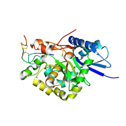
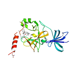
5AXI
 | | Crystal structure of Cbl-b TKB domain in complex with Cblin | | 分子名称: | CALCIUM ION, CHLORIDE ION, Cblin, ... | | 著者 | Ohno, A, Maita, N, Ochi, A, Nakao, R, Nikawa, T. | | 登録日 | 2015-07-29 | | 公開日 | 2016-03-02 | | 最終更新日 | 2023-11-15 | | 実験手法 | X-RAY DIFFRACTION (2.5 Å) | | 主引用文献 | Structural analysis of the TKB domain of ubiquitin ligase Cbl-b complexed with its small inhibitory peptide, Cblin
Arch.Biochem.Biophys., 594, 2016
|
|
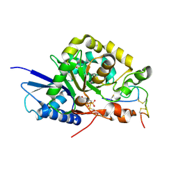
1G1M
 | | ALL-FERROUS NITROGENASE IRON PROTEIN FROM AZOTOBACTER VINELANDII | | 分子名称: | IRON/SULFUR CLUSTER, NITROGENASE IRON PROTEIN | | 著者 | Strop, P, Takahara, P.M, Chiu, H.-J, Angove, H.C, Burgess, B.K, Rees, D.C. | | 登録日 | 2000-10-12 | | 公開日 | 2001-01-31 | | 最終更新日 | 2024-02-07 | | 実験手法 | X-RAY DIFFRACTION (2.25 Å) | | 主引用文献 | Crystal structure of the all-ferrous [4Fe-4S]0 form of the nitrogenase iron protein from Azotobacter vinelandii.
Biochemistry, 40, 2001
|
|
1H2A
 | |
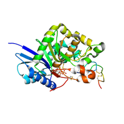
5AYF
 | | Crystal structure of SET7/9 in complex with cyproheptadine | | 分子名称: | 2-AMINO-2-HYDROXYMETHYL-PROPANE-1,3-DIOL, 4-(dibenzo[1,2-a:2',1'-d][7]annulen-11-ylidene)-1-methyl-piperidine, Histone-lysine N-methyltransferase SETD7, ... | | 著者 | Niwa, H, Handa, N, Takemoto, Y, Ito, A, Tomabechi, Y, Umehara, T, Shirouzu, M, Yoshida, M, Yokoyama, S. | | 登録日 | 2015-08-20 | | 公開日 | 2016-04-27 | | 最終更新日 | 2023-11-08 | | 実験手法 | X-RAY DIFFRACTION (2.005 Å) | | 主引用文献 | Identification of Cyproheptadine as an Inhibitor of SET Domain Containing Lysine Methyltransferase 7/9 (Set7/9) That Regulates Estrogen-Dependent Transcription
J.Med.Chem., 59, 2016
|
|
2GS0
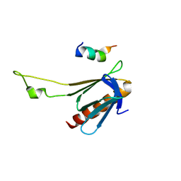 | | NMR structure of the complex between the PH domain of the Tfb1 subunit from TFIIH and the activation domain of p53 | | 分子名称: | Cellular tumor antigen p53, RNA polymerase II transcription factor B subunit 1 | | 著者 | Di Lello, P, Jones, T.N, Nguyen, B.D, Legault, P, Omichinski, J.G. | | 登録日 | 2006-04-25 | | 公開日 | 2006-10-31 | | 最終更新日 | 2024-05-29 | | 実験手法 | SOLUTION NMR | | 主引用文献 | Structure of the Tfb1/p53 complex: Insights into the interaction between the p62/Tfb1 subunit of TFIIH and the activation domain of p53.
Mol.Cell, 22, 2006
|
|
2RQ6
 | | Solution structure of the epsilon subunit of the F1-atpase from thermosynechococcus elongatus BP-1 | | 分子名称: | ATP synthase epsilon chain | | 著者 | Yagi, H, Konno, H, Murakami-Fuse, T, Oroguchi, H, Akutsu, T, Ikeguchi, M, Hisabori, T. | | 登録日 | 2009-03-03 | | 公開日 | 2010-01-12 | | 最終更新日 | 2024-05-29 | | 実験手法 | SOLUTION NMR | | 主引用文献 | Structural and functional analysis of the intrinsic inhibitor subunit epsilon of F1-ATPase from photosynthetic organisms.
Biochem.J., 425, 2010
|
|
8PWZ
 | | Crystal Structure of (3R)-hydroxyacyl-ACP dehydratase HadBD from Mycobacterium tuberculosis | | 分子名称: | (3R)-hydroxyacyl-ACP dehydratase subunit HadB, UPF0336 protein Rv0504c | | 著者 | Rima, J, Grimoire, Y, Bories, P, Bardou, F, Quemard, A, Bon, C, Mourey, L, Tranier, S. | | 登録日 | 2023-07-22 | | 公開日 | 2024-03-27 | | 実験手法 | X-RAY DIFFRACTION (2.00196719 Å) | | 主引用文献 | HadBD dehydratase from Mycobacterium tuberculosis fatty acid synthase type II: A singular structure for a unique function.
Protein Sci., 33, 2024
|
|
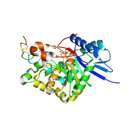
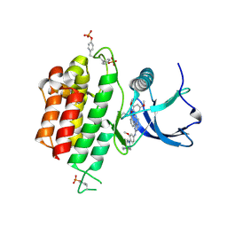
1FKV
 | | RECOMBINANT GOAT ALPHA-LACTALBUMIN T29I | | 分子名称: | ALPHA-LACTALBUMIN, CALCIUM ION | | 著者 | Horii, K, Matsushima, M, Tsumoto, K, Kumagai, I. | | 登録日 | 2000-08-10 | | 公開日 | 2001-02-14 | | 最終更新日 | 2021-11-03 | | 実験手法 | X-RAY DIFFRACTION (2 Å) | | 主引用文献 | Contribution of Thr29 to the thermodynamic stability of goat alpha-lactalbumin as determined by experimental and theoretical approaches.
Proteins, 45, 2001
|
|
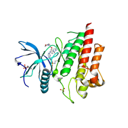
1FKQ
 | | RECOMBINANT GOAT ALPHA-LACTALBUMIN T29V | | 分子名称: | ALPHA-LACTALBUMIN, CALCIUM ION | | 著者 | Horii, K, Matsushima, M, Tsumoto, K, Kumagai, I. | | 登録日 | 2000-08-10 | | 公開日 | 2001-02-14 | | 最終更新日 | 2021-11-03 | | 実験手法 | X-RAY DIFFRACTION (1.8 Å) | | 主引用文献 | Contribution of Thr29 to the thermodynamic stability of goat alpha-lactalbumin as determined by experimental and theoretical approaches.
Proteins, 45, 2001
|
|


