6ZSD
 
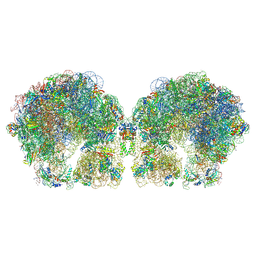
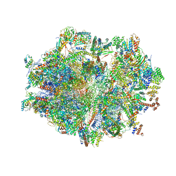 | | Human mitochondrial ribosome in complex with mRNA, P-site tRNA and E-site tRNA | | 分子名称: | 12S mitochondrial rRNA, 16S mitochondrial rRNA, 28S ribosomal protein S10, ... | | 著者 | Aibara, S, Singh, V, Modelska, A, Amunts, A. | | 登録日 | 2020-07-15 | | 公開日 | 2020-09-16 | | 最終更新日 | 2024-07-10 | | 実験手法 | ELECTRON MICROSCOPY (3.7 Å) | | 主引用文献 | Structural basis of mitochondrial translation.
Elife, 9, 2020
|
|
8VFO
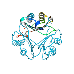 | | Crystal Structure of L117G Variant of D-Dopachrome Tautomerase (D-DT) | | 分子名称: | CITRIC ACID, D-dopachrome decarboxylase | | 著者 | Parkins, A, Pilien, A, Thompson, M.C, Pantouris, G. | | 登録日 | 2023-12-21 | | 公開日 | 2024-05-01 | | 最終更新日 | 2024-05-22 | | 実験手法 | X-RAY DIFFRACTION (1.35 Å) | | 主引用文献 | The C-terminal Region of D-DT Regulates Molecular Recognition for Protein-Ligand Complexes.
J.Med.Chem., 67, 2024
|
|
8VFW
 | | Crystal Structure of V113N D-Dopachrome Tautomerase (D-DT) | | 分子名称: | CITRIC ACID, D-dopachrome decarboxylase | | 著者 | Parkins, A, Wolff, A, Thompson, M.C, Pantouris, G. | | 登録日 | 2023-12-22 | | 公開日 | 2024-05-01 | | 最終更新日 | 2024-05-22 | | 実験手法 | X-RAY DIFFRACTION (1.31 Å) | | 主引用文献 | The C-terminal Region of D-DT Regulates Molecular Recognition for Protein-Ligand Complexes.
J.Med.Chem., 67, 2024
|
|
5JLV
 | | Receptor binding domain of Botulinum neurotoxin A in complex with human glycosylated SV2C | | 分子名称: | 2-acetamido-2-deoxy-beta-D-glucopyranose, 2-acetamido-2-deoxy-beta-D-glucopyranose-(1-4)-[alpha-L-fucopyranose-(1-6)]2-acetamido-2-deoxy-beta-D-glucopyranose, ACETATE ION, ... | | 著者 | Yao, G, Zhang, S, Mahrhold, S, Lam, K, Stern, D, Bagramyan, K, Perry, K, Kalkum, M, Rummel, A, Dong, M, Jin, R. | | 登録日 | 2016-04-27 | | 公開日 | 2016-06-15 | | 最終更新日 | 2023-09-27 | | 実験手法 | X-RAY DIFFRACTION (2 Å) | | 主引用文献 | N-linked glycosylation of SV2 is required for binding and uptake of botulinum neurotoxin A.
Nat.Struct.Mol.Biol., 23, 2016
|
|
5MNQ
 | | Cationic trypsin in complex with a derivative of N-amidinopiperidine | | 分子名称: | (2~{S})-1-[(2~{R})-2-azanyl-3-phenyl-propanoyl]-~{N}-[(1-carbamimidoylpiperidin-4-yl)methyl]pyrrolidine-2-carboxamide, CALCIUM ION, Cationic trypsin, ... | | 著者 | Schiebel, J, Ngo, K, Heine, A, Klebe, G. | | 登録日 | 2016-12-13 | | 公開日 | 2018-01-17 | | 最終更新日 | 2024-01-17 | | 実験手法 | X-RAY DIFFRACTION (1.337 Å) | | 主引用文献 | Intriguing role of water in protein-ligand binding studied by neutron crystallography on trypsin complexes.
Nat Commun, 9, 2018
|
|
6ZSC
 | | Human mitochondrial ribosome in complex with E-site tRNA | | 分子名称: | 12S mitochondrial rRNA, 16S mitochondrial rRNA, 28S ribosomal protein S10, ... | | 著者 | Aibara, S, Singh, V, Modelska, A, Amunts, A. | | 登録日 | 2020-07-15 | | 公開日 | 2020-09-16 | | 最終更新日 | 2024-07-10 | | 実験手法 | ELECTRON MICROSCOPY (3.5 Å) | | 主引用文献 | Structural basis of mitochondrial translation.
Elife, 9, 2020
|
|
8VG5
 | | Crystal Structure of V113N Variant of D-Dopachrome Tautomerase (D-DT) Bound with 4CPPC | | 分子名称: | 4-(3-carboxyphenyl)pyridine-2,5-dicarboxylic acid, CITRIC ACID, D-dopachrome decarboxylase | | 著者 | Parkins, A, Pilien, A, Thompson, M.C, Pantouris, G. | | 登録日 | 2023-12-22 | | 公開日 | 2024-05-01 | | 最終更新日 | 2024-05-22 | | 実験手法 | X-RAY DIFFRACTION (1.5 Å) | | 主引用文献 | The C-terminal Region of D-DT Regulates Molecular Recognition for Protein-Ligand Complexes.
J.Med.Chem., 67, 2024
|
|
3KFO
 | | Crystal structure of the C-terminal domain from the nuclear pore complex component NUP133 from Saccharomyces cerevisiae | | 分子名称: | GLYCEROL, Nucleoporin NUP133 | | 著者 | Sampathkumar, P, Bonanno, J.B, Miller, S, Bain, K, Dickey, M, Gheyi, T, Almo, S.C, Rout, M, Sali, A, Phillips, J, Pieper, U, Fernandez-Martinez, J, Franke, J.D, Atwell, S, Thompson, D.A, Emtage, J.S, Wasserman, S, Sauder, J.M, Burley, S.K, New York SGX Research Center for Structural Genomics (NYSGXRC) | | 登録日 | 2009-10-27 | | 公開日 | 2010-01-26 | | 最終更新日 | 2021-02-10 | | 実験手法 | X-RAY DIFFRACTION (1.9 Å) | | 主引用文献 | Structure of the C-terminal domain of Saccharomyces cerevisiae Nup133, a component of the nuclear pore complex.
Proteins, 79, 2011
|
|
5FEP
 | | HydE from T. maritima in complex with (2R,4R)-MeTDA | | 分子名称: | (2R,4R)-2-methyl-1,3-thiazolidine-2,4-dicarboxylic acid, 3-[(3-CHOLAMIDOPROPYL)DIMETHYLAMMONIO]-1-PROPANESULFONATE, CHLORIDE ION, ... | | 著者 | Rohac, R, Amara, P, Benjdia, A, Martin, L, Ruffie, P, Favier, A, Berteau, O, Mouesca, J.M, Fontecilla-Camps, J.C, Nicolet, Y. | | 登録日 | 2015-12-17 | | 公開日 | 2016-04-06 | | 最終更新日 | 2024-01-10 | | 実験手法 | X-RAY DIFFRACTION (1.45 Å) | | 主引用文献 | Carbon-sulfur bond-forming reaction catalysed by the radical SAM enzyme HydE.
Nat.Chem., 8, 2016
|
|
5JDA
 | | Bacillus cereus CotH kinase plus Mg2+/AMP | | 分子名称: | 1,2-ETHANEDIOL, ADENOSINE MONOPHOSPHATE, MAGNESIUM ION, ... | | 著者 | Tomchick, D.R, Tagliabracci, V.S, Sreelatha, A. | | 登録日 | 2016-04-15 | | 公開日 | 2016-05-18 | | 最終更新日 | 2023-09-27 | | 実験手法 | X-RAY DIFFRACTION (1.401 Å) | | 主引用文献 | Phosphorylation of spore coat proteins by a family of atypical protein kinases.
Proc.Natl.Acad.Sci.USA, 113, 2016
|
|
5J5X
 | | Complex of PKA with the bisubstrate protein kinase inhibitor ARC-1416 | | 分子名称: | 4-(piperazin-1-yl)-7H-pyrrolo[2,3-d]pyrimidine, 47P-AZ1-DAL-DAR-DAR-DAR-DAR, SULFATE ION, ... | | 著者 | Alam, K.A, Ivan, T, Uri, A, Engh, R.A. | | 登録日 | 2016-04-04 | | 公開日 | 2016-07-20 | | 最終更新日 | 2019-06-19 | | 実験手法 | X-RAY DIFFRACTION (2.6 Å) | | 主引用文献 | Bifunctional Ligands for Inhibition of Tight-Binding Protein-Protein Interactions.
Bioconjug.Chem., 27, 2016
|
|
5FT6
 | | Crystal structure of the cysteine desulfurase CsdA (S-sulfonic acid) from Escherichia coli at 2.050 Angstroem resolution | | 分子名称: | CYSTEINE DESULFURASE CSDA, DI(HYDROXYETHYL)ETHER, GLYCEROL, ... | | 著者 | Fernandez, F.J, Arda, A, Lopez-Estepa, M, Aranda, J, Penya-Soler, E, Garces, F, Round, A, Campos-Oliva, R, Bruix, M, Coll, M, Tunon, I, Jimenez-Barbero, J, Vega, M.C. | | 登録日 | 2016-01-11 | | 公開日 | 2016-11-23 | | 最終更新日 | 2024-01-10 | | 実験手法 | X-RAY DIFFRACTION (2.049 Å) | | 主引用文献 | Mechanism of Sulfur Transfer Across Protein-Protein Interfaces: The Cysteine Desulfurase Model System
Acs Catalysis, 6, 2016
|
|
5J6Q
 | | Cwp8 from Clostridium difficile | | 分子名称: | CHLORIDE ION, Cell wall binding protein cwp8, SULFATE ION | | 著者 | Renko, M, Usenik, A, Turk, D. | | 登録日 | 2016-04-05 | | 公開日 | 2017-02-08 | | 最終更新日 | 2024-05-08 | | 実験手法 | X-RAY DIFFRACTION (2.1 Å) | | 主引用文献 | The CWB2 Cell Wall-Anchoring Module Is Revealed by the Crystal Structures of the Clostridium difficile Cell Wall Proteins Cwp8 and Cwp6.
Structure, 25, 2017
|
|
5MWP
 | | The structure of MR in complex with AZD9977. | | 分子名称: | 2-[(3~{S})-7-fluoranyl-4-[(3-oxidanylidene-4~{H}-1,4-benzoxazin-6-yl)carbonyl]-2,3-dihydro-1,4-benzoxazin-3-yl]-~{N}-methyl-ethanamide, Mineralocorticoid receptor, NCOA1 peptide | | 著者 | Edman, K, Aagaard, A, Backstrom, S. | | 登録日 | 2017-01-19 | | 公開日 | 2018-03-07 | | 最終更新日 | 2024-01-17 | | 実験手法 | X-RAY DIFFRACTION (1.82 Å) | | 主引用文献 | Preclinical pharmacology of AZD9977: A novel mineralocorticoid receptor modulator separating organ protection from effects on electrolyte excretion.
PLoS ONE, 13, 2018
|
|
5MWY
 | |
8ARF
 | |
5MMG
 | |
5JBN
 | |
5MPN
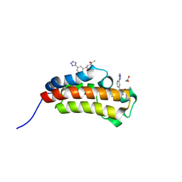 | | Crystal structure of CREBBP bromodomain complexed with FA26 | | 分子名称: | 1,2-ETHANEDIOL, 1-[4-ethoxy-3-[3-(2~{H}-1,2,3,4-tetrazol-5-yl)phenyl]phenyl]ethanone, CREB-binding protein | | 著者 | Zhu, J, Caflisch, A. | | 登録日 | 2016-12-16 | | 公開日 | 2018-01-17 | | 最終更新日 | 2024-05-08 | | 実験手法 | X-RAY DIFFRACTION (1.23 Å) | | 主引用文献 | Binding Motifs in the CBP Bromodomain: An Analysis of 20 Crystal Structures of Complexes with Small Molecules.
ACS Med Chem Lett, 9, 2018
|
|
4LLW
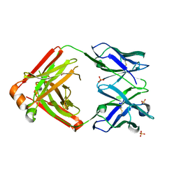 | | Crystal structure of Pertuzumab Clambda Fab with variable domain redesign (VRD2) at 1.95A | | 分子名称: | SULFATE ION, light chain Clambda, mutated Pertuzumab Fab heavy chain | | 著者 | Pustilnik, A, Lewis, S.M, Wu, X, Sereno, A, Huang, F, Guntas, G, Leaver-Fay, A, Smith, E.M, Ho, C, Hansen-Estruch, C, Chamberlain, A.K, Truhlar, S.M, Kuhlman, B, Demarest, S.J, Atwell, S. | | 登録日 | 2013-07-09 | | 公開日 | 2014-01-29 | | 最終更新日 | 2019-06-26 | | 実験手法 | X-RAY DIFFRACTION (1.95 Å) | | 主引用文献 | Generation of bispecific IgG antibodies by structure-based design of an orthogonal Fab interface.
Nat.Biotechnol., 32, 2014
|
|
4E3A
 | | CRYSTAL STRUCTURE OF probable sugar kinase protein from Rhizobium etli CFN 42 | | 分子名称: | ADENOSINE, sugar kinase protein | | 著者 | Malashkevich, V.N, Bhosle, R, Toro, R, Hillerich, B, Gizzi, A, Garforth, S, Kar, A, Chan, M.K, Lafluer, J, Patel, H, Matikainen, B, Chamala, S, Lim, S, Celikgil, A, Villegas, G, Evans, B, Zenchek, W, Love, J, Fiser, A, Khafizov, K, Seidel, R, Bonanno, J.B, Almo, S.C, New York Structural Genomics Research Consortium (NYSGRC) | | 登録日 | 2012-03-09 | | 公開日 | 2012-03-21 | | 最終更新日 | 2023-12-06 | | 実験手法 | X-RAY DIFFRACTION (1.63 Å) | | 主引用文献 | CRYSTAL STRUCTURE OF probable sugar kinase protein from Rhizobium etli CFN 42
To be Published
|
|
5FTN
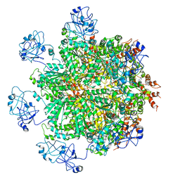 | | Cryo-EM structure of human p97 bound to ATPgS (Conformation III) | | 分子名称: | MAGNESIUM ION, PHOSPHOTHIOPHOSPHORIC ACID-ADENYLATE ESTER, TRANSITIONAL ENDOPLASMIC RETICULUM ATPASE | | 著者 | Banerjee, S, Bartesaghi, A, Merk, A, Rao, P, Bulfer, S.L, Yan, Y, Green, N, Mroczkowski, B, Neitz, R.J, Wipf, P, Falconieri, V, Deshaies, R.J, Milne, J.L.S, Huryn, D, Arkin, M, Subramaniam, S. | | 登録日 | 2016-01-14 | | 公開日 | 2016-01-27 | | 最終更新日 | 2024-05-08 | | 実験手法 | ELECTRON MICROSCOPY (3.3 Å) | | 主引用文献 | 2.3 A Resolution Cryo-Em Structure of Human P97 and Mechanism of Allosteric Inhibition
Science, 351, 2016
|
|
6ZSE
 | | Human mitochondrial ribosome in complex with mRNA, A/P-tRNA and P/E-tRNA | | 分子名称: | 12S mitochondrial rRNA, 16S mitochondrial rRNA, 28S ribosomal protein S10, ... | | 著者 | Aibara, S, Singh, V, Modelska, A, Amunts, A. | | 登録日 | 2020-07-15 | | 公開日 | 2020-09-16 | | 最終更新日 | 2024-07-10 | | 実験手法 | ELECTRON MICROSCOPY (5 Å) | | 主引用文献 | Structural basis of mitochondrial translation.
Elife, 9, 2020
|
|
6FXC
 | | The cryo-EM structure of hibernating 100S ribosome dimer from pathogenic Staphylococcus aureus | | 分子名称: | 16S ribosomal RNA, 23S ribosomal RNA, 30S ribosomal protein S10, ... | | 著者 | Matzov, D, Aibara, S, Zimmerman, E, Bashan, A, Kidmose, R, Amunts, A, Yonath, A. | | 登録日 | 2018-03-08 | | 公開日 | 2018-03-21 | | 最終更新日 | 2018-11-21 | | 実験手法 | ELECTRON MICROSCOPY (6.76 Å) | | 主引用文献 | The cryo-EM structure of hibernating 100S ribosome dimer from pathogenic Staphylococcus aureus.
Nat Commun, 8, 2017
|
|
5FZE
 | | Crystal structure of the catalytic domain of human JARID1B in complex with MC3960 | | 分子名称: | 1,2-ETHANEDIOL, CHLORIDE ION, DIMETHYL SULFOXIDE, ... | | 著者 | Nowak, R, Krojer, T, Johansson, C, Gileadi, C, Kupinska, K, Strain-Damerell, C, Szykowska, A, von Delft, F, Burgess-Brown, N.A, Arrowsmith, C.H, Bountra, C, Edwards, A.M, Rotili, D, Mai, A, Oppermann, U. | | 登録日 | 2016-03-14 | | 公開日 | 2017-03-29 | | 最終更新日 | 2024-01-10 | | 実験手法 | X-RAY DIFFRACTION (2.02 Å) | | 主引用文献 | Crystal Structure of the Catalytic Domain of Human Jarid1B in Complex with Mc3960
To be Published
|
|


