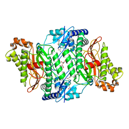
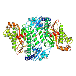
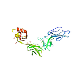
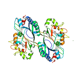
7B80
 
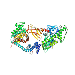 | | DeAMPylation complex of monomeric FICD and AMPylated BiP (state 2) | | 分子名称: | 3,6,9,12,15,18-HEXAOXAICOSANE-1,20-DIOL, ADENOSINE MONOPHOSPHATE, DI(HYDROXYETHYL)ETHER, ... | | 著者 | Perera, L.A, Ron, D. | | 登録日 | 2020-12-12 | | 公開日 | 2021-07-07 | | 最終更新日 | 2024-01-31 | | 実験手法 | X-RAY DIFFRACTION (1.87 Å) | | 主引用文献 | Structures of a deAMPylation complex rationalise the switch between antagonistic catalytic activities of FICD.
Nat Commun, 12, 2021
|
|
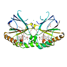
5MC3
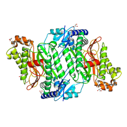
 | | Crystal Structure of Glu412Lys mutant of Human Prolidase with Mn ions and GlyPro ligand | | 分子名称: | GLYCEROL, GLYCINE, HYDROXIDE ION, ... | | 著者 | Wilk, P, Mueller, U, Dobbek, H, Weiss, M.S. | | 登録日 | 2016-11-09 | | 公開日 | 2017-12-20 | | 最終更新日 | 2024-01-17 | | 実験手法 | X-RAY DIFFRACTION (1.52 Å) | | 主引用文献 | Structural basis for prolidase deficiency disease mechanisms.
FEBS J., 285, 2018
|
|
5MC4
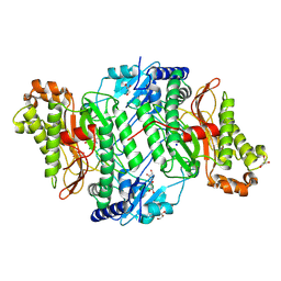
 | | Crystal Structure of Gly448Arg mutant of Human Prolidase with Mn ions and GlyPro ligand | | 分子名称: | GLYCEROL, GLYCINE, HYDROXIDE ION, ... | | 著者 | Wilk, P, Mueller, U, Dobbek, H, Weiss, M.S. | | 登録日 | 2016-11-09 | | 公開日 | 2017-12-20 | | 最終更新日 | 2024-01-17 | | 実験手法 | X-RAY DIFFRACTION (1.8 Å) | | 主引用文献 | Structural basis for prolidase deficiency disease mechanisms.
FEBS J., 285, 2018
|
|
5MC0
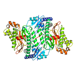
 | | Crystal Structure of delTyr231 mutant of Human Prolidase with Mn ions and GlyPro ligand | | 分子名称: | CHLORIDE ION, GLYCEROL, GLYCINE, ... | | 著者 | Wilk, P, Mueller, U, Dobbek, H, Weiss, M.S. | | 登録日 | 2016-11-09 | | 公開日 | 2017-12-20 | | 最終更新日 | 2024-01-17 | | 実験手法 | X-RAY DIFFRACTION (1.56 Å) | | 主引用文献 | Structural basis for prolidase deficiency disease mechanisms.
FEBS J., 285, 2018
|
|
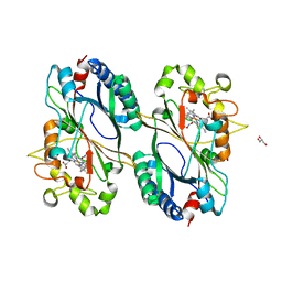
5M4G
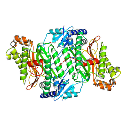
 | | Crystal Structure of Wild-Type Human Prolidase with Mn ions | | 分子名称: | GLYCEROL, HYDROXIDE ION, MANGANESE (II) ION, ... | | 著者 | Wilk, P, Weiss, M.S, Mueller, U, Dobbek, H. | | 登録日 | 2016-10-18 | | 公開日 | 2017-07-12 | | 最終更新日 | 2024-01-17 | | 実験手法 | X-RAY DIFFRACTION (1.48 Å) | | 主引用文献 | Substrate specificity and reaction mechanism of human prolidase.
FEBS J., 284, 2017
|
|
5MY4
 | |
5MYK
 | |
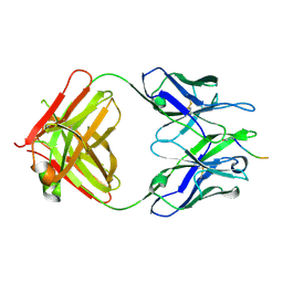
5MYO
 | | Structure of Pyroglutamate-Abeta-specific Fab c#6 in complex with human Abeta-pE3-12-PEGb | | 分子名称: | Amyloid beta A4 protein, Fab c#6 heavy chain, Fab c#6 light chain, ... | | 著者 | Parthier, C, Piechotta, A, Stubbs, M.T. | | 登録日 | 2017-01-27 | | 公開日 | 2017-06-28 | | 最終更新日 | 2024-01-17 | | 実験手法 | X-RAY DIFFRACTION (1.59 Å) | | 主引用文献 | Structural and functional analyses of pyroglutamate-amyloid-beta-specific antibodies as a basis for Alzheimer immunotherapy.
J. Biol. Chem., 292, 2017
|
|
5MC2
 | | Crystal Structure of Gly278Asp mutant of Human Prolidase with Mn ions and GlyPro ligand | | 分子名称: | GLYCEROL, GLYCINE, MANGANESE (II) ION, ... | | 著者 | Wilk, P, Mueller, U, Dobbek, H, Weiss, M.S. | | 登録日 | 2016-11-09 | | 公開日 | 2017-12-20 | | 最終更新日 | 2024-01-17 | | 実験手法 | X-RAY DIFFRACTION (1.7 Å) | | 主引用文献 | Structural basis for prolidase deficiency disease mechanisms.
FEBS J., 285, 2018
|
|
5M4L
 | | Crystal Structure of Wild-Type Human Prolidase with Mg ions and LeuPro ligand | | 分子名称: | GLYCEROL, HYDROXIDE ION, LEUCINE, ... | | 著者 | Wilk, P, Weiss, M.S, Mueller, U, Dobbek, H. | | 登録日 | 2016-10-18 | | 公開日 | 2017-07-12 | | 最終更新日 | 2020-04-22 | | 実験手法 | X-RAY DIFFRACTION (1.49 Å) | | 主引用文献 | Substrate specificity and reaction mechanism of human prolidase.
FEBS J., 284, 2017
|
|
5MBY
 | | Crystal Structure of Arg184Gln mutant of Human Prolidase with Mn ions and GlyPro ligand | | 分子名称: | GLYCEROL, GLYCINE, HYDROXIDE ION, ... | | 著者 | Wilk, P, Piwowarczyk, R, Mueller, U, Dobbek, H, Weiss, M.S. | | 登録日 | 2016-11-09 | | 公開日 | 2017-12-20 | | 最終更新日 | 2024-01-17 | | 実験手法 | X-RAY DIFFRACTION (1.55 Å) | | 主引用文献 | Structural basis for prolidase deficiency disease mechanisms.
FEBS J., 285, 2018
|
|
5MBZ
 | | Crystal Structure of Ser202Phe mutant of Human Prolidase with Mn ions and GlyPro ligand | | 分子名称: | CHLORIDE ION, GLYCEROL, GLYCINE, ... | | 著者 | Wilk, P, Mueller, U, Dobbek, H, Weiss, M.S. | | 登録日 | 2016-11-09 | | 公開日 | 2017-12-20 | | 最終更新日 | 2024-01-17 | | 実験手法 | X-RAY DIFFRACTION (1.5 Å) | | 主引用文献 | Structural basis for prolidase deficiency disease mechanisms.
FEBS J., 285, 2018
|
|
5MC1
 | | Crystal Structure of Asp276Asn mutant of Human Prolidase with Mn ions and GlyPro ligand | | 分子名称: | GLYCEROL, GLYCINE, MANGANESE (II) ION, ... | | 著者 | Wilk, P, Mueller, U, Dobbek, H, Weiss, M.S. | | 登録日 | 2016-11-09 | | 公開日 | 2017-12-20 | | 最終更新日 | 2024-01-17 | | 実験手法 | X-RAY DIFFRACTION (1.43 Å) | | 主引用文献 | Structural basis for prolidase deficiency disease mechanisms.
FEBS J., 285, 2018
|
|
5MYX
 | |
2UKD
 | |
6E61
 | | Bacteroides ovatus mixed-linkage glucan utilization locus (MLGUL) SGBP-A in complex with mixed-linkage heptasaccharide | | 分子名称: | 1,2-ETHANEDIOL, MAGNESIUM ION, beta-D-glucopyranose-(1-4)-beta-D-glucopyranose-(1-4)-beta-D-glucopyranose-(1-3)-beta-D-glucopyranose-(1-4)-beta-D-glucopyranose-(1-4)-beta-D-glucopyranose-(1-3)-beta-D-glucopyranose, ... | | 著者 | Tamura, K, Gardill, B.R, Brumer, H, Van Petegem, F. | | 登録日 | 2018-07-23 | | 公開日 | 2019-05-15 | | 最終更新日 | 2023-10-11 | | 実験手法 | X-RAY DIFFRACTION (2.51 Å) | | 主引用文献 | Surface glycan-binding proteins are essential for cereal beta-glucan utilization by the human gut symbiont Bacteroides ovatus.
Cell.Mol.Life Sci., 76, 2019
|
|
6E9B
 | | Bacteroides ovatus mixed-linkage glucan utilization locus (MLGUL) SGBP-B in complex with mixed-linkage heptasaccharide | | 分子名称: | Mixed-linkage glucan utilization locus (MLGUL) SGBP-B, SULFATE ION, beta-D-glucopyranose-(1-4)-beta-D-glucopyranose-(1-3)-beta-D-glucopyranose-(1-4)-beta-D-glucopyranose-(1-4)-beta-D-glucopyranose-(1-4)-beta-D-glucopyranose-(1-3)-beta-D-glucopyranose | | 著者 | Tamura, K, Gardill, B.R, Brumer, H, Van Petegem, F. | | 登録日 | 2018-07-31 | | 公開日 | 2019-05-15 | | 最終更新日 | 2023-10-11 | | 実験手法 | X-RAY DIFFRACTION (3.15 Å) | | 主引用文献 | Surface glycan-binding proteins are essential for cereal beta-glucan utilization by the human gut symbiont Bacteroides ovatus.
Cell.Mol.Life Sci., 76, 2019
|
|
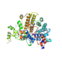
6FKS
 | | Crystal structure of a dye-decolorizing peroxidase from Klebsiella pneumoniae (KpDyP) | | 分子名称: | GLYCEROL, Iron-dependent peroxidase, MAGNESIUM ION, ... | | 著者 | Pfanzagl, V, Hofbauer, S, Mlynek, G. | | 登録日 | 2018-01-24 | | 公開日 | 2018-08-08 | | 最終更新日 | 2024-01-17 | | 実験手法 | X-RAY DIFFRACTION (1.60000467 Å) | | 主引用文献 | Roles of distal aspartate and arginine of B-class dye-decolorizing peroxidase in heterolytic hydrogen peroxide cleavage.
J. Biol. Chem., 293, 2018
|
|
6FL2
 | | Crystal structure of a dye-decolorizing peroxidase D143A variant from Klebsiella pneumoniae (KpDyP) | | 分子名称: | GLYCEROL, Iron-dependent peroxidase, MAGNESIUM ION, ... | | 著者 | Pfanzagl, V, Hofbauer, S, Mlynek, G. | | 登録日 | 2018-01-25 | | 公開日 | 2018-08-08 | | 最終更新日 | 2024-01-17 | | 実験手法 | X-RAY DIFFRACTION (1.270001 Å) | | 主引用文献 | Roles of distal aspartate and arginine of B-class dye-decolorizing peroxidase in heterolytic hydrogen peroxide cleavage.
J. Biol. Chem., 293, 2018
|
|
6E60
 | | Bacteroides ovatus mixed-linkage glucan utilization locus (MLGUL) SGBP-A | | 分子名称: | 1,2-ETHANEDIOL, MAGNESIUM ION, mixed-linkage glucan utilization locus (MLGUL) SGBP-B | | 著者 | Tamura, K, Gardill, B.R, Brumer, H, Van Petegem, F. | | 登録日 | 2018-07-23 | | 公開日 | 2019-05-15 | | 最終更新日 | 2024-04-03 | | 実験手法 | X-RAY DIFFRACTION (1.5 Å) | | 主引用文献 | Surface glycan-binding proteins are essential for cereal beta-glucan utilization by the human gut symbiont Bacteroides ovatus.
Cell.Mol.Life Sci., 76, 2019
|
|
6FIY
 | | Crystal structure of a dye-decolorizing peroxidase D143AR232A variant from Klebsiella pneumoniae (KpDyP) | | 分子名称: | GLYCEROL, Iron-dependent peroxidase, MAGNESIUM ION, ... | | 著者 | Pfanzagl, V, Hofbauer, S, Mlynek, G. | | 登録日 | 2018-01-19 | | 公開日 | 2018-08-08 | | 最終更新日 | 2024-01-17 | | 実験手法 | X-RAY DIFFRACTION (1.09000432 Å) | | 主引用文献 | Roles of distal aspartate and arginine of B-class dye-decolorizing peroxidase in heterolytic hydrogen peroxide cleavage.
J. Biol. Chem., 293, 2018
|
|
6FKT
 | |
5UKD
 | |
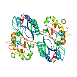
5NKV
 | | Crystal structure of dimeric chlorite dismutase from Cyanothece sp. PCC7425 at pH 9.0 and 293 K. | | 分子名称: | CHLORIDE ION, Chlorite Dismutase, GLYCEROL, ... | | 著者 | Puehringer, D, Schaffner, I, Mlynek, G, Obinger, C, Djinovic-Carugo, K. | | 登録日 | 2017-04-03 | | 公開日 | 2018-01-31 | | 最終更新日 | 2024-05-08 | | 実験手法 | X-RAY DIFFRACTION (2 Å) | | 主引用文献 | Molecular Mechanism of Enzymatic Chlorite Detoxification: Insights from Structural and Kinetic Studies.
ACS Catal, 7, 2017
|
|
5NKU
 | | Joint neutron/X-ray structure of dimeric chlorite dismutase from Cyanothece sp. PCC7425 | | 分子名称: | CHLORIDE ION, Chlorite Dismutase, GLYCEROL, ... | | 著者 | Puehringer, D, Schaffner, I, Mlynek, G, Obinger, C, Djinovic-Carugo, K. | | 登録日 | 2017-04-03 | | 公開日 | 2018-02-28 | | 最終更新日 | 2024-05-01 | | 実験手法 | NEUTRON DIFFRACTION (2 Å), X-RAY DIFFRACTION | | 主引用文献 | Molecular Mechanism of Enzymatic Chlorite Detoxification: Insights from Structural and Kinetic Studies.
ACS Catal, 7, 2017
|
|


