5XDP
 
 | |
1S5X
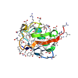
 | | The crystal structure of Trematomus bernacchii hemoglobin oxidized by air | | 分子名称: | Hemoglobin alpha chain, Hemoglobin beta chain, PROTOPORPHYRIN IX CONTAINING FE | | 著者 | Vitagliano, L, Bonomi, G, Riccio, A, di Prisco, G, Smulevich, G, Mazzarella, L. | | 登録日 | 2004-01-22 | | 公開日 | 2004-05-04 | | 最終更新日 | 2011-07-13 | | 実験手法 | X-RAY DIFFRACTION (2.4 Å) | | 主引用文献 | The oxidation process of Antarctic fish hemoglobins
Eur.J.Biochem., 271, 2004
|
|
1S5Y
 | | The crystal structure of Trematomus bernacchii hemoglobin oxidized by ferricyanide | | 分子名称: | Hemoglobin alpha chain, Hemoglobin beta chain, PROTOPORPHYRIN IX CONTAINING FE | | 著者 | Vitagliano, L, Bonomi, G, Riccio, A, di Prisco, G, Smulevich, G, Mazzarella, L. | | 登録日 | 2004-01-22 | | 公開日 | 2004-05-04 | | 最終更新日 | 2017-10-11 | | 実験手法 | X-RAY DIFFRACTION (2.5 Å) | | 主引用文献 | The oxidation process of Antarctic fish hemoglobins
Eur.J.Biochem., 271, 2004
|
|
6KTO
 | |
6MIW
 | | WWE domain of human HUWE1 | | 分子名称: | 2-(N-MORPHOLINO)-ETHANESULFONIC ACID, E3 ubiquitin-protein ligase HUWE1, UNKNOWN ATOM OR ION | | 著者 | Halabelian, L, Loppnau, P, Tempel, W, Wong, F, Bountra, C, Arrowsmith, C.H, Edwards, A.M, Tong, Y, Structural Genomics Consortium (SGC) | | 登録日 | 2018-09-20 | | 公開日 | 2018-10-17 | | 最終更新日 | 2023-10-11 | | 実験手法 | X-RAY DIFFRACTION (2 Å) | | 主引用文献 | WWE domain of human HUWE1
To Be Published
|
|
6N31
 | | WD repeats of human WDR12 | | 分子名称: | Ribosome biogenesis protein WDR12, UNKNOWN ATOM OR ION | | 著者 | Halabelian, L, Zeng, H, Tempel, W, Li, Y, Seitova, A, Hutchinson, A, Bountra, C, Edwards, A.M, Arrowsmith, C.H, Structural Genomics Consortium (SGC) | | 登録日 | 2018-11-14 | | 公開日 | 2018-12-12 | | 最終更新日 | 2023-10-11 | | 実験手法 | X-RAY DIFFRACTION (2.6 Å) | | 主引用文献 | WD repeats of human WDR12
To Be Published
|
|
3F7O
 | | Crystal structure of Cuticle-Degrading Protease from Paecilomyces lilacinus (PL646) | | 分子名称: | (MSU)(ALA)(ALA)(PRO)(VAL), CALCIUM ION, Serine protease | | 著者 | Liang, L, Lou, Z, Meng, Z, Rao, Z, Zhang, K. | | 登録日 | 2008-11-10 | | 公開日 | 2009-11-17 | | 最終更新日 | 2024-01-31 | | 実験手法 | X-RAY DIFFRACTION (2.2 Å) | | 主引用文献 | The crystal structures of two cuticle-degrading proteases from nematophagous fungi and their contribution to infection against nematodes.
Faseb J., 24, 2010
|
|
6JOI
 | |
6JOJ
 | |
6JOK
 | |
6JOL
 | |
3F7M
 | | Crystal structure of apo Cuticle-Degrading Protease (ver112) from Verticillium psalliotae | | 分子名称: | Alkaline serine protease ver112 | | 著者 | Liang, L, Lou, Z, Ye, F, Meng, Z, Rao, Z, Zhang, K. | | 登録日 | 2008-11-09 | | 公開日 | 2009-11-17 | | 最終更新日 | 2023-11-01 | | 実験手法 | X-RAY DIFFRACTION (1.6 Å) | | 主引用文献 | The crystal structures of two cuticle-degrading proteases from nematophagous fungi and their contribution to infection against nematodes.
Faseb J., 24, 2010
|
|
6VAH
 | | Crystal structure of human TEAD2 transcription factor in complex with Flufenamic acid derivative | | 分子名称: | 2-fluoro-6-[(3-hexylphenyl)amino]benzoic acid, Transcriptional enhancer factor TEF-4, UNKNOWN ATOM OR ION | | 著者 | Halabelian, L, Zeng, H, Dong, A, Li, Y, Melin, L, Gagnon, A, Bountra, C, Edwards, A.M, Arrowsmith, C.H, Santhakumar, V, Structural Genomics Consortium (SGC) | | 登録日 | 2019-12-17 | | 公開日 | 2020-04-15 | | 最終更新日 | 2023-10-11 | | 実験手法 | X-RAY DIFFRACTION (2.11 Å) | | 主引用文献 | Crystal structure of human TEAD2 transcription factor in complex with Flufenamic acid derivative
to be published
|
|
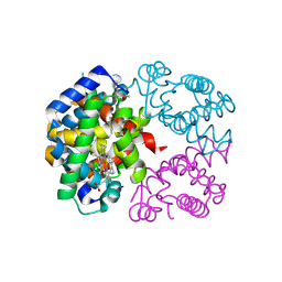
1SKQ
 | | The crystal structure of Sulfolobus solfataricus elongation factor 1-alpha in complex with magnesium and GDP | | 分子名称: | Elongation factor 1-alpha, GUANOSINE-5'-DIPHOSPHATE, MAGNESIUM ION | | 著者 | Vitagliano, L, Ruggiero, A, Masullo, M, Cantiello, P, Arcari, P, Zagari, A. | | 登録日 | 2004-03-05 | | 公開日 | 2004-07-20 | | 最終更新日 | 2023-08-23 | | 実験手法 | X-RAY DIFFRACTION (1.8 Å) | | 主引用文献 | The crystal structure of Sulfolobus solfataricus elongation factor 1alpha in complex with magnesium and GDP.
Biochemistry, 43, 2004
|
|
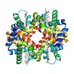
6VO5
 | | Crystal structure of Human histone acetytransferas 1 (HAT1) in complex with isobutryl-COA and K12A mutant variant of histone H4 | | 分子名称: | ACETATE ION, GLYCEROL, Histone H4, ... | | 著者 | Halabelian, L, Zeng, H, Dong, A, Loppnau, P, Bountra, C, Edwards, A.M, Arrowsmith, C.H, Structural Genomics Consortium (SGC) | | 登録日 | 2020-01-30 | | 公開日 | 2020-03-11 | | 実験手法 | X-RAY DIFFRACTION (1.6 Å) | | 主引用文献 | Crystal structure of Human histone acetytransferas 1 (HAT1) in complex with isobutryl-COA and K12A mutant variant of histone H4
to be published
|
|
6WAD
 | | Crystal Structure of Human Protein arginine N-methyltransferase 6 (PRMT6) in complex with MT2739 inhibitor | | 分子名称: | 5-bromo-N-(diphenylmethyl)-N-methylthiophene-2-carboxamide, Protein arginine N-methyltransferase 6, S-ADENOSYL-L-HOMOCYSTEINE | | 著者 | Halabelian, L, Zeng, H, Dong, A, Schapira, M, De Freitas, R.F, Hutchinson, A, Seitova, A, Bountra, C, Edwards, A.M, Arrowsmith, C.H, Brown, P.J, Structural Genomics Consortium (SGC) | | 登録日 | 2020-03-25 | | 公開日 | 2020-09-02 | | 最終更新日 | 2023-10-18 | | 実験手法 | X-RAY DIFFRACTION (2.45 Å) | | 主引用文献 | Crystal Structure of Human Protein arginine N-methyltransferase 6 (PRMT6) in complex with MT2739 inhibitor
to be published
|
|
6WAJ
 | | Crystal structure of the UBL domain of human NLE1 | | 分子名称: | NLE1, UNKNOWN ATOM OR ION | | 著者 | Halabelian, L, Zeng, H, Li, Y, Bountra, C, Edwards, A.M, Arrowsmith, C.H, Structural Genomics Consortium (SGC) | | 登録日 | 2020-03-25 | | 公開日 | 2020-04-22 | | 最終更新日 | 2023-10-18 | | 実験手法 | X-RAY DIFFRACTION (1.9 Å) | | 主引用文献 | Crystal structure of the UBL domain of human NLE1
To be Published
|
|
7WM2
 | | Cryo-EM structure of AKT1 | | 分子名称: | (2S)-3-(hexadecanoyloxy)-2-[(9Z)-octadec-9-enoyloxy]propyl 2-(trimethylammonio)ethyl phosphate, POTASSIUM ION, Potassium channel AKT1 | | 著者 | Dongliang, L, Zijie, Z, Yannan, Q, Yuyue, T, Huaizong, S. | | 登録日 | 2022-01-14 | | 公開日 | 2023-01-25 | | 最終更新日 | 2024-06-26 | | 実験手法 | ELECTRON MICROSCOPY (2.69 Å) | | 主引用文献 | Structural basis for the regulation mechanism of a hyperpolarization-activated K+ channel AKT1 by AtKC1
To Be Published
|
|
7WM1
 | | Cryo-EM structure of AKT1-AtKC1 | | 分子名称: | (2S)-3-(hexadecanoyloxy)-2-[(9Z)-octadec-9-enoyloxy]propyl 2-(trimethylammonio)ethyl phosphate, POTASSIUM ION, Potassium channel AKT1, ... | | 著者 | Dongliang, L, Zijie, Z, Yannan, Q, Yuyue, T, Huaizong, S. | | 登録日 | 2022-01-14 | | 公開日 | 2023-01-25 | | 最終更新日 | 2024-06-26 | | 実験手法 | ELECTRON MICROSCOPY (2.8 Å) | | 主引用文献 | Structural basis for the regulation mechanism of a hyper polarization-activated K+ channel AKT1 by AtKC1
To Be Published
|
|
4RA3
 | | Crystal structure of dimeric S33C beta-2 microglobulin mutant in complex with Thioflavin (ThT) at 2.8 Angstrom resolution | | 分子名称: | 2-[4-(dimethylamino)phenyl]-3,6-dimethyl-1,3-benzothiazol-3-ium, Beta-2-microglobulin | | 著者 | Halabelian, L, Bolognesi, M, Ricagno, S. | | 登録日 | 2014-09-09 | | 公開日 | 2015-09-09 | | 最終更新日 | 2023-09-20 | | 実験手法 | X-RAY DIFFRACTION (2.8 Å) | | 主引用文献 | A covalent homodimer probing early oligomers along amyloid aggregation.
Sci Rep, 5, 2015
|
|
4R9H
 | |
4RAH
 | |
4RZK
 | | Crystal structure of Sulfolobus solfataricus Hsp20.1 ACD | | 分子名称: | Small heat shock protein hsp20 family | | 著者 | Liang, L, Yun, C.H. | | 登録日 | 2014-12-22 | | 公開日 | 2015-12-23 | | 最終更新日 | 2024-02-28 | | 実験手法 | X-RAY DIFFRACTION (2.677 Å) | | 主引用文献 | Crystal structure and function of an unusual dimeric Hsp20.1 provide insight into the thermal protection mechanism of small heat shock proteins.
Biochem.Biophys.Res.Commun., 2015
|
|
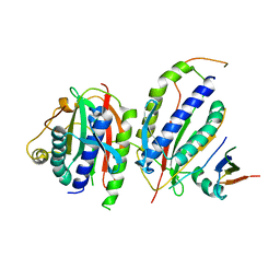
5WCJ
 | | Crystal Structure of Human Methyltransferase-like protein 13 in complex with SAH | | 分子名称: | Methyltransferase-like protein 13, S-ADENOSYL-L-HOMOCYSTEINE, UNKNOWN ATOM OR ION | | 著者 | Halabelian, L, Loppnau, P, Seitova, A, Hutchinson, A, Hunt, B, Dong, A, Bountra, C, Edwards, A.M, Arrowsmith, C.H, Structural Genomics Consortium (SGC) | | 登録日 | 2017-06-30 | | 公開日 | 2017-07-19 | | 最終更新日 | 2023-10-04 | | 実験手法 | X-RAY DIFFRACTION (1.7 Å) | | 主引用文献 | The dual methyltransferase METTL13 targets N terminus and Lys55 of eEF1A and modulates codon-specific translation rates.
Nat Commun, 9, 2018
|
|
6SVF
 | | Crystal structure of the P235GK mutant of ArgBP from T. maritima | | 分子名称: | ARGININE, Amino acid ABC transporter, periplasmic amino acid-binding protein | | 著者 | Vitagliano, L, Berisio, R, Esposito, L, Balasco, N, Smaldone, G, Ruggiero, A. | | 登録日 | 2019-09-18 | | 公開日 | 2019-11-20 | | 最終更新日 | 2024-01-24 | | 実験手法 | X-RAY DIFFRACTION (1.6 Å) | | 主引用文献 | The non-swapped monomeric structure of the arginine-binding protein from Thermotoga maritima.
Acta Crystallogr.,Sect.F, 75, 2019
|
|


