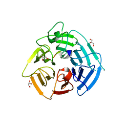
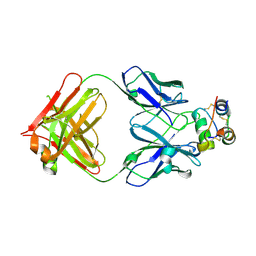
5O6C
 
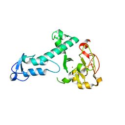 | | Crystal Structure of a threonine-selective RCR E3 ligase | | 分子名称: | E3 ubiquitin-protein ligase MYCBP2, ZINC ION | | 著者 | Pao, K.-C, Rafie, K.Z, van Aalten, D, Virdee, S. | | 登録日 | 2017-06-06 | | 公開日 | 2018-04-18 | | 最終更新日 | 2024-05-08 | | 実験手法 | X-RAY DIFFRACTION (1.75 Å) | | 主引用文献 | Activity-based E3 ligase profiling uncovers an E3 ligase with esterification activity.
Nature, 556, 2018
|
|
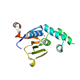
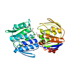
1HTT
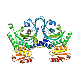 | | HISTIDYL-TRNA SYNTHETASE | | 分子名称: | ADENOSINE MONOPHOSPHATE, HISTIDINE, HISTIDYL-TRNA SYNTHETASE | | 著者 | Arnez, J.G, Harris, D.C, Mitschler, A, Rees, B, Francklyn, C.S, Moras, D. | | 登録日 | 1996-03-09 | | 公開日 | 1997-01-27 | | 最終更新日 | 2024-02-07 | | 実験手法 | X-RAY DIFFRACTION (2.6 Å) | | 主引用文献 | Crystal structure of histidyl-tRNA synthetase from Escherichia coli complexed with histidyl-adenylate.
EMBO J., 14, 1995
|
|
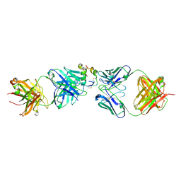
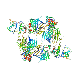
1J9M
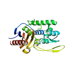 | | K38H mutant of Streptomyces K15 DD-transpeptidase | | 分子名称: | CHLORIDE ION, DD-transpeptidase, SODIUM ION | | 著者 | Fonze, E, Rhazi, N, Nguyen-Disteche, M, Charlier, P. | | 登録日 | 2001-05-28 | | 公開日 | 2001-06-13 | | 最終更新日 | 2024-02-07 | | 実験手法 | X-RAY DIFFRACTION (1.65 Å) | | 主引用文献 | Catalytic mechanism of the Streptomyces K15 DD-transpeptidase/penicillin-binding protein probed by site-directed mutagenesis and structural analysis.
Biochemistry, 42, 2003
|
|
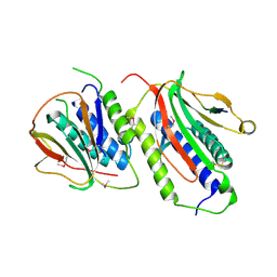
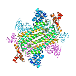
8BEO
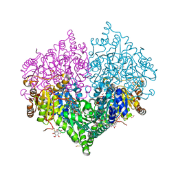 | | Crystal structure of E. coli glyoxylate carboligase mutant I393A with MAP | | 分子名称: | (2R,3S)-1,4-DIMERCAPTOBUTANE-2,3-DIOL, 2,3-DIHYDROXY-1,4-DITHIOBUTANE, 2,3-DIMETHOXY-5-METHYL-1,4-BENZOQUINONE, ... | | 著者 | Shaanan, B, Binshtein, E. | | 登録日 | 2022-10-21 | | 公開日 | 2023-11-08 | | 最終更新日 | 2023-11-15 | | 実験手法 | X-RAY DIFFRACTION (1.96 Å) | | 主引用文献 | Crystal structure of E. coli glyoxylate carboligase mutant I393A with MAP
To Be Published
|
|
7QV1
 | |
7QV2
 | |
7QV3
 | |
8SGE
 | | KLHDC2 Kelch Domain with ligand KDRLKZ-1 | | 分子名称: | GLYCEROL, Kelch domain-containing protein 2, [(5P)-5-{3-[(2R)-butan-2-yl]-7-[(2-methoxyethoxy)carbonyl]-2-oxo-5,6,7,8-tetrahydro-1,7-naphthyridin-1(2H)-yl}-2-oxopyridin-1(2H)-yl]acetic acid | | 著者 | Digianantonio, K.M, Bekes, M, Langley, D.R, Zimmerman, K. | | 登録日 | 2023-04-12 | | 公開日 | 2024-01-03 | | 最終更新日 | 2024-02-28 | | 実験手法 | X-RAY DIFFRACTION (1.509 Å) | | 主引用文献 | Co-opting the E3 ligase KLHDC2 for targeted protein degradation by small molecules.
Nat.Struct.Mol.Biol., 31, 2024
|
|
8SGF
 | | KLHDC2 Kelch Domain with KLHDC2 c-terminal peptide bound | | 分子名称: | GLYCEROL, HIS-SER-VAL-ASN-GLN-ARG-PHE-GLY-SER-ASN-ASN-THR-SER-GLY-SER, Kelch domain-containing protein 2 | | 著者 | Digianantonio, K.M, Bekes, M, Langley, D.R, Zimmerman, K. | | 登録日 | 2023-04-12 | | 公開日 | 2024-01-03 | | 最終更新日 | 2024-02-28 | | 実験手法 | X-RAY DIFFRACTION (1.418 Å) | | 主引用文献 | Co-opting the E3 ligase KLHDC2 for targeted protein degradation by small molecules.
Nat.Struct.Mol.Biol., 31, 2024
|
|
6W9O
 | |
6W9R
 | |
8ONK
 | |
2QYF
 | |
8ONI
 | | Human insulin in complex with the analytical antibody S1 Fab | | 分子名称: | 2-acetamido-2-deoxy-beta-D-glucopyranose, Heavy chain of analytical antibody S1 Fab, Insulin A chain, ... | | 著者 | Johansson, E. | | 登録日 | 2023-04-03 | | 公開日 | 2024-04-10 | | 実験手法 | X-RAY DIFFRACTION (2.3 Å) | | 主引用文献 | Macromolecular structure of insulin and IgE clonality impacts IgE-mediated activation of sensitized basophils
To Be Published
|
|
3ZH4
 | |
8SH2
 | | KLHDC2 in complex with EloB and EloC | | 分子名称: | Elongin-B, Elongin-C, Kelch domain-containing protein 2 | | 著者 | Digianantonio, K.M, Bekes, M. | | 登録日 | 2023-04-13 | | 公開日 | 2024-01-03 | | 最終更新日 | 2024-02-28 | | 実験手法 | ELECTRON MICROSCOPY (3.74 Å) | | 主引用文献 | Co-opting the E3 ligase KLHDC2 for targeted protein degradation by small molecules.
Nat.Struct.Mol.Biol., 31, 2024
|
|
7LUB
 | |
7MBH
 | | Structure of Human Enolase 2 in complex with phosphoserine | | 分子名称: | 1,2-ETHANEDIOL, ACETATE ION, Gamma-enolase, ... | | 著者 | Leonard, P.G, Hicks, K.G, Rutter, J. | | 登録日 | 2021-03-31 | | 公開日 | 2022-11-09 | | 最終更新日 | 2023-10-25 | | 実験手法 | X-RAY DIFFRACTION (2.1 Å) | | 主引用文献 | Protein-metabolite interactomics of carbohydrate metabolism reveal regulation of lactate dehydrogenase.
Science, 379, 2023
|
|
7NB0
 | |
5FGK
 | | CDK8-CYCC IN COMPLEX WITH 8-[3-(3-Amino-1H-indazol-6-yl)-5-chloro- pyridine-4-yl]-2,8-diaza-spiro[4.5]decan-1-one | | 分子名称: | 1,2-ETHANEDIOL, 8-[3-(3-azanyl-2~{H}-indazol-6-yl)-5-chloranyl-pyridin-4-yl]-2,8-diazaspiro[4.5]decan-1-one, Cyclin-C, ... | | 著者 | Musil, D, Blagg, J, Mallinger, A. | | 登録日 | 2015-12-20 | | 公開日 | 2016-02-03 | | 最終更新日 | 2024-05-08 | | 実験手法 | X-RAY DIFFRACTION (2.36 Å) | | 主引用文献 | Discovery of Potent, Selective, and Orally Bioavailable Small-Molecule Modulators of the Mediator Complex-Associated Kinases CDK8 and CDK19.
J.Med.Chem., 59, 2016
|
|
4DZO
 | |
6FK0
 | |
8RPH
 | | JanthE from Janthinobacterium sp. HH01,ketobutyryl-ThDP | | 分子名称: | (2~{S})-2-[3-[(4-azanyl-2-methyl-pyrimidin-5-yl)methyl]-4-methyl-5-[2-[oxidanyl(phosphonooxy)phosphoryl]oxyethyl]-1,3-thiazol-2-yl]-2-oxidanyl-butanoic acid, FLAVIN-ADENINE DINUCLEOTIDE, MAGNESIUM ION, ... | | 著者 | Lanza, L, Leogrande, C, Rabe von Pappenheim, F, Tittmann, K, Mueller, M. | | 登録日 | 2024-01-16 | | 公開日 | 2024-06-12 | | 最終更新日 | 2024-06-19 | | 実験手法 | X-RAY DIFFRACTION (2.96 Å) | | 主引用文献 | JhE from Janthinobacterium sp. HH01
To Be Published
|
|
8RPJ
 | | JanthE from Janthinobacterium sp. HH01 | | 分子名称: | ACETATE ION, FLAVIN-ADENINE DINUCLEOTIDE, GLYCEROL, ... | | 著者 | Lanza, L, Leogrande, C, Rabe von Pappenheim, F, Tittmann, K, Mueller, M. | | 登録日 | 2024-01-16 | | 公開日 | 2024-06-12 | | 最終更新日 | 2024-06-19 | | 実験手法 | X-RAY DIFFRACTION (1.9 Å) | | 主引用文献 | JhE from Janthinobacterium sp. HH01
To Be Published
|
|
8RPI
 | | JanthE from Janthinobacterium sp. HH01, lactyl-ThDP | | 分子名称: | 3-[(4-AMINO-2-METHYLPYRIMIDIN-5-YL)METHYL]-2-(1-CARBOXY-1-HYDROXYETHYL)-5-(2-{[HYDROXY(PHOSPHONOOXY)PHOSPHORYL]OXY}ETHYL)-4-METHYL-1,3-THIAZOL-3-IUM, FLAVIN-ADENINE DINUCLEOTIDE, MAGNESIUM ION, ... | | 著者 | Lanza, L, Leogrande, C, Rabe von Pappenheim, F, Tittmann, K, Mueller, M. | | 登録日 | 2024-01-16 | | 公開日 | 2024-06-12 | | 最終更新日 | 2024-06-19 | | 実験手法 | X-RAY DIFFRACTION (2.71 Å) | | 主引用文献 | JhE from Janthinobacterium sp. HH01
To Be Published
|
|


