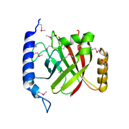
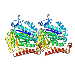
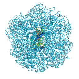
2OC8
 
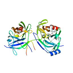 | | Structure of Hepatitis C Viral NS3 protease domain complexed with NS4A peptide and ketoamide SCH503034 | | 分子名称: | BETA-MERCAPTOETHANOL, Hepatitis C virus, ZINC ION, ... | | 著者 | Prongay, A.J, Guo, Z, Yao, N, Fischmann, T, Strickland, C, Myers, J, Weber, P.C, Malcolm, B, Beyer, B.M, Ingram, R, Pichardo, J, Hong, Z, Prosise, W.W, Ramanathan, L, Taremi, S.S, Yarosh-Tomaine, T, Zhang, R, Senior, M, Yang, R.S, Arasappan, A, Bennett, F, Bogen, S.L, Chen, K, Jao, E, Liu, Y.T, Lovey, R.G, Saksena, A.K, Venkatraman, S, Girijavallabhan, V, Njoroge, F.G, Madison, V. | | 登録日 | 2006-12-20 | | 公開日 | 2007-07-31 | | 最終更新日 | 2023-08-30 | | 実験手法 | X-RAY DIFFRACTION (2.66 Å) | | 主引用文献 | Discovery of the HCV NS3/4A protease inhibitor (1R,5S)-N-[3-amino-1-(cyclobutylmethyl)-2,3-dioxopropyl]-3- [2(S)-[[[(1,1-dimethylethyl)amino]carbonyl]amino]-3,3-dimethyl-1-oxobutyl]- 6,6-dimethyl-3-azabicyclo[3.1.0]hexan-2(S)-carboxamide (Sch 503034) II. Key steps in structure-based optimization.
J.Med.Chem., 50, 2007
|
|
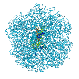
6U42
 | | Natively decorated ciliary doublet microtubule | | 分子名称: | DC1, DC2, DC3, ... | | 著者 | Ma, M, Stoyanova, M, Rademacher, G, Dutcher, S.K, Brown, A, Zhang, R. | | 登録日 | 2019-08-22 | | 公開日 | 2019-11-13 | | 最終更新日 | 2020-01-08 | | 実験手法 | ELECTRON MICROSCOPY (3.4 Å) | | 主引用文献 | Structure of the Decorated Ciliary Doublet Microtubule.
Cell, 179, 2019
|
|
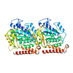
2I5U
 | | Crystal structure of DnaD domain protein from Enterococcus faecalis. Structural genomics target APC85179 | | 分子名称: | DnaD domain protein, MAGNESIUM ION | | 著者 | Wu, R, Zhang, R, Bargassa, M, Joachimiak, A, Midwest Center for Structural Genomics (MCSG) | | 登録日 | 2006-08-25 | | 公開日 | 2006-11-07 | | 最終更新日 | 2011-07-13 | | 実験手法 | X-RAY DIFFRACTION (1.5 Å) | | 主引用文献 | 1.5 A crystal structure of a DnaD domain protein from Enterococcus Faecalis
To be Published, 2006
|
|
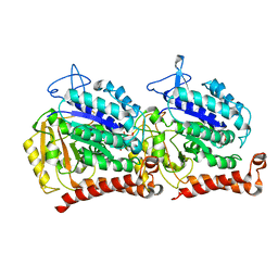
3USB
 | | Crystal Structure of Bacillus anthracis Inosine Monophosphate Dehydrogenase in the complex with IMP | | 分子名称: | CHLORIDE ION, GLYCEROL, INOSINIC ACID, ... | | 著者 | Kim, Y, Zhang, R, Wu, R, Gu, M, Anderson, W.F, Joachimiak, A, CSGID, Center for Structural Genomics of Infectious Diseases (CSGID) | | 登録日 | 2011-11-23 | | 公開日 | 2011-12-07 | | 最終更新日 | 2019-08-14 | | 実験手法 | X-RAY DIFFRACTION (2.38 Å) | | 主引用文献 | Bacillus anthracis inosine 5'-monophosphate dehydrogenase in action: the first bacterial series of structures of phosphate ion-, substrate-, and product-bound complexes.
Biochemistry, 51, 2012
|
|
2OQW
 | | The crystal structure of sortase B from B.anthracis in complex with AAEK1 | | 分子名称: | Sortase B | | 著者 | Wu, R, Zhang, R, Marresso, A.W, Schneewind, O, Joachimiak, A. | | 登録日 | 2007-02-01 | | 公開日 | 2007-06-19 | | 最終更新日 | 2023-12-27 | | 実験手法 | X-RAY DIFFRACTION (2.1 Å) | | 主引用文献 | Activation of inhibitors by sortase triggers irreversible modification of the active site.
J.Biol.Chem., 282, 2007
|
|
2OQZ
 | | The crystal structure of sortase B from B.anthracis in complex with AAEK2 | | 分子名称: | ACETIC ACID, Sortase B | | 著者 | Wu, R, Zhang, R, Maresso, A.W, Schneewind, O, Joachimiak, A. | | 登録日 | 2007-02-01 | | 公開日 | 2007-06-19 | | 最終更新日 | 2018-01-24 | | 実験手法 | X-RAY DIFFRACTION (1.6 Å) | | 主引用文献 | Activation of inhibitors by sortase triggers irreversible modification of the active site.
J.Biol.Chem., 282, 2007
|
|
2PPW
 | |
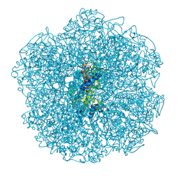
8OTZ
 | | 48-nm repeat of the native axonemal doublet microtubule from bovine sperm | | 分子名称: | ATP6V1F neighbor, CFAP97 domain containing 1, Chromosome 13 C20orf85 homolog, ... | | 著者 | Leung, M.R, Zeng, J, Zhang, R, Zeev-Ben-Mordehai, T. | | 登録日 | 2023-04-21 | | 公開日 | 2023-11-22 | | 実験手法 | ELECTRON MICROSCOPY (3.6 Å) | | 主引用文献 | Structural specializations of the sperm tail.
Cell, 186, 2023
|
|
3CM8
 | | A RNA polymerase subunit structure from virus | | 分子名称: | Polymerase acidic protein, peptide from RNA-directed RNA polymerase catalytic subunit | | 著者 | He, X, Zhou, J, Zeng, Z, Ma, J, Zhang, R, Rao, Z, Liu, Y. | | 登録日 | 2008-03-21 | | 公開日 | 2008-07-15 | | 最終更新日 | 2024-03-13 | | 実験手法 | X-RAY DIFFRACTION (2.899 Å) | | 主引用文献 | Crystal structure of the polymerase PAC-PB1N complex from an avian influenza H5N1 virus
Nature, 454, 2008
|
|
8JMY
 | |
2G5C
 | | Crystal Structure of Prephenate Dehydrogenase from Aquifex aeolicus | | 分子名称: | NICOTINAMIDE-ADENINE-DINUCLEOTIDE, prephenate dehydrogenase | | 著者 | Sun, W, Singh, S, Zhang, R, Turnbull, J.L, Christendat, D. | | 登録日 | 2006-02-22 | | 公開日 | 2006-03-07 | | 最終更新日 | 2011-07-13 | | 実験手法 | X-RAY DIFFRACTION (1.9 Å) | | 主引用文献 | Crystal Structure of Prephenate Dehydrogenase from Aquifex aeolicus: Insights into the Catalytic Mechanism
J.Biol.Chem., 281, 2006
|
|
2HMC
 | | The Crystal Structure of Dihydrodipicolinate Synthase DapA from Agrobacterium tumefaciens | | 分子名称: | Dihydrodipicolinate synthase, MAGNESIUM ION | | 著者 | Kim, Y, Zhang, R, Xu, X, Zheng, H, Savchenko, A, Joachimiak, A, Midwest Center for Structural Genomics (MCSG) | | 登録日 | 2006-07-11 | | 公開日 | 2006-09-05 | | 最終更新日 | 2011-07-13 | | 実験手法 | X-RAY DIFFRACTION (1.9 Å) | | 主引用文献 | The Crystal Structure of Dihydrodipicolinate Synthase DapA from Agrobacterium tumefaciens
To be Published, 2006
|
|
5W3H
 | | Yeast microtubule stabilized with epothilone | | 分子名称: | EPOTHILONE A, GUANOSINE-5'-DIPHOSPHATE, GUANOSINE-5'-TRIPHOSPHATE, ... | | 著者 | Howes, S.C, Geyer, E.A, LaFrance, B, Zhang, R, Kellogg, E.H, Westermann, S, Rice, L.M, Nogales, E. | | 登録日 | 2017-06-07 | | 公開日 | 2017-07-19 | | 最終更新日 | 2024-03-13 | | 実験手法 | ELECTRON MICROSCOPY (4 Å) | | 主引用文献 | Structural differences between yeast and mammalian microtubules revealed by cryo-EM.
J. Cell Biol., 216, 2017
|
|
5W3J
 | | Yeast microtubule stabilized with Taxol assembled from mutated tubulin | | 分子名称: | GUANOSINE-5'-DIPHOSPHATE, GUANOSINE-5'-TRIPHOSPHATE, MAGNESIUM ION, ... | | 著者 | Howes, S.C, Geyer, E.A, LaFrance, B, Zhang, R, Kellogg, E.H, Westermann, S, Rice, L.M, Nogales, E. | | 登録日 | 2017-06-07 | | 公開日 | 2017-07-19 | | 最終更新日 | 2024-03-13 | | 実験手法 | ELECTRON MICROSCOPY (4 Å) | | 主引用文献 | Structural differences between yeast and mammalian microtubules revealed by cryo-EM.
J. Cell Biol., 216, 2017
|
|
5W3F
 | | Yeast tubulin polymerized with GTP in vitro | | 分子名称: | GUANOSINE-5'-DIPHOSPHATE, GUANOSINE-5'-TRIPHOSPHATE, MAGNESIUM ION, ... | | 著者 | Howes, S.C, Geyer, E.A, LaFrance, B, Zhang, R, Kellogg, E.H, Westermann, S, Rice, L.M, Nogales, E. | | 登録日 | 2017-06-07 | | 公開日 | 2017-07-19 | | 最終更新日 | 2024-03-13 | | 実験手法 | ELECTRON MICROSCOPY (3.7 Å) | | 主引用文献 | Structural differences between yeast and mammalian microtubules revealed by cryo-EM.
J. Cell Biol., 216, 2017
|
|
5WTL
 | | Crystal structure of the periplasmic portion of outer membrane protein A (OmpA) from Capnocytophaga gingivalis | | 分子名称: | 2-(N-MORPHOLINO)-ETHANESULFONIC ACID, CALCIUM ION, OmpA family protein, ... | | 著者 | Dai, S, Tan, K, Ye, S, Zhang, R. | | 登録日 | 2016-12-13 | | 公開日 | 2017-12-13 | | 最終更新日 | 2023-11-08 | | 実験手法 | X-RAY DIFFRACTION (2.298 Å) | | 主引用文献 | Structure of thrombospondin type 3 repeats in bacterial outer membrane protein A reveals its intra-repeat disulfide bond-dependent calcium-binding capability.
Cell Calcium, 66, 2017
|
|
5WTP
 | | Crystal structure of the C-terminal domain of outer membrane protein A (OmpA) from Capnocytophaga gingivalis | | 分子名称: | OmpA family protein, SULFATE ION | | 著者 | Dai, S, Tan, K, Ye, S, Zhang, R. | | 登録日 | 2016-12-13 | | 公開日 | 2017-12-13 | | 最終更新日 | 2023-11-08 | | 実験手法 | X-RAY DIFFRACTION (2.15 Å) | | 主引用文献 | Structure of thrombospondin type 3 repeats in bacterial outer membrane protein A reveals its intra-repeat disulfide bond-dependent calcium-binding capability.
Cell Calcium, 66, 2017
|
|
3DOB
 | | Peptide-binding domain of Heat shock 70 kDa protein F44E5.5 from C.elegans. | | 分子名称: | BETA-MERCAPTOETHANOL, Heat shock 70 kDa protein F44E5.5 | | 著者 | Osipiuk, J, Hatzos, C, Gu, M, Zhang, R, Voisine, C, Morimoto, R.I, Joachimiak, A, Midwest Center for Structural Genomics (MCSG) | | 登録日 | 2008-07-03 | | 公開日 | 2008-07-22 | | 最終更新日 | 2023-08-30 | | 実験手法 | X-RAY DIFFRACTION (2.39 Å) | | 主引用文献 | X-ray crystal structure of Peptide-binding domain of Heat shock 70 kDa protein F44E5.5 from C.elegans.
To be Published
|
|
3ECR
 | | Structure of human porphobilinogen deaminase | | 分子名称: | 3-[5-{[3-(2-carboxyethyl)-4-(carboxymethyl)-5-methyl-1H-pyrrol-2-yl]methyl}-4-(carboxymethyl)-1H-pyrrol-3-yl]propanoic acid, Porphobilinogen deaminase | | 著者 | Song, G, Li, Y, Cheng, C, Zhao, Y, Gao, A, Zhang, R, Joachimiak, A, Shaw, N, Liu, Z.J. | | 登録日 | 2008-09-01 | | 公開日 | 2008-09-30 | | 最終更新日 | 2024-03-20 | | 実験手法 | X-RAY DIFFRACTION (2.182 Å) | | 主引用文献 | Structural insight into acute intermittent porphyria.
Faseb J., 23, 2009
|
|
9B4H
 | | Chlamydomonas reinhardtii mastigoneme filament | | 分子名称: | 2-acetamido-2-deoxy-beta-D-glucopyranose-(1-4)-2-acetamido-2-deoxy-beta-D-glucopyranose, C-type lectin domain-containing protein, Tyrosine-protein kinase ephrin type A/B receptor-like domain-containing protein, ... | | 著者 | Dai, J, Ma, M, Zhang, R, Brown, A. | | 登録日 | 2024-03-20 | | 公開日 | 2024-04-10 | | 実験手法 | ELECTRON MICROSCOPY (3.1 Å) | | 主引用文献 | Mastigoneme structure reveals insights into the O-linked glycosylation code of native hydroxyproline-rich helices.
Cell, 2024
|
|
3D6K
 | | The crystal structure of a putative aminotransferase from Corynebacterium diphtheriae | | 分子名称: | 1,2-ETHANEDIOL, CHLORIDE ION, Putative aminotransferase, ... | | 著者 | Tan, K, Zhang, R, Duggan, E, Clancy, S, Joachimiak, A, Midwest Center for Structural Genomics (MCSG) | | 登録日 | 2008-05-19 | | 公開日 | 2008-07-15 | | 最終更新日 | 2011-07-13 | | 実験手法 | X-RAY DIFFRACTION (2 Å) | | 主引用文献 | The crystal structure of a putative aminotransferase from Corynebacterium diphtheriae
To be Published
|
|
3CQV
 | | Crystal structure of Reverb beta in complex with heme | | 分子名称: | Nuclear receptor subfamily 1 group D member 2, PROTOPORPHYRIN IX CONTAINING FE | | 著者 | Xu, X, Dong, A, Pardee, K.I, Reinking, J, Krause, H, Schuetz, A, Zhang, R, Cui, H, Edwards, A, Arrowsmith, C.H, Weigelt, J, Bountra, C, Savchenko, A, Botchkarev, A, Structural Genomics Consortium (SGC) | | 登録日 | 2008-04-03 | | 公開日 | 2008-08-05 | | 最終更新日 | 2011-07-13 | | 実験手法 | X-RAY DIFFRACTION (1.9 Å) | | 主引用文献 | The structural basis of gas-responsive transcription by the human nuclear hormone receptor REV-ERBbeta.
Plos Biol., 7, 2009
|
|
8Q20
 | | Crystal structure of Vanadium-dependent haloperoxidase R425D mutant (A. marina) | | 分子名称: | PHOSPHATE ION, SULFATE ION, Vanadium-dependent bromoperoxidase, ... | | 著者 | Zeides, P, Bellmannn-Sickert, K, Zhang, R, Seel, C.J, Most, V, Schroeder, C.T, Groll, M, Gulder, T. | | 登録日 | 2023-08-01 | | 公開日 | 2024-08-14 | | 実験手法 | X-RAY DIFFRACTION (3.5 Å) | | 主引用文献 | Unraveling the Molecular Basis of Substrate Specificity and Halogen Activation in Vanadium-Dependent Haloperoxidases
to be published
|
|
8Q21
 | | Crystal structure of Vanadium-dependent haloperoxidase R425S mutant (A. marina) | | 分子名称: | PHOSPHATE ION, SODIUM ION, Vanadium-dependent bromoperoxidase, ... | | 著者 | Zeides, P, Bellmannn-Sickert, K, Zhang, R, Seel, C.J, Most, V, Schroeder, C.T, Groll, M, Gulder, T. | | 登録日 | 2023-08-01 | | 公開日 | 2024-08-14 | | 実験手法 | X-RAY DIFFRACTION (3.3 Å) | | 主引用文献 | Unraveling the Molecular Basis of Substrate Specificity and Halogen Activation in Vanadium-Dependent Haloperoxidases
to be published
|
|
8Q22
 | | Crystal structure of Vanadium-dependent haloperoxidase R425S mutant in complex with 1,3,5-trimethoxybenzene (A. marina) | | 分子名称: | 1,3,5-trimethoxybenzene, PHOSPHATE ION, SODIUM ION, ... | | 著者 | Zeides, P, Bellmannn-Sickert, K, Zhang, R, Seel, C.J, Most, V, Schroeder, C.T, Groll, M, Gulder, T. | | 登録日 | 2023-08-01 | | 公開日 | 2024-08-14 | | 実験手法 | X-RAY DIFFRACTION (3.3 Å) | | 主引用文献 | Unraveling the Molecular Basis of Substrate Specificity and Halogen Activation in Vanadium-Dependent Haloperoxidases
to be published
|
|


