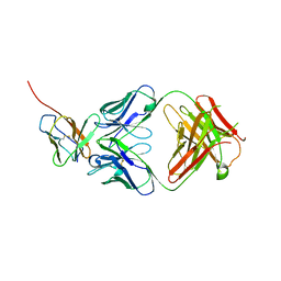
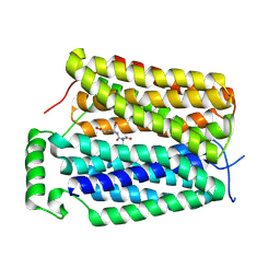
4OU7
 
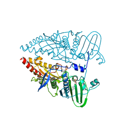
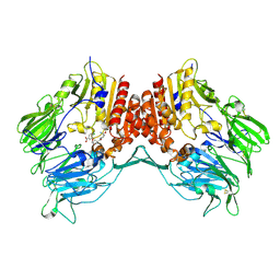
 | | Crystal structure of DnaT84-153-dT10 ssDNA complex reveals a novel single-stranded DNA binding mode | | 分子名称: | DNA (5'-D(P*TP*TP*TP*TP*TP*TP*TP*TP*TP*T)-3'), Primosomal protein 1 | | 著者 | Liu, Z, Chen, P, Niu, L, Teng, M, Li, X. | | 登録日 | 2014-02-15 | | 公開日 | 2014-08-13 | | 最終更新日 | 2024-05-29 | | 実験手法 | X-RAY DIFFRACTION (2.83 Å) | | 主引用文献 | Crystal structure of DnaT84-153-dT10 ssDNA complex reveals a novel single-stranded DNA binding mode.
Nucleic Acids Res., 42, 2014
|
|
8KGT
 | |
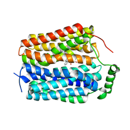
3HVN
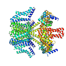
 | | Crystal structure of cytotoxin protein suilysin from Streptococcus suis | | 分子名称: | 1,1,1,3,3,3-hexafluoropropan-2-ol, HEPTANE-1,2,3-TRIOL, Hemolysin | | 著者 | Xu, L, Huang, B, Du, H, Zhang, C.X, Xu, J, Li, X, Rao, Z. | | 登録日 | 2009-06-16 | | 公開日 | 2010-03-02 | | 最終更新日 | 2024-05-29 | | 実験手法 | X-RAY DIFFRACTION (2.852 Å) | | 主引用文献 | Crystal structure of cytotoxin protein suilysin from Streptococcus suis.
Protein Cell, 1, 2010
|
|
4JGY
 | | Crystal structure of human coxsackievirus A16 uncoating intermediate (space group P4232) | | 分子名称: | Polyprotein, capsid protein VP1, capsid protein VP2, ... | | 著者 | Ren, J, Wang, X, Hu, Z, Gao, Q, Sun, Y, Li, X, Porta, C, Walter, T.S, Gilbert, R.J, Zhao, Y, Axford, D, Williams, M, Mcauley, K, Rowlands, D.J, Yin, W, Wang, J, Stuart, D.I, Rao, Z, Fry, E.E. | | 登録日 | 2013-03-04 | | 公開日 | 2013-06-05 | | 最終更新日 | 2023-09-20 | | 実験手法 | X-RAY DIFFRACTION (3 Å) | | 主引用文献 | Picornavirus uncoating intermediate captured in atomic detail.
Nat Commun, 4, 2013
|
|
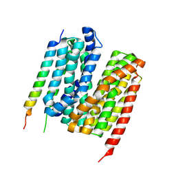
6AYF
 | | TRPML3/ML-SA1 complex at pH 7.4 | | 分子名称: | 2-acetamido-2-deoxy-beta-D-glucopyranose, Mucolipin-3 | | 著者 | Zhou, X, Li, M, Su, D, Jia, Q, Li, H, Li, X, Yang, J. | | 登録日 | 2017-09-08 | | 公開日 | 2017-11-08 | | 最終更新日 | 2024-03-13 | | 実験手法 | ELECTRON MICROSCOPY (3.62 Å) | | 主引用文献 | Cryo-EM structures of the human endolysosomal TRPML3 channel in three distinct states.
Nat. Struct. Mol. Biol., 24, 2017
|
|
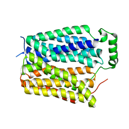
6AYG
 | | Human Apo-TRPML3 channel at pH 4.8 | | 分子名称: | Mucolipin-3 | | 著者 | Zhou, X, Li, M, Su, D, Jia, Q, Li, H, Li, X, Yang, J. | | 登録日 | 2017-09-08 | | 公開日 | 2017-11-08 | | 最終更新日 | 2024-03-13 | | 実験手法 | ELECTRON MICROSCOPY (4.65 Å) | | 主引用文献 | Cryo-EM structures of the human endolysosomal TRPML3 channel in three distinct states.
Nat. Struct. Mol. Biol., 24, 2017
|
|
4JGZ
 | | Crystal structure of human coxsackievirus A16 uncoating intermediate (space group I222) | | 分子名称: | Polyprotein, capsid protein VP1, capsid protein VP2, ... | | 著者 | Ren, J, Wang, X, Hu, Z, Gao, Q, Sun, Y, Li, X, Porta, C, Walter, T.S, Gilbert, R.J, Zhao, Y, Axford, D, Williams, M, McAuley, K, Rowlands, D.J, Yin, W, Wang, J, Stuart, D.I, Rao, Z, Fry, E.E. | | 登録日 | 2013-03-04 | | 公開日 | 2013-06-05 | | 最終更新日 | 2023-09-20 | | 実験手法 | X-RAY DIFFRACTION (3 Å) | | 主引用文献 | Picornavirus uncoating intermediate captured in atomic detail.
Nat Commun, 4, 2013
|
|
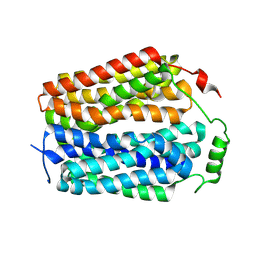
6AYE
 | | Human apo-TRPML3 channel at pH 7.4 | | 分子名称: | Mucolipin-3 | | 著者 | Zhou, X, Li, M, Su, D, Jia, Q, Li, H, Li, X, Yang, J. | | 登録日 | 2017-09-08 | | 公開日 | 2017-11-08 | | 最終更新日 | 2024-03-13 | | 実験手法 | ELECTRON MICROSCOPY (4.06 Å) | | 主引用文献 | Cryo-EM structures of the human endolysosomal TRPML3 channel in three distinct states.
Nat. Struct. Mol. Biol., 24, 2017
|
|
2KBV
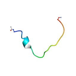
 | | Structural and functional analysis of TM XI of the NHE1 isoform of thE NA+/H+ exchanger | | 分子名称: | Sodium/hydrogen exchanger 1 | | 著者 | Lee, B.L, Li, X, Liu, Y, Sykes, B.D, Fliegel, L. | | 登録日 | 2008-12-09 | | 公開日 | 2009-01-27 | | 最終更新日 | 2023-06-14 | | 実験手法 | SOLUTION NMR | | 主引用文献 | Structural and Functional Analysis of Transmembrane XI of the NHE1 Isoform of the Na+/H+ Exchanger
J.Biol.Chem., 284, 2009
|
|
8W4U
 | | human KCNQ2-CaM in complex with PIP2 and HN37 | | 分子名称: | Calmodulin-1, Potassium voltage-gated channel subfamily KQT member 2, [(2R)-2-octanoyloxy-3-[oxidanyl-[(1R,2R,3S,4R,5R,6S)-2,3,6-tris(oxidanyl)-4,5-diphosphonooxy-cyclohexyl]oxy-phosphoryl]oxy-propyl] octanoate, ... | | 著者 | Ma, D, Li, X, Guo, J. | | 登録日 | 2023-08-25 | | 公開日 | 2023-12-13 | | 実験手法 | ELECTRON MICROSCOPY (3.3 Å) | | 主引用文献 | Ligand activation mechanisms of human KCNQ2 channel.
Nat Commun, 14, 2023
|
|
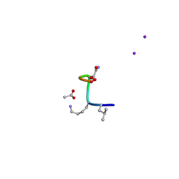
2K3C
 | | Structural and Functional Characterization of TM IX of the NHE1 Isoform of the Na+/H+ Exchanger | | 分子名称: | TMIX peptide | | 著者 | Reddy, T, Ding, J, Li, X, Sykes, B.D, Fliegel, L, Rainey, J.K. | | 登録日 | 2008-05-01 | | 公開日 | 2008-06-03 | | 最終更新日 | 2020-02-19 | | 実験手法 | SOLUTION NMR | | 主引用文献 | Structural and Functional Characterization of Transmembrane Segment IX of the NHE1 Isoform of the Na+/H+ Exchanger.
J.Biol.Chem., 283, 2008
|
|
8CUF
 | | Synthetic epi-Novo29 (2R,3S), X-ray diffractometer structure | | 分子名称: | ACETATE ION, IODIDE ION, Synthetic epi-Novo29 (2R,3S) | | 著者 | Kreutzer, A.G, Li, X, Krumberger, M, Nowick, J.S. | | 登録日 | 2022-05-17 | | 公開日 | 2023-01-11 | | 最終更新日 | 2023-11-15 | | 実験手法 | X-RAY DIFFRACTION (1.68 Å) | | 主引用文献 | Synthesis and Stereochemical Determination of the Peptide Antibiotic Novo29.
J.Org.Chem., 88, 2023
|
|
8CUG
 | | Synthetic epi-Novo29 (2R,3S), synchrotron structure | | 分子名称: | ACETATE ION, Synthetic epi-Novo29 (2R,3S) | | 著者 | Kreutzer, A.G, Li, X, Krumberger, M, Nowick, J.S. | | 登録日 | 2022-05-17 | | 公開日 | 2023-01-11 | | 最終更新日 | 2023-11-15 | | 実験手法 | X-RAY DIFFRACTION (1.131 Å) | | 主引用文献 | Synthesis and Stereochemical Determination of the Peptide Antibiotic Novo29.
J.Org.Chem., 88, 2023
|
|
7Y1Q
 | | 5.0 angstrom cryo-EM structure of transmembrane regions of mouse Basigin/MCT1 in complex with antibody 6E7F1 | | 分子名称: | Isoform 2 of Basigin, Monocarboxylate transporter 1 | | 著者 | Zhang, H, Yang, X, Xue, Y, Huang, Y, Mo, X, Zhang, H, Li, N, Gao, N, Li, X, Wang, S, Gao, Y, Liao, J. | | 登録日 | 2022-06-08 | | 公開日 | 2023-06-14 | | 実験手法 | ELECTRON MICROSCOPY (5.03 Å) | | 主引用文献 | Allosteric modulation of monocarboxylate transporters 1 and 4 by targeting their chaperon Basigin-2
To Be Published
|
|
7Y1B
 | | 3.2 angstrom cryo-EM structure of extracellular region of mouse Basigin-2 in complex with the Fab fragment of antibody 6E7F1 | | 分子名称: | Heavy chain of 6E7F1, Isoform 2 of Basigin, Light chain of 6E7F1 | | 著者 | Zhang, H, Yang, X, Xue, Y, Huang, Y, Mo, X, Zhang, H, Li, N, Gao, N, Li, X, Wang, S, Gao, Y, Liao, J. | | 登録日 | 2022-06-08 | | 公開日 | 2023-06-14 | | 実験手法 | ELECTRON MICROSCOPY (3.23 Å) | | 主引用文献 | Allosteric modulation of monocarboxylate transporters 1 and 4 by targeting their chaperon Basigin
To Be Published
|
|
5ZZ3
 | | Crystal structure of intracellular B30.2 domain of BTN3A3 | | 分子名称: | Butyrophilin, subfamily 3, member A3 isoform b variant | | 著者 | Yang, Y.Y, Li, X, Liu, W.D, Chen, C.C, Guo, R.T, Zhang, Y.H. | | 登録日 | 2018-05-30 | | 公開日 | 2019-04-03 | | 最終更新日 | 2023-11-22 | | 実験手法 | X-RAY DIFFRACTION (3 Å) | | 主引用文献 | A Structural Change in Butyrophilin upon Phosphoantigen Binding Underlies Phosphoantigen-Mediated V gamma 9V delta 2 T Cell Activation.
Immunity, 50, 2019
|
|
8U3E
 | |
8U3D
 | |
8U3G
 | |
8U3F
 | |
8U3H
 | |
2G5P
 | | Crystal structure of human dipeptidyl peptidase IV (DPPIV) complexed with cyanopyrrolidine (C5-pro-pro) inhibitor 21ac | | 分子名称: | 4-{[(2R,5S)-5-{[(2S)-2-(AMINOMETHYL)PYRROLIDIN-1-YL]CARBONYL}PYRROLIDIN-2-YL]METHOXY}-3-TERT-BUTYLBENZOIC ACID, Dipeptidyl peptidase 4 | | 著者 | Longenecker, K.L, Fry, E.H, Lake, M.R, Solomon, L.R, Pei, Z, Li, X. | | 登録日 | 2006-02-23 | | 公開日 | 2006-07-04 | | 最終更新日 | 2017-10-18 | | 実験手法 | X-RAY DIFFRACTION (2.4 Å) | | 主引用文献 | Discovery, structure-activity relationship, and pharmacological evaluation of (5-substituted-pyrrolidinyl-2-carbonyl)-2-cyanopyrrolidines as potent dipeptidyl peptidase IV inhibitors.
J.Med.Chem., 49, 2006
|
|
2G63
 | | Crystal structure of human dipeptidyl peptidase IV (DPPIV) complexed with cyanopyrrolidine (C5-pro-pro) inhibitor 24b | | 分子名称: | Dipeptidyl peptidase 4, METHYL 4-{[({[(2R,5S)-5-{[(2S)-2-(AMINOMETHYL)PYRROLIDIN-1-YL]CARBONYL}PYRROLIDIN-2-YL]METHYL}AMINO)CARBONYL]AMINO}BENZOATE | | 著者 | Longenecker, K.L, Fry, E.H, Lake, M.R, Solomon, L.R, Pei, Z, Li, X. | | 登録日 | 2006-02-24 | | 公開日 | 2006-07-04 | | 最終更新日 | 2017-10-18 | | 実験手法 | X-RAY DIFFRACTION (2 Å) | | 主引用文献 | Discovery, structure-activity relationship, and pharmacological evaluation of (5-substituted-pyrrolidinyl-2-carbonyl)-2-cyanopyrrolidines as potent dipeptidyl peptidase IV inhibitors.
J.Med.Chem., 49, 2006
|
|
2G5T
 | | Crystal structure of human dipeptidyl peptidase IV (DPPIV) complexed with cyanopyrrolidine (C5-pro-pro) inhibitor 21ag | | 分子名称: | 3-{[(2R,5S)-5-{[(2S)-2-(AMINOMETHYL)PYRROLIDIN-1-YL]CARBONYL}PYRROLIDIN-2-YL]METHOXY}-4-CHLOROBENZOIC ACID, Dipeptidyl peptidase 4 | | 著者 | Longenecker, K.L, Fry, E.H, Lake, M.R, Solomon, L.R, Pei, Z, Li, X. | | 登録日 | 2006-02-23 | | 公開日 | 2006-07-04 | | 最終更新日 | 2017-10-18 | | 実験手法 | X-RAY DIFFRACTION (2.3 Å) | | 主引用文献 | Discovery, structure-activity relationship, and pharmacological evaluation of (5-substituted-pyrrolidinyl-2-carbonyl)-2-cyanopyrrolidines as potent dipeptidyl peptidase IV inhibitors.
J.Med.Chem., 49, 2006
|
|
2GVE
 | | Time-of-Flight Neutron Diffraction Structure of D-Xylose Isomerase | | 分子名称: | COBALT (II) ION, Xylose isomerase | | 著者 | Katz, A.K, Li, X, Carrell, H.L, Hanson, B.L, Langan, P, Coates, L, Schoenborn, B.P, Glusker, J.P, Bunick, G.J. | | 登録日 | 2006-05-02 | | 公開日 | 2006-05-16 | | 最終更新日 | 2023-08-30 | | 実験手法 | NEUTRON DIFFRACTION (2.2 Å) | | 主引用文献 | Locating active-site hydrogen atoms in D-xylose isomerase: Time-of-flight neutron diffraction.
Proc.Natl.Acad.Sci.Usa, 103, 2006
|
|


