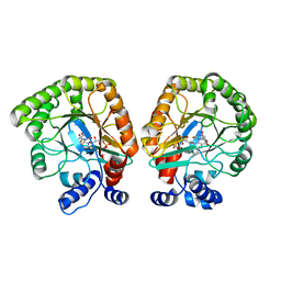
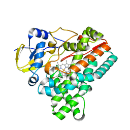
3X2M
 
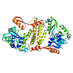
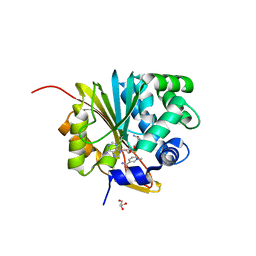
 | | X-ray structure of PcCel45A with cellopentaose at 0.64 angstrom resolution. | | 分子名称: | Endoglucanase V-like protein, beta-D-glucopyranose-(1-4)-beta-D-glucopyranose-(1-4)-beta-D-glucopyranose-(1-4)-beta-D-glucopyranose-(1-4)-alpha-D-glucopyranose, beta-D-glucopyranose-(1-4)-beta-D-glucopyranose-(1-4)-beta-D-glucopyranose-(1-4)-beta-D-glucopyranose-(1-4)-beta-D-glucopyranose | | 著者 | Nakamura, A, Ishida, T, Samejima, M, Igarashi, K. | | 登録日 | 2014-12-22 | | 公開日 | 2015-10-14 | | 最終更新日 | 2020-07-29 | | 実験手法 | X-RAY DIFFRACTION (0.64 Å) | | 主引用文献 | "Newton's cradle" proton relay with amide-imidic acid tautomerization in inverting cellulase visualized by neutron crystallography.
Sci Adv, 1, 2015
|
|
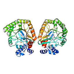
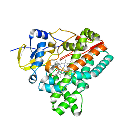
3X2P
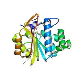
 | | Neutron and X-ray joint refined structure of PcCel45A with cellopentaose at 298K. | | 分子名称: | Endoglucanase V-like protein, beta-D-glucopyranose-(1-4)-beta-D-glucopyranose-(1-4)-beta-D-glucopyranose-(1-4)-beta-D-glucopyranose-(1-4)-beta-D-glucopyranose | | 著者 | Nakamura, A, Ishida, T, Kusaka, K, Yamada, T, Tanaka, I, Niimura, N, Samejima, M, Igarashi, K. | | 登録日 | 2014-12-22 | | 公開日 | 2015-10-14 | | 最終更新日 | 2020-07-29 | | 実験手法 | NEUTRON DIFFRACTION (1.518 Å), X-RAY DIFFRACTION | | 主引用文献 | "Newton's cradle" proton relay with amide-imidic acid tautomerization in inverting cellulase visualized by neutron crystallography.
Sci Adv, 1, 2015
|
|
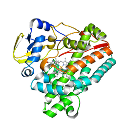
3X2K
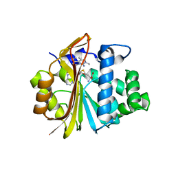
 | | X-ray structure of PcCel45A D114N with cellopentaose at 95K. | | 分子名称: | 2-AMINO-2-HYDROXYMETHYL-PROPANE-1,3-DIOL, 3-methylpentane-1,5-diol, Endoglucanase V-like protein, ... | | 著者 | Nakamura, A, Ishida, T, Samejima, M, Igarashi, K. | | 登録日 | 2014-12-22 | | 公開日 | 2015-10-07 | | 最終更新日 | 2020-07-29 | | 実験手法 | X-RAY DIFFRACTION (1.182 Å) | | 主引用文献 | "Newton's cradle" proton relay with amide-imidic acid tautomerization in inverting cellulase visualized by neutron crystallography.
Sci Adv, 1, 2015
|
|
3X2I
 | | X-ray structure of PcCel45A N92D apo form at 298K. | | 分子名称: | 3-methylpentane-1,5-diol, Endoglucanase V-like protein | | 著者 | Nakamura, A, Ishida, T, Samejima, M, Igarashi, K. | | 登録日 | 2014-12-22 | | 公開日 | 2015-10-07 | | 最終更新日 | 2019-12-18 | | 実験手法 | X-RAY DIFFRACTION (1.6 Å) | | 主引用文献 | "Newton's cradle" proton relay with amide-imidic acid tautomerization in inverting cellulase visualized by neutron crystallography.
Sci Adv, 1, 2015
|
|
3X2H
 | | X-ray structure of PcCel45A N92D with cellopentaose at 95K. | | 分子名称: | 3-methylpentane-1,5-diol, Endoglucanase V-like protein, beta-D-glucopyranose-(1-4)-beta-D-glucopyranose-(1-4)-beta-D-glucopyranose-(1-4)-beta-D-glucopyranose-(1-4)-alpha-D-glucopyranose, ... | | 著者 | Nakamura, A, Ishida, T, Samejima, M, Igarashi, K. | | 登録日 | 2014-12-22 | | 公開日 | 2015-10-07 | | 最終更新日 | 2020-07-29 | | 実験手法 | X-RAY DIFFRACTION (0.99 Å) | | 主引用文献 | "Newton's cradle" proton relay with amide-imidic acid tautomerization in inverting cellulase visualized by neutron crystallography.
Sci Adv, 1, 2015
|
|
3X2J
 | | X-ray structure of PcCel45A D114N apo form at 95K. | | 分子名称: | 3-methylpentane-1,5-diol, Endoglucanase V-like protein | | 著者 | Nakamura, A, Ishida, T, Samejima, M, Igarashi, K. | | 登録日 | 2014-12-22 | | 公開日 | 2015-10-07 | | 最終更新日 | 2019-12-18 | | 実験手法 | X-RAY DIFFRACTION (1.301 Å) | | 主引用文献 | "Newton's cradle" proton relay with amide-imidic acid tautomerization in inverting cellulase visualized by neutron crystallography.
Sci Adv, 1, 2015
|
|
3X2G
 | | X-ray structure of PcCel45A N92D apo form at 100K | | 分子名称: | 2-AMINO-2-HYDROXYMETHYL-PROPANE-1,3-DIOL, 3-methylpentane-1,5-diol, Endoglucanase V-like protein | | 著者 | Nakamura, A, Ishida, T, Samejima, M, Igarashi, K. | | 登録日 | 2014-12-22 | | 公開日 | 2015-10-07 | | 最終更新日 | 2019-12-18 | | 実験手法 | X-RAY DIFFRACTION (1 Å) | | 主引用文献 | "Newton's cradle" proton relay with amide-imidic acid tautomerization in inverting cellulase visualized by neutron crystallography.
Sci Adv, 1, 2015
|
|
3X2L
 | | X-ray structure of PcCel45A apo form at 95K. | | 分子名称: | 2-AMINO-2-HYDROXYMETHYL-PROPANE-1,3-DIOL, 3-methylpentane-1,5-diol, Endoglucanase V-like protein | | 著者 | Nakamura, A, Ishida, T, Ohta, K, Tanaka, H, Inaka, K, Samejima, M, Igarashi, K. | | 登録日 | 2014-12-22 | | 公開日 | 2015-10-14 | | 最終更新日 | 2019-12-18 | | 実験手法 | X-RAY DIFFRACTION (0.83 Å) | | 主引用文献 | "Newton's cradle" proton relay with amide-imidic acid tautomerization in inverting cellulase visualized by neutron crystallography.
Sci Adv, 1, 2015
|
|
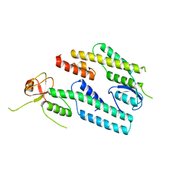
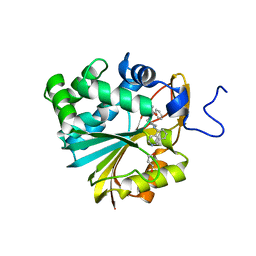
5D1P
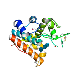
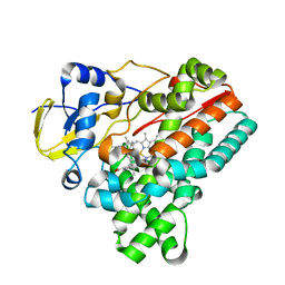
 | | Archaeal ATP-dependent RNA ligase - form 2 | | 分子名称: | ATP-dependent RNA ligase, MAGNESIUM ION, SULFATE ION | | 著者 | Murakami, K.S. | | 登録日 | 2015-08-04 | | 公開日 | 2016-03-09 | | 最終更新日 | 2016-03-30 | | 実験手法 | X-RAY DIFFRACTION (2.199 Å) | | 主引用文献 | Structural and mutational analysis of archaeal ATP-dependent RNA ligase identifies amino acids required for RNA binding and catalysis.
Nucleic Acids Res., 44, 2016
|
|
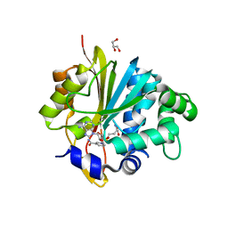
4IJ4
 | | Crystal Structure of a Family GH19 chitinase from Bryum coronatum in complex with (GlcNAc)4 | | 分子名称: | 1,2-ETHANEDIOL, 2-acetamido-2-deoxy-beta-D-glucopyranose-(1-4)-2-acetamido-2-deoxy-beta-D-glucopyranose-(1-4)-2-acetamido-2-deoxy-beta-D-glucopyranose-(1-4)-2-acetamido-2-deoxy-beta-D-glucopyranose, Chitinase A | | 著者 | Numata, T, Umemoto, N, Ohnuma, T, Fukamizo, T. | | 登録日 | 2012-12-21 | | 公開日 | 2014-03-26 | | 最終更新日 | 2022-08-24 | | 実験手法 | X-RAY DIFFRACTION (1.58 Å) | | 主引用文献 | Crystal structure of a "loopless" GH19 chitinase in complex with chitin tetrasaccharide spanning the catalytic center.
Biochim.Biophys.Acta, 1844, 2014
|
|
7T6C
 | | E. coli dihydroorotate dehydrogenase bound to the ubiquinone surrogate DCIP | | 分子名称: | 2,6-bis(chloranyl)-4-[(4-hydroxyphenyl)amino]phenol, 2-{2-[2-(2-{2-[2-(2-ETHOXY-ETHOXY)-ETHOXY]-ETHOXY}-ETHOXY)-ETHOXY]-ETHOXY}-ETHANOL, Dihydroorotate dehydrogenase (quinone), ... | | 著者 | Horwitz, S.M, Ambarian, J.A, Davis, K.M. | | 登録日 | 2021-12-13 | | 公開日 | 2022-03-02 | | 最終更新日 | 2023-10-18 | | 実験手法 | X-RAY DIFFRACTION (2.53 Å) | | 主引用文献 | Structural insights into inhibition of the drug target dihydroorotate dehydrogenase by bacterial hydroxyalkylquinolines.
Rsc Chem Biol, 3, 2022
|
|
7T5K
 | | E. coli dihydroorotate dehydrogenase bound to the inhibitor HQNO | | 分子名称: | 2-HEPTYL-4-HYDROXY QUINOLINE N-OXIDE, Dihydroorotate dehydrogenase (quinone), FLAVIN MONONUCLEOTIDE, ... | | 著者 | Horwitz, S.M, Ambarian, J.A, Davis, K.M. | | 登録日 | 2021-12-12 | | 公開日 | 2022-03-02 | | 最終更新日 | 2023-10-18 | | 実験手法 | X-RAY DIFFRACTION (2.25 Å) | | 主引用文献 | Structural insights into inhibition of the drug target dihydroorotate dehydrogenase by bacterial hydroxyalkylquinolines.
Rsc Chem Biol, 3, 2022
|
|
7T6H
 | | E. coli dihydroorotate dehydrogenase | | 分子名称: | Dihydroorotate dehydrogenase (quinone), FLAVIN MONONUCLEOTIDE, OROTIC ACID | | 著者 | Horwitz, S.M, Ambarian, J.A, Davis, K.M. | | 登録日 | 2021-12-13 | | 公開日 | 2022-03-02 | | 最終更新日 | 2024-02-28 | | 実験手法 | X-RAY DIFFRACTION (2.42 Å) | | 主引用文献 | Structural insights into inhibition of the drug target dihydroorotate dehydrogenase by bacterial hydroxyalkylquinolines.
Rsc Chem Biol, 3, 2022
|
|
7T5Y
 | | E. coli dihydroorotate dehydrogenase bound to the inhibitor HMNQ | | 分子名称: | 3-methyl-2-[(2E)-non-2-en-1-yl]quinolin-4(1H)-one, Dihydroorotate dehydrogenase (quinone), FLAVIN MONONUCLEOTIDE, ... | | 著者 | Horwitz, S.M, Ambarian, J.A, Davis, K.M. | | 登録日 | 2021-12-13 | | 公開日 | 2022-03-02 | | 最終更新日 | 2023-10-18 | | 実験手法 | X-RAY DIFFRACTION (2.62 Å) | | 主引用文献 | Structural insights into inhibition of the drug target dihydroorotate dehydrogenase by bacterial hydroxyalkylquinolines.
Rsc Chem Biol, 3, 2022
|
|
3ANW
 | | A protein complex essential initiation of DNA replication | | 分子名称: | Putative uncharacterized protein | | 著者 | Oyama, T, Shirai, T, Nagasawa, N, Ishino, Y, Morikawa, K. | | 登録日 | 2010-09-10 | | 公開日 | 2011-07-27 | | 最終更新日 | 2024-03-13 | | 実験手法 | X-RAY DIFFRACTION (2.65 Å) | | 主引用文献 | Architectures of archaeal GINS complexes, essential DNA replication initiation factors
Bmc Biol., 9, 2011
|
|
5XVQ
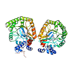
 | | Crystal structure of monkey Nicotinamide N-methyltransferase (NNMT) bound with end product, 1-methyl Nicotinamide (MNA) | | 分子名称: | 3-carbamoyl-1-methylpyridin-1-ium, GLYCEROL, Nicotinamide N-methyltransferase (NNMT), ... | | 著者 | Birudukota, S, Swaminathan, S, Thakur, M.K, Parveen, R, Kandan, S, Kannt, A, Gosu, R. | | 登録日 | 2017-06-28 | | 公開日 | 2017-08-02 | | 最終更新日 | 2023-11-22 | | 実験手法 | X-RAY DIFFRACTION (2.29 Å) | | 主引用文献 | Crystal structures of monkey and mouse nicotinamide N-methyltransferase (NNMT) bound with end product, 1-methyl nicotinamide
Biochem. Biophys. Res. Commun., 491, 2017
|
|
5XVK
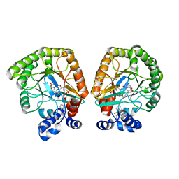
 | | Crystal structure of mouse Nicotinamide N-methyltransferase (NNMT) bound with end product, 1-methyl Nicotinamide (MNA) | | 分子名称: | 3-carbamoyl-1-methylpyridin-1-ium, GLYCEROL, Nicotinamide N-methyltransferase, ... | | 著者 | Swaminathan, S, Birudukota, S, Thakur, M.K, Parveen, R, Kandan, S, Kannt, A, Gosu, R. | | 登録日 | 2017-06-28 | | 公開日 | 2017-08-02 | | 最終更新日 | 2023-11-22 | | 実験手法 | X-RAY DIFFRACTION (1.88 Å) | | 主引用文献 | Crystal structures of monkey and mouse nicotinamide N-methyltransferase (NNMT) bound with end product, 1-methyl nicotinamide
Biochem. Biophys. Res. Commun., 491, 2017
|
|
7EU5
 | | Co-crystal structure of Human Nicotinamide N-methyltransferase (NNMT) with tricyclic small molecule inhibitor JBSNF-000107 | | 分子名称: | 6-fluoranyl-10-methyl-1,10-diazatricyclo[6.3.1.0^{4,12}]dodeca-4,6,8(12)-trien-11-imine, Nicotinamide N-methyltransferase, S-ADENOSYL-L-HOMOCYSTEINE | | 著者 | Swaminathan, S, Gosu, R, Birudukota, S, Kandan, S, Vaithilingam, K. | | 登録日 | 2021-05-16 | | 公開日 | 2022-05-18 | | 最終更新日 | 2023-11-29 | | 実験手法 | X-RAY DIFFRACTION (2.731 Å) | | 主引用文献 | Novel tricyclic small molecule inhibitors of Nicotinamide N-methyltransferase for the treatment of metabolic disorders.
Sci Rep, 12, 2022
|
|
7ET7
 | | Co-crystal structure of Human Nicotinamide N-methyltransferase (NNMT) with tricyclic small molecule inhibitor JBSNF-000028 | | 分子名称: | 10-methyl-1,10-diazatricyclo[6.3.1.0^{4,12}]dodeca-4,6,8(12)-trien-11-imine, Nicotinamide N-methyltransferase, S-ADENOSYL-L-HOMOCYSTEINE | | 著者 | Swaminathan, S, Gosu, R, Birudukota, S, Kandan, S, Vaithilingam, K. | | 登録日 | 2021-05-12 | | 公開日 | 2022-05-18 | | 最終更新日 | 2023-11-29 | | 実験手法 | X-RAY DIFFRACTION (2.61 Å) | | 主引用文献 | Novel tricyclic small molecule inhibitors of Nicotinamide N-methyltransferase for the treatment of metabolic disorders.
Sci Rep, 12, 2022
|
|
6J82
 | | Crystal structure of TleB apo | | 分子名称: | Cytochrome P-450, PROTOPORPHYRIN IX CONTAINING FE | | 著者 | Alblova, M, Nakamura, H, Mori, T, Abe, I. | | 登録日 | 2019-01-18 | | 公開日 | 2019-08-07 | | 最終更新日 | 2023-11-22 | | 実験手法 | X-RAY DIFFRACTION (2.202 Å) | | 主引用文献 | Molecular basis for the P450-catalyzed C-N bond formation in indolactam biosynthesis.
Nat.Chem.Biol., 15, 2019
|
|
6J88
 | | Crystal structure of HinD with benzo[b]thiophen analog | | 分子名称: | N-[(2S)-1-(1-benzothiophen-3-yl)-3-hydroxypropan-2-yl]-N~2~-methyl-L-valinamide, Nocardicin N-oxygenase, PROTOPORPHYRIN IX CONTAINING FE | | 著者 | Fei, H, Mori, T, Abe, I. | | 登録日 | 2019-01-18 | | 公開日 | 2019-08-07 | | 最終更新日 | 2023-11-22 | | 実験手法 | X-RAY DIFFRACTION (2.35 Å) | | 主引用文献 | Molecular basis for the P450-catalyzed C-N bond formation in indolactam biosynthesis.
Nat.Chem.Biol., 15, 2019
|
|
6J85
 | | Crystal structure of HinD apo | | 分子名称: | Nocardicin N-oxygenase, PROTOPORPHYRIN IX CONTAINING FE | | 著者 | Fei, H, Mori, T, Abe, I. | | 登録日 | 2019-01-18 | | 公開日 | 2019-08-07 | | 最終更新日 | 2023-11-22 | | 実験手法 | X-RAY DIFFRACTION (2.2 Å) | | 主引用文献 | Molecular basis for the P450-catalyzed C-N bond formation in indolactam biosynthesis.
Nat.Chem.Biol., 15, 2019
|
|
6J86
 | | Crystal structure of HinD with NMFT | | 分子名称: | N-[(2S)-1-hydroxy-3-(1H-indol-3-yl)propan-2-yl]-Nalpha-methyl-L-phenylalaninamide, Nocardicin N-oxygenase, PROTOPORPHYRIN IX CONTAINING FE | | 著者 | Fei, H, Mori, T, Abe, I. | | 登録日 | 2019-01-18 | | 公開日 | 2019-08-07 | | 最終更新日 | 2023-11-22 | | 実験手法 | X-RAY DIFFRACTION (2.6 Å) | | 主引用文献 | Molecular basis for the P450-catalyzed C-N bond formation in indolactam biosynthesis.
Nat.Chem.Biol., 15, 2019
|
|
6J83
 | | Crystal structure of TleB with NMVT | | 分子名称: | Cytochrome P-450, N-[(2S)-1-hydroxy-3-(1H-indol-3-yl)propan-2-yl]-N~2~-methyl-L-valinamide, PROTOPORPHYRIN IX CONTAINING FE | | 著者 | Fei, H, Mori, T, Abe, I. | | 登録日 | 2019-01-18 | | 公開日 | 2019-08-07 | | 最終更新日 | 2023-11-22 | | 実験手法 | X-RAY DIFFRACTION (1.9 Å) | | 主引用文献 | Molecular basis for the P450-catalyzed C-N bond formation in indolactam biosynthesis.
Nat.Chem.Biol., 15, 2019
|
|
7NBJ
 | |


