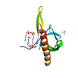
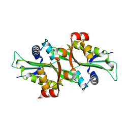
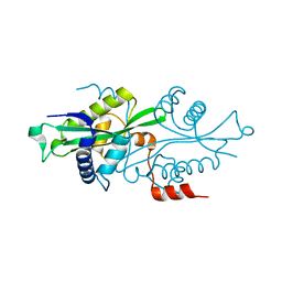
7DZJ
 
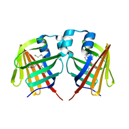 | | Fabp protein before hv | | 分子名称: | Fatty acid-binding protein, liver, PALMITIC ACID | | 著者 | Li, H, Yu, L.-J, Liu, X, Shen, J.-R, Wang, J. | | 登録日 | 2021-01-25 | | 公開日 | 2022-07-27 | | 最終更新日 | 2023-11-29 | | 実験手法 | X-RAY DIFFRACTION (1.63 Å) | | 主引用文献 | Excited-state intermediates in a designer protein encoding a phototrigger caught by an X-ray free-electron laser.
Nat.Chem., 14, 2022
|
|
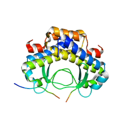
7DZK
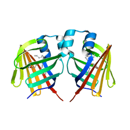 | | Fabp protein after hv | | 分子名称: | Fatty acid-binding protein, liver, PALMITIC ACID | | 著者 | Li, H, Yu, L.-J, Liu, X, Shen, J.-R, Wang, J. | | 登録日 | 2021-01-25 | | 公開日 | 2022-07-27 | | 最終更新日 | 2023-11-29 | | 実験手法 | X-RAY DIFFRACTION (1.54 Å) | | 主引用文献 | Excited-state intermediates in a designer protein encoding a phototrigger caught by an X-ray free-electron laser.
Nat.Chem., 14, 2022
|
|
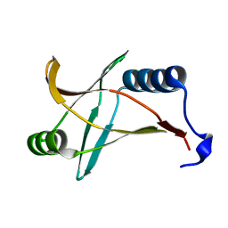
7DZL
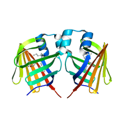 | | A69C-M71L mutant of Fabp protein | | 分子名称: | Fatty acid-binding protein, liver, PALMITIC ACID | | 著者 | Li, H, Yu, L.-J, Liu, X, Shen, J.-R, Wang, J. | | 登録日 | 2021-01-25 | | 公開日 | 2022-07-27 | | 最終更新日 | 2023-11-29 | | 実験手法 | X-RAY DIFFRACTION (1.64 Å) | | 主引用文献 | Excited-state intermediates in a designer protein encoding a phototrigger caught by an X-ray free-electron laser.
Nat.Chem., 14, 2022
|
|
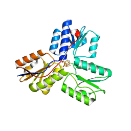
6IYV
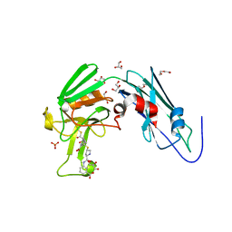 | | Crystal sturucture of L,D-transpeptidase LdtMt2 from Mycobacterium tuberculosis in complex with ertapenem adduct | | 分子名称: | (2S,3R,4S)-4-({(3S,5S)-5-[(3-carboxyphenyl)carbamoyl]pyrrolidin-3-yl}sulfanyl)-2-[(1S,2R)-1-formyl-2-hydroxypropyl]-3-methyl-3,4-dihydro-2H-pyrrole-5-carboxylic acid, 2-AMINO-2-HYDROXYMETHYL-PROPANE-1,3-DIOL, DI(HYDROXYETHYL)ETHER, ... | | 著者 | Zhao, F, Li, D.F, Wang, D.C. | | 登録日 | 2018-12-17 | | 公開日 | 2019-02-27 | | 最終更新日 | 2023-11-22 | | 実験手法 | X-RAY DIFFRACTION (1.5 Å) | | 主引用文献 | The 1-beta-methyl group confers a lower affinity of l,d-transpeptidase LdtMt2for ertapenem than for imipenem.
Biochem. Biophys. Res. Commun., 510, 2019
|
|
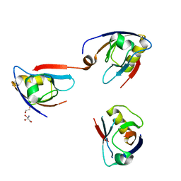
5Y6G
 | | PilZ domain with c-di-GMP of YcgR from Escherichia coli | | 分子名称: | 9,9'-[(2R,3R,3aS,5S,7aR,9R,10R,10aS,12S,14aR)-3,5,10,12-tetrahydroxy-5,12-dioxidooctahydro-2H,7H-difuro[3,2-d:3',2'-j][1,3,7,9,2,8]tetraoxadiphosphacyclododecine-2,9-diyl]bis(2-amino-1,9-dihydro-6H-purin-6-one), Flagellar brake protein YcgR, SULFATE ION | | 著者 | Hou, Y.J, Wang, D.C, Li, D.F. | | 登録日 | 2017-08-11 | | 公開日 | 2018-07-18 | | 最終更新日 | 2023-11-22 | | 実験手法 | X-RAY DIFFRACTION (2.3 Å) | | 主引用文献 | Structural insights into the mechanism of c-di-GMP-bound YcgR regulating flagellar motility inEscherichia coli.
J.Biol.Chem., 295, 2020
|
|
2H0D
 | |
5Y21
 | |
5Y6H
 | |
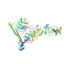
7EXD
 | | Lasmiditan-bound serotonin 1F (5-HT1F) receptor-Gi protein complex | | 分子名称: | 2,4,6-tris(fluoranyl)-N-[6-(1-methylpiperidin-4-yl)carbonylpyridin-2-yl]benzamide, Guanine nucleotide-binding protein G(I)/G(S)/G(O) subunit gamma-2, Guanine nucleotide-binding protein G(I)/G(S)/G(T) subunit beta-1, ... | | 著者 | Huang, S, Xu, P, Jiang, Y, Xu, H.E. | | 登録日 | 2021-05-27 | | 公開日 | 2021-08-04 | | 最終更新日 | 2021-10-06 | | 実験手法 | ELECTRON MICROSCOPY (3.4 Å) | | 主引用文献 | Structural basis for recognition of anti-migraine drug lasmiditan by the serotonin receptor 5-HT 1F -G protein complex.
Cell Res., 31, 2021
|
|
2F5I
 | |
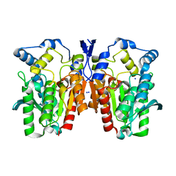
6N4X
 | | Metabotropic Glutamate Receptor 5 Apo Form Ligand Binding Domain | | 分子名称: | 2-acetamido-2-deoxy-beta-D-glucopyranose, MAGNESIUM ION, Metabotropic glutamate receptor 5 | | 著者 | Koehl, A, Hu, H, Feng, D, Sun, B, Weis, W.I, Skiniotis, G.S, Mathiesen, J.M, Kobilka, B.K. | | 登録日 | 2018-11-20 | | 公開日 | 2019-01-23 | | 最終更新日 | 2023-10-11 | | 実験手法 | X-RAY DIFFRACTION (4 Å) | | 主引用文献 | Structural insights into the activation of metabotropic glutamate receptors.
Nature, 566, 2019
|
|
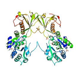
4Q05
 | | Crystal structure of an esterase E25 | | 分子名称: | SODIUM ION, esterase E25 | | 著者 | Li, P.Y, Li, C.Y, Zhang, Y.Z. | | 登録日 | 2014-03-31 | | 公開日 | 2014-06-04 | | 最終更新日 | 2024-04-03 | | 実験手法 | X-RAY DIFFRACTION (2.05 Å) | | 主引用文献 | Structural basis for dimerization and catalysis of a novel esterase from the GTSAG motif subfamily of the bacterial hormone-sensitive lipase family
J.Biol.Chem., 289, 2014
|
|
4Q2P
 | |
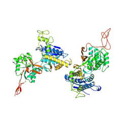
6BX3
 | | Structure of histone H3k4 methyltransferase | | 分子名称: | COMPASS component BRE2, COMPASS component SDC1, COMPASS component SPP1, ... | | 著者 | Skiniotis, G, Qu, Q.H. | | 登録日 | 2017-12-16 | | 公開日 | 2018-09-05 | | 最終更新日 | 2024-03-13 | | 実験手法 | ELECTRON MICROSCOPY (4.3 Å) | | 主引用文献 | Structure and Conformational Dynamics of a COMPASS Histone H3K4 Methyltransferase Complex.
Cell, 174, 2018
|
|
6MNZ
 | | Crystal structure of RibBX, a two domain 3,4-dihydroxy-2-butanone 4-phosphate synthase from A. baumannii. | | 分子名称: | 3,4-dihydroxy-2-butanone 4-phosphate synthase, CHLORIDE ION, SULFATE ION | | 著者 | Wang, J, Gonzalez-Gutierrez, G, Giedroc, D.P. | | 登録日 | 2018-10-03 | | 公開日 | 2019-04-17 | | 最終更新日 | 2024-03-13 | | 実験手法 | X-RAY DIFFRACTION (2.66 Å) | | 主引用文献 | Multi-metal Restriction by Calprotectin Impacts De Novo Flavin Biosynthesis in Acinetobacter baumannii.
Cell Chem Biol, 26, 2019
|
|
4MV5
 | | IspH in complex with 6-chloropyridin-3-ylmethyl diphosphate | | 分子名称: | (6-chloropyridin-3-yl)methyl trihydrogen diphosphate, 4-hydroxy-3-methylbut-2-enyl diphosphate reductase, FE3-S4 CLUSTER | | 著者 | Span, I, Wang, K, Song, Y, Eisenreich, W, Bacher, A, Oldfield, E, Groll, M. | | 登録日 | 2013-09-23 | | 公開日 | 2014-06-18 | | 最終更新日 | 2023-09-20 | | 実験手法 | X-RAY DIFFRACTION (1.9 Å) | | 主引用文献 | Insights into the Binding of Pyridines to the Iron-Sulfur Enzyme IspH.
J.Am.Chem.Soc., 136, 2014
|
|
3HMH
 | | Crystal structure of the second bromodomain of human TBP-associated factor RNA polymerase 1-like (TAF1L) | | 分子名称: | Transcription initiation factor TFIID 210 kDa subunit | | 著者 | Filippakopoulos, P, Picaud, S, Keates, T, Zhang, Y, Pike, A.C.W, von Delft, F, Arrowsmith, C.H, Edwards, A, Weigelt, J, Bountra, C, Knapp, S, Structural Genomics Consortium (SGC) | | 登録日 | 2009-05-29 | | 公開日 | 2009-06-23 | | 最終更新日 | 2023-11-01 | | 実験手法 | X-RAY DIFFRACTION (2.05 Å) | | 主引用文献 | Histone recognition and large-scale structural analysis of the human bromodomain family.
Cell(Cambridge,Mass.), 149, 2012
|
|
2G3T
 | |
4KPS
 | | Structure and receptor binding specificity of the hemagglutinin H13 from avian influenza A virus H13N6 | | 分子名称: | 2-acetamido-2-deoxy-beta-D-glucopyranose, Hemagglutinin, N-acetyl-alpha-neuraminic acid-(2-3)-beta-D-galactopyranose, ... | | 著者 | Lu, X, Qi, J, Shi, Y, Gao, G. | | 登録日 | 2013-05-14 | | 公開日 | 2013-07-03 | | 最終更新日 | 2023-11-08 | | 実験手法 | X-RAY DIFFRACTION (2.587 Å) | | 主引用文献 | Structure and Receptor Binding Specificity of Hemagglutinin H13 from Avian Influenza A Virus H13N6
J.Virol., 87, 2013
|
|
4O9S
 | | Crystal structure of Retinol-Binding Protein 4 (RBP4)in complex with a non-retinoid ligand | | 分子名称: | 1,2-ETHANEDIOL, 1-[4-(7-thia-9,11-diazatricyclo[6.4.0.0^{2,6}]dodeca-1(12),2(6),8,10-tetraen-12-yl)piperazin-1-yl]-2-[2-(trifluoromethyl)phenyl]ethanone, CHLORIDE ION, ... | | 著者 | Wang, Z, Johnstone, S, Walker, N.P. | | 登録日 | 2014-01-02 | | 公開日 | 2014-07-02 | | 最終更新日 | 2020-02-19 | | 実験手法 | X-RAY DIFFRACTION (2.3 Å) | | 主引用文献 | Structure-assisted discovery of the first non-retinoid ligands for Retinol-Binding Protein 4.
Bioorg.Med.Chem.Lett., 24, 2014
|
|
4Q2N
 | |
7D32
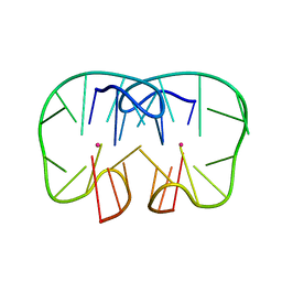 | | The TBA-Pb2+ complex in P41212 space group | | 分子名称: | DNA (5'-D(*GP*GP*TP*TP*GP*GP*TP*GP*TP*GP*GP*TP*TP*GP*G)-3'), LEAD (II) ION | | 著者 | Liu, H.H, Gao, Y.Q, Sheng, J, Gan, J.H. | | 登録日 | 2020-09-18 | | 公開日 | 2021-09-22 | | 最終更新日 | 2023-11-29 | | 実験手法 | X-RAY DIFFRACTION (1.707 Å) | | 主引用文献 | Structure-guided development of Pb 2+ -binding DNA aptamers.
Sci Rep, 12, 2022
|
|
7D33
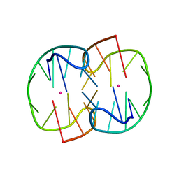 | | The Pb2+ complexed structure of TBA G8C mutant | | 分子名称: | DNA (5'-D(*GP*GP*TP*TP*GP*GP*TP*CP*TP*GP*GP*TP*TP*GP*G)-3'), LEAD (II) ION | | 著者 | Liu, H.H, Gao, Y.Q, Sheng, J, Gan, J.H. | | 登録日 | 2020-09-18 | | 公開日 | 2021-09-22 | | 最終更新日 | 2023-11-29 | | 実験手法 | X-RAY DIFFRACTION (2.117 Å) | | 主引用文献 | Structure-guided development of Pb 2+ -binding DNA aptamers.
Sci Rep, 12, 2022
|
|
7D31
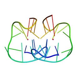 | | The TBA-Pb2+ complex in P41212 space group | | 分子名称: | DNA (5'-D(*GP*GP*TP*TP*GP*GP*TP*GP*TP*GP*GP*TP*TP*GP*G)-3'), LEAD (II) ION | | 著者 | Liu, H.H, Gao, Y.Q, Sheng, J, Gan, J.H. | | 登録日 | 2020-09-18 | | 公開日 | 2021-09-22 | | 最終更新日 | 2024-05-29 | | 実験手法 | X-RAY DIFFRACTION (1.396 Å) | | 主引用文献 | Structure-guided development of Pb 2+ -binding DNA aptamers.
Sci Rep, 12, 2022
|
|
4QEI
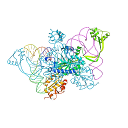 | | Two distinct conformational states of GlyRS captured in crystal lattice | | 分子名称: | ADENOSINE MONOPHOSPHATE, Glycine--tRNA ligase, tRNA-Gly-CCC-2-2 | | 著者 | Xie, W, Qin, X, Deng, X, Zhang, Q, Li, Q. | | 登録日 | 2014-05-16 | | 公開日 | 2015-05-20 | | 最終更新日 | 2023-11-08 | | 実験手法 | X-RAY DIFFRACTION (2.875 Å) | | 主引用文献 | Large Conformational Changes of Insertion 3 in Human Glycyl-tRNA Synthetase (hGlyRS) during Catalysis
J.Biol.Chem., 291, 2016
|
|


