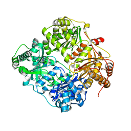
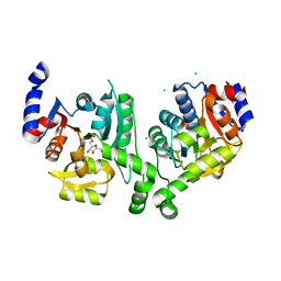
1YOX
 
 | | Structure of the conserved Protein of Unknown Function PA3696 from Pseudomonas aeruginosa | | 分子名称: | hypothetical protein PA3696 | | 著者 | Walker, J.R, Xu, X, Gu, J, Joachimiak, A, Edwards, A, Savchenko, A, Midwest Center for Structural Genomics (MCSG) | | 登録日 | 2005-01-28 | | 公開日 | 2005-04-26 | | 最終更新日 | 2017-10-11 | | 実験手法 | X-RAY DIFFRACTION (2.3 Å) | | 主引用文献 | X-ray structure of the conserved hypothetical protein PA3696
To be Published
|
|
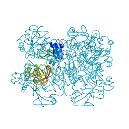
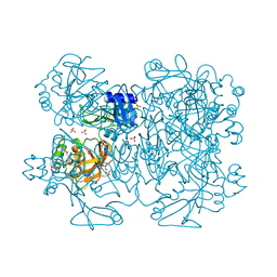
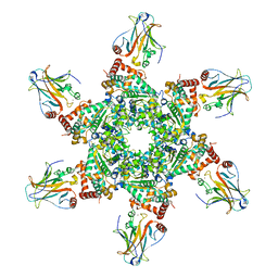
1GR5
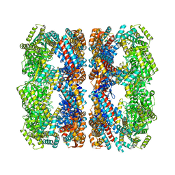 | | Solution Structure of apo GroEL by Cryo-Electron microscopy | | 分子名称: | 60 KDA CHAPERONIN | | 著者 | Ranson, N.A, Farr, G.W, Roseman, A.M, Gowen, B, Fenton, W.A, Horwich, A.L, Saibil, H.R. | | 登録日 | 2001-12-14 | | 公開日 | 2002-01-28 | | 最終更新日 | 2024-05-08 | | 実験手法 | ELECTRON MICROSCOPY (7.9 Å) | | 主引用文献 | ATP-Bound States of Groel Captured by Cryo-Electron Microscopy.
Cell(Cambridge,Mass.), 107, 2001
|
|
3BXG
 | |
2Q06
 | | Crystal structure of Influenza A Virus H5N1 Nucleoprotein | | 分子名称: | Nucleoprotein | | 著者 | Ng, A.K.L, Zhang, H, Tan, K, Wang, J, Shaw, P.C. | | 登録日 | 2007-05-21 | | 公開日 | 2008-05-27 | | 最終更新日 | 2023-08-30 | | 実験手法 | X-RAY DIFFRACTION (3.3 Å) | | 主引用文献 | Structure of the influenza virus A H5N1 nucleoprotein: implications for RNA binding, oligomerization, and vaccine design.
Faseb J., 22, 2008
|
|
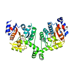
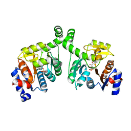
2ID3
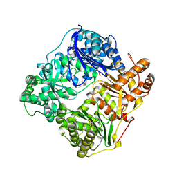
 | | Crystal structure of transcriptional regulator SCO5951 from Streptomyces coelicolor A3(2) | | 分子名称: | CALCIUM ION, CHLORIDE ION, Putative transcriptional regulator | | 著者 | Grabowski, M, Chruszcz, M, Koclega, K.D, Cymborowski, M, Gu, J, Xu, X, Savchenko, A, Edwards, A, Joachimiak, A, Minor, W, Midwest Center for Structural Genomics (MCSG) | | 登録日 | 2006-09-14 | | 公開日 | 2006-10-17 | | 最終更新日 | 2022-04-13 | | 実験手法 | X-RAY DIFFRACTION (1.7 Å) | | 主引用文献 |
|
|
3BXE
 | |
2G54
 | |
1L5X
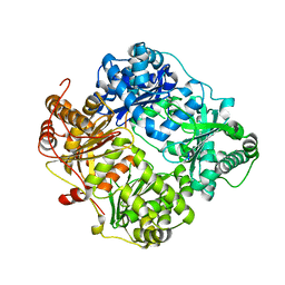
 | | The 2.0-Angstrom resolution crystal structure of a survival protein E (SurE) homolog from Pyrobaculum aerophilum | | 分子名称: | ACETIC ACID, GLYCEROL, Survival protein E | | 著者 | Mura, C, Katz, J.E, Clarke, S.G, Eisenberg, D. | | 登録日 | 2002-03-08 | | 公開日 | 2003-02-25 | | 最終更新日 | 2011-07-13 | | 実験手法 | X-RAY DIFFRACTION (2 Å) | | 主引用文献 | Structure and Function of an Archaeal Homolog of Survival
Protein E (SurE-alpha): An Acid Phosphatase with Purine
Nucleotide Specificity
J.Mol.Biol., 326, 2003
|
|
7KAG
 | | Crystal structure of the ubiquitin-like domain 1 (Ubl1) of Nsp3 from SARS-CoV-2 | | 分子名称: | 1,2-ETHANEDIOL, Non-structural protein 3, SULFATE ION | | 著者 | Stogios, P.J, Skarina, T, Chang, C, Kim, Y, Di Leo, R, Savchenko, A, Joachimiak, A, Satchell, K.J.F, Center for Structural Genomics of Infectious Diseases (CSGID) | | 登録日 | 2020-09-30 | | 公開日 | 2020-10-14 | | 最終更新日 | 2023-10-18 | | 実験手法 | X-RAY DIFFRACTION (3.21 Å) | | 主引用文献 | Crystal structure of the ubiquitin-like domain 1 (Ubl1) of Nsp3 from SARS-CoV-2
To Be Published
|
|
7JM1
 | | Crystal structure of aminoglycoside resistance enzyme ApmA, complex with acetyl-CoA | | 分子名称: | ACETYL COENZYME *A, Aminocyclitol acetyltransferase ApmA | | 著者 | Stogios, P.J, Evdokimova, E, Di Leo, R, Bordeleau, E, Wright, G.D, Savchenko, A, Joachimiak, A, Satchell, K.J.F, Center for Structural Genomics of Infectious Diseases (CSGID) | | 登録日 | 2020-07-30 | | 公開日 | 2020-09-16 | | 実験手法 | X-RAY DIFFRACTION (2.31 Å) | | 主引用文献 | Crystal structure of aminoglycoside resistance enzyme ApmA, complex with acetyl-CoA
To Be Published
|
|
2G47
 | |
2G48
 | |
2G56
 | |
7JM2
 | | Crystal structure of aminoglycoside resistance enzyme ApmA, complex with apramycin | | 分子名称: | APRAMYCIN, Aminocyclitol acetyltransferase ApmA, CHLORIDE ION | | 著者 | Stogios, P.J, Evdokimova, E, Di Leo, R, Bordeleau, E, Wright, G.D, Savchenko, A, Joachimiak, A, Satchell, K.J.F, Center for Structural Genomics of Infectious Diseases (CSGID) | | 登録日 | 2020-07-30 | | 公開日 | 2020-09-16 | | 最終更新日 | 2023-10-18 | | 実験手法 | X-RAY DIFFRACTION (1.85 Å) | | 主引用文献 | Crystal structure of aminoglycoside resistance enzyme ApmA, complex with apramycin
To Be Published
|
|
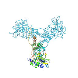
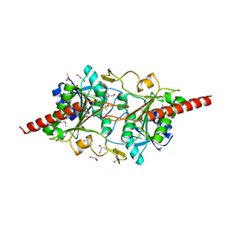
3CM8
 | | A RNA polymerase subunit structure from virus | | 分子名称: | Polymerase acidic protein, peptide from RNA-directed RNA polymerase catalytic subunit | | 著者 | He, X, Zhou, J, Zeng, Z, Ma, J, Zhang, R, Rao, Z, Liu, Y. | | 登録日 | 2008-03-21 | | 公開日 | 2008-07-15 | | 最終更新日 | 2024-03-13 | | 実験手法 | X-RAY DIFFRACTION (2.899 Å) | | 主引用文献 | Crystal structure of the polymerase PAC-PB1N complex from an avian influenza H5N1 virus
Nature, 454, 2008
|
|
2G9T
 | | Crystal structure of the SARS coronavirus nsp10 at 2.1A | | 分子名称: | ZINC ION, orf1a polyprotein | | 著者 | Su, D, Lou, Z, Yang, H, Sun, F, Rao, Z. | | 登録日 | 2006-03-07 | | 公開日 | 2006-08-15 | | 最終更新日 | 2024-03-13 | | 実験手法 | X-RAY DIFFRACTION (2.1 Å) | | 主引用文献 | Dodecamer Structure of Severe Acute Respiratory Syndrome Coronavirus Nonstructural Protein nsp10
J.Virol., 80, 2006
|
|
1NAQ
 | | Crystal structure of CUTA1 from E.coli at 1.7 A resolution | | 分子名称: | MERCURIBENZOIC ACID, MERCURY (II) ION, Periplasmic divalent cation tolerance protein cutA | | 著者 | Calderone, V, Mangani, S, Benvenuti, M, Viezzoli, M.S, Banci, L, Bertini, I, Structural Proteomics in Europe (SPINE) | | 登録日 | 2002-11-28 | | 公開日 | 2003-11-25 | | 最終更新日 | 2024-02-14 | | 実験手法 | X-RAY DIFFRACTION (1.7 Å) | | 主引用文献 | The evolutionarily conserved trimeric structure of CutA1 proteins suggests a role in signal transduction.
J.Biol.Chem., 278, 2003
|
|
2G49
 | |
2GA6
 | | The crystal structure of SARS nsp10 without zinc ion as additive | | 分子名称: | ZINC ION, orf1a polyprotein | | 著者 | Su, D, Lou, Z, Sun, F, Zhai, Y, Yang, H, Rao, Z. | | 登録日 | 2006-03-08 | | 公開日 | 2006-08-15 | | 最終更新日 | 2023-10-25 | | 実験手法 | X-RAY DIFFRACTION (2.7 Å) | | 主引用文献 | Dodecamer Structure of Severe Acute Respiratory Syndrome Coronavirus Nonstructural Protein nsp10
J.Virol., 80, 2006
|
|
2GTH
 | | crystal structure of the wildtype MHV coronavirus non-structural protein nsp15 | | 分子名称: | Replicase polyprotein 1ab | | 著者 | Xu, X, Zhai, Y, Sun, F, Lou, Z, Su, D, Rao, Z. | | 登録日 | 2006-04-28 | | 公開日 | 2006-08-15 | | 最終更新日 | 2023-10-25 | | 実験手法 | X-RAY DIFFRACTION (2.7 Å) | | 主引用文献 | New Antiviral Target Revealed by the Hexameric Structure of Mouse Hepatitis Virus Nonstructural Protein nsp15
J.Virol., 80, 2006
|
|
7JM0
 | | Crystal structure of aminoglycoside resistance enzyme ApmA, apoenzyme | | 分子名称: | Aminocyclitol acetyltransferase ApmA, SULFATE ION | | 著者 | Stogios, P.J, Evdokimova, E, Di Leo, R, Bordeleau, E, Wright, G.D, Savchenko, A, Joachimiak, A, Satchell, K.J.F, Center for Structural Genomics of Infectious Diseases (CSGID) | | 登録日 | 2020-07-30 | | 公開日 | 2020-09-16 | | 最終更新日 | 2023-10-18 | | 実験手法 | X-RAY DIFFRACTION (2.08 Å) | | 主引用文献 | Crystal structure of aminoglycoside resistance enzyme ApmA, apoenzyme
To Be Published
|
|
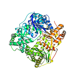
7KZW
 | | Crystal structure of FTT_1639c from Francisella tularensis str. tularensis SCHU S4 | | 分子名称: | CHLORIDE ION, FTT_1639c | | 著者 | Stogios, P.J, Skarina, T, Osipiuk, J, Di Leo, R, Savchenko, A, Joachimiak, A, Satchell, K.J.F, Center for Structural Genomics of Infectious Diseases (CSGID) | | 登録日 | 2020-12-10 | | 公開日 | 2020-12-30 | | 実験手法 | X-RAY DIFFRACTION (1.34 Å) | | 主引用文献 | Crystal structure of FTT_1639c from Francisella tularensis str. tularensis SCHU S4
To Be Published
|
|
2GTI
 | | mutation of MHV coronavirus non-structural protein nsp15 (F307L) | | 分子名称: | GLYCEROL, Replicase polyprotein 1ab, SULFATE ION | | 著者 | Xu, X, Zhai, Y, Sun, F, Lou, Z, Su, D, Rao, Z. | | 登録日 | 2006-04-28 | | 公開日 | 2006-08-15 | | 最終更新日 | 2021-11-10 | | 実験手法 | X-RAY DIFFRACTION (2.15 Å) | | 主引用文献 | New Antiviral Target Revealed by the Hexameric Structure of Mouse Hepatitis Virus Nonstructural Protein nsp15
J.Virol., 80, 2006
|
|
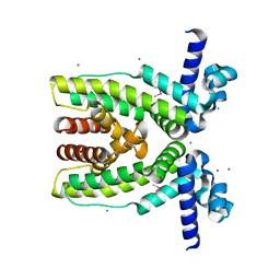
3BXF
 | | Crystal structure of effector binding domain of central glycolytic gene regulator (CggR) from Bacillus subtilis in complex with effector fructose-1,6-bisphosphate | | 分子名称: | 1,3-DIHYDROXYACETONEPHOSPHATE, 1,6-di-O-phosphono-beta-D-fructofuranose, CHLORIDE ION, ... | | 著者 | Rezacova, P, Otwinowski, Z. | | 登録日 | 2008-01-13 | | 公開日 | 2008-07-01 | | 最終更新日 | 2023-08-30 | | 実験手法 | X-RAY DIFFRACTION (1.7 Å) | | 主引用文献 | Crystal structures of the effector-binding domain of repressor Central glycolytic gene Regulator from Bacillus subtilis reveal ligand-induced structural changes upon binding of several glycolytic intermediates.
Mol.Microbiol., 69, 2008
|
|
2H1L
 | |


