6CU7
 
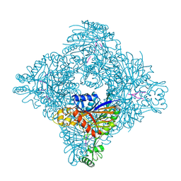
 | | Alpha Synuclein fibril formed by full length protein - Rod Polymorph | | 分子名称: | Alpha-synuclein | | 著者 | Li, B, Hatami, A, Ge, P, Murray, K.A, Sheth, P, Zhang, M, Nair, G, Sawaya, M.R, Zhu, C, Broad, M, Shin, W.S, Ye, S, John, V, Eisenberg, D.S, Zhou, Z.H, Jiang, L. | | 登録日 | 2018-03-23 | | 公開日 | 2018-09-12 | | 最終更新日 | 2024-03-13 | | 実験手法 | ELECTRON MICROSCOPY (3.5 Å) | | 主引用文献 | Cryo-EM of full-length alpha-synuclein reveals fibril polymorphs with a common structural kernel.
Nat Commun, 9, 2018
|
|
3ETY
 | | Crystal structure of bacterial adhesin FadA L14A mutant | | 分子名称: | Adhesin A | | 著者 | Nithianantham, S, Xu, M, Wu, N, Shoham, M, Han, Y.W. | | 登録日 | 2008-10-08 | | 公開日 | 2008-12-02 | | 最終更新日 | 2023-09-06 | | 実験手法 | X-RAY DIFFRACTION (2.9 Å) | | 主引用文献 | Crystal Structure of FadA Adhesin from Fusobacterium nucleatum Reveals a Novel Oligomerization Motif, the Leucine Chain.
J.Biol.Chem., 284, 2009
|
|
7AMP
 | | Crystal structure of the complex of HuJovi-1 Fab with the human A6 T-cell receptor TRBC1 | | 分子名称: | Alpha chain of A6 T-cell receptor, Beta chain 1 of A6 T-cell receptor TRBC1, CHLORIDE ION, ... | | 著者 | Ferrari, M, Bulek, A, Bughda, R, Jha, R, Welin, M, Logan, D.T, Sewell, A, Onuoha, S, Pule, M. | | 登録日 | 2020-10-09 | | 公開日 | 2022-04-20 | | 最終更新日 | 2024-01-31 | | 実験手法 | X-RAY DIFFRACTION (2.64 Å) | | 主引用文献 | Structure-Guided Engineering of Immunotherapies Targeting TRBC1 and TRBC2 in T Cell Malignancies
Res Sq, 2022
|
|
7AMQ
 | | Crystal structure of the complex of HuJovi-1 Fab with the human TRBC2 | | 分子名称: | CHLORIDE ION, GLYCEROL, Human A6 T-cell receptor alpha chain, ... | | 著者 | Ferrari, M, Bulek, A, Bughda, R, Jha, R, Welin, M, Logan, D.T, Sewell, A, Onuoha, S, Pule, M. | | 登録日 | 2020-10-09 | | 公開日 | 2022-04-20 | | 最終更新日 | 2022-04-27 | | 実験手法 | X-RAY DIFFRACTION (2.353 Å) | | 主引用文献 | Structure-Guided Engineering of Immunotherapies Targeting TRBC1 and TRBC2 in T Cell Malignancies
Res Sq, 2022
|
|
7AMS
 | | Crystal structure of the complex of the KFN mutant of HuJovi-1 Fab with human TRBC2 | | 分子名称: | CHLORIDE ION, GLYCEROL, Human A6 T-cell receptor alpha chain, ... | | 著者 | Ferrari, M, Bulek, A, Bughda, R, Jha, R, Welin, M, Svensson, A, Logan, D.T, Sewell, A, Onuoha, S, Pule, M. | | 登録日 | 2020-10-09 | | 公開日 | 2022-04-20 | | 最終更新日 | 2022-04-27 | | 実験手法 | X-RAY DIFFRACTION (2.419 Å) | | 主引用文献 | Structure-Guided Engineering of Immunotherapies Targeting TRBC1 and TRBC2 in T Cell Malignancies
Res Sq, 2022
|
|
4V60
 | | The structure of rat liver vault at 3.5 angstrom resolution | | 分子名称: | Major vault protein | | 著者 | Kato, K, Zhou, Y, Tanaka, H, Yao, M, Yamashita, E, Yoshimura, M, Tsukihara, T. | | 登録日 | 2008-10-24 | | 公開日 | 2014-07-09 | | 最終更新日 | 2024-04-03 | | 実験手法 | X-RAY DIFFRACTION (3.5 Å) | | 主引用文献 | The structure of rat liver vault at 3.5 angstrom resolution
Science, 323, 2009
|
|
7AMR
 | | Crystal structure of the complex of the KFN mutant of Jovi-1 Fab with human TRBC1 | | 分子名称: | CHLORIDE ION, GLYCEROL, Human Jovi-1 Fab fragment, ... | | 著者 | Ferrari, M, Bulek, A, Bughda, R, Jha, R, Welin, M, Logan, D.T, Sewell, A, Onuoha, S, Pule, M. | | 登録日 | 2020-10-09 | | 公開日 | 2022-04-20 | | 最終更新日 | 2022-04-27 | | 実験手法 | X-RAY DIFFRACTION (1.949 Å) | | 主引用文献 | Structure-Guided Engineering of Immunotherapies Targeting TRBC1 and TRBC2 in T Cell Malignancies
Res Sq, 2022
|
|
3ETW
 | | Crystal Structure of bacterial adhesin FadA | | 分子名称: | Adhesin A, THIOCYANATE ION | | 著者 | Nithianantham, S, Xu, M, Wu, N, Shoham, M, Han, Y.W. | | 登録日 | 2008-10-08 | | 公開日 | 2008-12-02 | | 最終更新日 | 2023-12-27 | | 実験手法 | X-RAY DIFFRACTION (2 Å) | | 主引用文献 | Crystal Structure of FadA Adhesin from Fusobacterium nucleatum Reveals a Novel Oligomerization Motif, the Leucine Chain.
J.Biol.Chem., 284, 2009
|
|
7TBM
 | | Composite structure of the dilated human nuclear pore complex (NPC) generated with a 37A in situ cryo-ET map of CD4+ T cell NPC | | 分子名称: | DDX19, NUP107 CTD, NUP107 NTD, ... | | 著者 | Bley, C.J, Nie, S, Mobbs, G.W, Petrovic, S, Gres, A.T, Liu, X, Mukherjee, S, Harvey, S, Huber, F.M, Lin, D.H, Brown, B, Tang, A.W, Rundlet, E.J, Correia, A.R, Chen, S, Regmi, S.G, Stevens, T.A, Jette, C.A, Dasso, M, Patke, A, Palazzo, A.F, Kossiakoff, A.A, Hoelz, A. | | 登録日 | 2021-12-22 | | 公開日 | 2022-06-15 | | 最終更新日 | 2022-06-22 | | 実験手法 | ELECTRON MICROSCOPY (37 Å) | | 主引用文献 | Architecture of the cytoplasmic face of the nuclear pore.
Science, 376, 2022
|
|
3ETX
 | | Crystal structure of bacterial adhesin FadA L14A mutant | | 分子名称: | Adhesin A | | 著者 | Nithianantham, S, Xu, M, Wu, N, Shoham, M, Han, Y.W. | | 登録日 | 2008-10-08 | | 公開日 | 2008-12-02 | | 最終更新日 | 2023-09-06 | | 実験手法 | X-RAY DIFFRACTION (3 Å) | | 主引用文献 | Crystal Structure of FadA Adhesin from Fusobacterium nucleatum Reveals a Novel Oligomerization Motif, the Leucine Chain.
J.Biol.Chem., 284, 2009
|
|
3ETZ
 | | Crystal structure of bacterial adhesin FadA L76A mutant | | 分子名称: | Adhesin A | | 著者 | Nithianantham, S, Xu, M, Wu, N, Shoham, M, Han, Y.W. | | 登録日 | 2008-10-08 | | 公開日 | 2008-12-02 | | 最終更新日 | 2023-09-06 | | 実験手法 | X-RAY DIFFRACTION (2 Å) | | 主引用文献 | Crystal Structure of FadA Adhesin from Fusobacterium nucleatum Reveals a Novel Oligomerization Motif, the Leucine Chain.
J.Biol.Chem., 284, 2009
|
|
8ITG
 | |
8IEJ
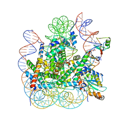 | | RNF20-RNF40/hRad6A-Ub/nucleosome complex | | 分子名称: | DNA (147-MER), E3 ubiquitin-protein ligase BRE1A, E3 ubiquitin-protein ligase BRE1B, ... | | 著者 | Ai, H, Deng, Z, Sun, M, Du, Y, Pan, M, Liu, L. | | 登録日 | 2023-02-15 | | 公開日 | 2023-09-06 | | 最終更新日 | 2023-09-20 | | 実験手法 | ELECTRON MICROSCOPY (3.12 Å) | | 主引用文献 | Mechanistic insights into nucleosomal H2B monoubiquitylation mediated by yeast Bre1-Rad6 and its human homolog RNF20/RNF40-hRAD6A.
Mol.Cell, 83, 2023
|
|
3ELU
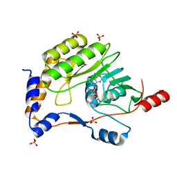 | | Wesselsbron virus Methyltransferase in complex with AdoMet | | 分子名称: | Methyltransferase, S-ADENOSYLMETHIONINE, SULFATE ION | | 著者 | Bollati, M, Ricagno, S, Milani, M, Mastrangelo, E, Bolognesi, M. | | 登録日 | 2008-09-23 | | 公開日 | 2008-11-18 | | 最終更新日 | 2023-11-01 | | 実験手法 | X-RAY DIFFRACTION (2 Å) | | 主引用文献 | Recognition of RNA Cap in the Wesselsbron Virus NS5 Methyltransferase Domain: Implications for RNA-Capping Mechanisms in Flavivirus
J.Mol.Biol., 385, 2009
|
|
3EMD
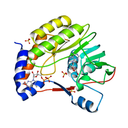 | | Wesselsbron virus Methyltransferase in complex with Sinefungin and 7MeGpppA | | 分子名称: | P1-7-METHYLGUANOSINE-P3-ADENOSINE-5',5'-TRIPHOSPHATE, SINEFUNGIN, SULFATE ION, ... | | 著者 | Bollati, M, Milani, M, Ricagno, S, Mastrangelo, E, Bolognesi, M. | | 登録日 | 2008-09-24 | | 公開日 | 2008-11-18 | | 最終更新日 | 2023-11-01 | | 実験手法 | X-RAY DIFFRACTION (2 Å) | | 主引用文献 | Recognition of RNA Cap in the Wesselsbron Virus NS5 Methyltransferase Domain: Implications for RNA-Capping Mechanisms in Flavivirus
J.Mol.Biol., 385, 2009
|
|
2I3R
 | | Engineered catalytic domain of protein tyrosine phosphatase HPTPbeta | | 分子名称: | CHLORIDE ION, Receptor-type tyrosine-protein phosphatase beta | | 著者 | Evdokimov, A.G, Pokross, M.E, Walter, R.L, Mekel, M. | | 登録日 | 2006-08-20 | | 公開日 | 2006-08-29 | | 最終更新日 | 2023-08-30 | | 実験手法 | X-RAY DIFFRACTION (1.85 Å) | | 主引用文献 | Engineering the catalytic domain of human protein tyrosine phosphatase beta for structure-based drug discovery.
Acta Crystallogr.,Sect.D, 62, 2006
|
|
2UZ5
 | | Solution structure of the fkbp-domain of Legionella pneumophila Mip | | 分子名称: | MACROPHAGE INFECTIVITY POTENTIATOR | | 著者 | Ceymann, A, Horstmann, M, Ehses, P, Schweimer, K, Steinert, M, Kamphausen, T, Fischer, G, Hacker, J, Rosch, P, Faber, C. | | 登録日 | 2007-04-25 | | 公開日 | 2007-06-12 | | 最終更新日 | 2024-05-15 | | 実験手法 | SOLUTION NMR | | 主引用文献 | Domain Motions of the Mip Protein from Legionella Pneumophila
Biochemistry, 45, 2006
|
|
2IBN
 | | Crystal structure of Human myo-Inositol Oxygenase (MIOX) | | 分子名称: | (2S,3R,4R,5S,6S)-2,3,4,5,6-PENTAHYDROXYCYCLOHEXANONE, CYSTEINE, FE (III) ION, ... | | 著者 | Hallberg, B.M, Busam, R.D, Arrowsmith, C, Berglund, H, Collins, R, Edwards, A, Ehn, M, Flodin, S, Flores, A, Graslund, S, Hammarstrom, M, Hogbom, M, Holmberg-Schiavone, L, Johansson, I, Karlberg, T, Kotenyova, T, Nilsson-Ehle, P, Nordlund, P, Nyman, T, Ogg, D, Sagemark, J, Stenmark, P, Sundstrom, M, Uppenberg, J, Van Den Berg, S, Weigelt, J, Thorsell, A.G, Persson, C, Structural Genomics Consortium (SGC) | | 登録日 | 2006-09-11 | | 公開日 | 2006-10-17 | | 最終更新日 | 2023-12-13 | | 実験手法 | X-RAY DIFFRACTION (1.5 Å) | | 主引用文献 | Structural and Biophysical Characterization of Human myo-Inositol Oxygenase
J.Biol.Chem., 283, 2008
|
|
7AEN
 | | Galectin-8 N-terminal carbohydrate recognition domain in complex with methyl 3-O-((7-carboxy)quinolin-2-yl)-methoxy)-beta-D-galactopyranoside | | 分子名称: | CHLORIDE ION, GLYCEROL, Isoform 2 of Galectin-8, ... | | 著者 | Hassan, M, Klavern, V.S, Hakansson, M, Anderluh, M, Tomasic, T, Jakopin, Z, Nilsson, J.U, Kovacic, R, Walse, B, Diehl, C. | | 登録日 | 2020-09-17 | | 公開日 | 2021-07-28 | | 最終更新日 | 2024-01-31 | | 実験手法 | X-RAY DIFFRACTION (1.6 Å) | | 主引用文献 | Structure-Guided Design of d-Galactal Derivatives with High Affinity and Selectivity for the Galectin-8 N-Terminal Domain
Acs Med.Chem.Lett., 12, 2021
|
|
4IJ6
 | | Crystal Structure of a Novel-type Phosphoserine Phosphatase Mutant (H9A) from Hydrogenobacter thermophilus TK-6 in Complex with L-phosphoserine | | 分子名称: | 1,2-ETHANEDIOL, CHLORIDE ION, PHOSPHOSERINE, ... | | 著者 | Chiba, Y, Horita, S, Ohtsuka, J, Arai, H, Nagata, K, Igarashi, Y, Tanokura, M, Ishii, M. | | 登録日 | 2012-12-21 | | 公開日 | 2013-03-20 | | 最終更新日 | 2023-11-08 | | 実験手法 | X-RAY DIFFRACTION (1.8 Å) | | 主引用文献 | Structural units important for activity of a novel-type phosphoserine phosphatase from Hydrogenobacter thermophilus TK-6 revealed by crystal structure analysis
J.Biol.Chem., 288, 2013
|
|
2I3U
 | | Structural studies of protein tyrosine phosphatase beta catalytic domain in complex with inhibitors | | 分子名称: | Receptor-type tyrosine-protein phosphatase beta | | 著者 | Evdokimov, A.G, Pokross, M.E, Walter, R.L, Mekel, M. | | 登録日 | 2006-08-20 | | 公開日 | 2006-08-29 | | 最終更新日 | 2023-08-30 | | 実験手法 | X-RAY DIFFRACTION (1.85 Å) | | 主引用文献 | Engineering the catalytic domain of human protein tyrosine phosphatase beta for structure-based drug discovery.
Acta Crystallogr.,Sect.D, 62, 2006
|
|
5DCS
 | |
2I4E
 | | Structural studies of protein tyrosine phosphatase beta catalytic domain in complex with inhibitors | | 分子名称: | Receptor-type tyrosine-protein phosphatase beta, VANADATE ION | | 著者 | Evdokimov, A.G, Pokross, M.E, Walter, R.L, Mekel, M. | | 登録日 | 2006-08-21 | | 公開日 | 2006-08-29 | | 最終更新日 | 2023-08-30 | | 実験手法 | X-RAY DIFFRACTION (1.75 Å) | | 主引用文献 | Engineering the catalytic domain of human protein tyrosine phosphatase beta for structure-based drug discovery.
Acta Crystallogr.,Sect.D, 62, 2006
|
|
4IOH
 | | Crystal Structure of the Tll1086 protein from Thermosynechococcus elongatus, Northeast Structural Genomics Consortium Target TeR258 | | 分子名称: | Tll1086 protein | | 著者 | Vorobiev, S, Su, M, Seetharaman, J, Maglaqui, M, Xiao, X, Kohan, E, Wang, D, Everett, J.K, Acton, T.B, Montelione, G.T, Tong, L, Hunt, J.F, Northeast Structural Genomics Consortium (NESG) | | 登録日 | 2013-01-07 | | 公開日 | 2013-01-30 | | 最終更新日 | 2013-11-20 | | 実験手法 | X-RAY DIFFRACTION (2.53 Å) | | 主引用文献 | Crystal Structure of the Tll1086 protein from Thermosynechococcus elongatus.
To be Published
|
|
4R15
 | | High-resolution crystal structure of Z-DNA in complex with Cr3+ cations | | 分子名称: | CHROMIUM ION, DNA (5'-D(*CP*GP*CP*GP*CP*G)-3') | | 著者 | Drozdzal, P, Gilski, M, Kierzek, R, Lomozik, L, Jaskolski, M. | | 登録日 | 2014-08-04 | | 公開日 | 2015-03-04 | | 最終更新日 | 2023-09-20 | | 実験手法 | X-RAY DIFFRACTION (0.97 Å) | | 主引用文献 | High-resolution crystal structure of Z-DNA in complex with Cr(3+) cations.
J.Biol.Inorg.Chem., 20, 2015
|
|


