Loading
PDBj
MenuPDBj@FacebookPDBj@TwitterPDBj@YouTubewwPDB FoundationwwPDB
RCSB PDBPDBeBMRBAdv. SearchSearch help
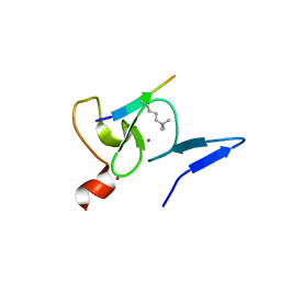
Search by PDB author
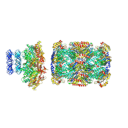
7LHI
DownloadVisualize
BU of 7lhi by Molmil
Cryo-EM structure of E. coli P pilus tip assembly intermediate PapC-PapD-PapK-PapF-PapG
Descriptor: Chaperone protein PapD, Fimbrial adapter PapK, Fimbrial protein, ...
Authors:Du, M, Yuan, Z, Werneburg, G, Henderson, N, Chauhan, H, Kovach, A, Zhao, G, Johl, J, Li, H, Thanassi, D.
Deposit date:2021-01-23
Release date:2021-08-11
Last modified:2024-05-29
Method:ELECTRON MICROSCOPY (7.6 Å)
Cite:Processive dynamics of the usher assembly platform during uropathogenic Escherichia coli P pilus biogenesis.
Nat Commun, 12, 2021
6AYF
DownloadVisualize
BU of 6ayf by Molmil
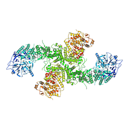
TRPML3/ML-SA1 complex at pH 7.4
Descriptor: 2-acetamido-2-deoxy-beta-D-glucopyranose, Mucolipin-3
Authors:Zhou, X, Li, M, Su, D, Jia, Q, Li, H, Li, X, Yang, J.
Deposit date:2017-09-08
Release date:2017-11-08
Last modified:2024-03-13
Method:ELECTRON MICROSCOPY (3.62 Å)
Cite:Cryo-EM structures of the human endolysosomal TRPML3 channel in three distinct states.
Nat. Struct. Mol. Biol., 24, 2017
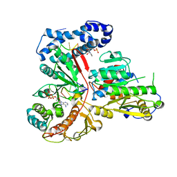
2RFK
DownloadVisualize
BU of 2rfk by Molmil
Substrate RNA Positioning in the Archaeal H/ACA Ribonucleoprotein Complex
Descriptor: Probable tRNA pseudouridine synthase B, Ribosome biogenesis protein Nop10, Small nucleolar rnp similar to gar1, ...
Authors:Liang, B, Xue, S, Terns, R.M, Terns, M.P, Li, H, Southeast Collaboratory for Structural Genomics (SECSG)
Deposit date:2007-09-30
Release date:2008-02-05
Last modified:2023-08-30
Method:X-RAY DIFFRACTION (2.87 Å)
Cite:Substrate RNA positioning in the archaeal H/ACA ribonucleoprotein complex.
Nat.Struct.Mol.Biol., 14, 2007
6AYG
DownloadVisualize
BU of 6ayg by Molmil
Human Apo-TRPML3 channel at pH 4.8
Descriptor: Mucolipin-3
Authors:Zhou, X, Li, M, Su, D, Jia, Q, Li, H, Li, X, Yang, J.
Deposit date:2017-09-08
Release date:2017-11-08
Last modified:2024-03-13
Method:ELECTRON MICROSCOPY (4.65 Å)
Cite:Cryo-EM structures of the human endolysosomal TRPML3 channel in three distinct states.
Nat. Struct. Mol. Biol., 24, 2017
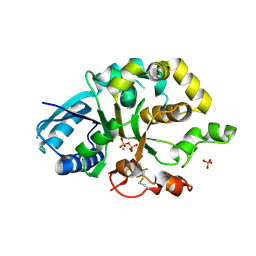
2RK5
DownloadVisualize
BU of 2rk5 by Molmil
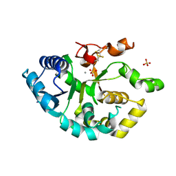
Crystal structure of a domain of the putative hemolysin from Streptococcus mutans UA159
Descriptor: Putative hemolysin
Authors:Zhang, R, Li, H, Clancy, S, Joachimiak, A, Midwest Center for Structural Genomics (MCSG)
Deposit date:2007-10-16
Release date:2007-11-06
Last modified:2024-02-21
Method:X-RAY DIFFRACTION (1.5 Å)
Cite:The crystal structure of a domain of the putative hemolysin from Streptococcus mutans UA159.
To be Published
6AYE
DownloadVisualize
BU of 6aye by Molmil
Human apo-TRPML3 channel at pH 7.4
Descriptor: Mucolipin-3
Authors:Zhou, X, Li, M, Su, D, Jia, Q, Li, H, Li, X, Yang, J.
Deposit date:2017-09-08
Release date:2017-11-08
Last modified:2024-03-13
Method:ELECTRON MICROSCOPY (4.06 Å)
Cite:Cryo-EM structures of the human endolysosomal TRPML3 channel in three distinct states.
Nat. Struct. Mol. Biol., 24, 2017
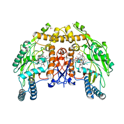
1RS6
DownloadVisualize
BU of 1rs6 by Molmil
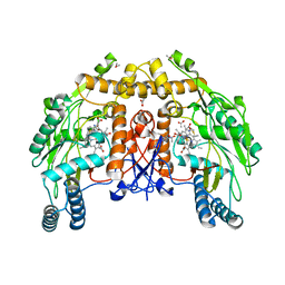
Rat neuronal NOS heme domain with D-lysine-D-nitroarginine amide bound
Descriptor: 5,6,7,8-TETRAHYDROBIOPTERIN, ACETATE ION, D-MANNITOL, ...
Authors:Flinspach, M, Li, H, Jamal, J, Yang, W, Huang, H, Silverman, R.B, Poulos, T.L.
Deposit date:2003-12-09
Release date:2004-05-18
Last modified:2024-02-14
Method:X-RAY DIFFRACTION (1.95 Å)
Cite:Structures of the Neuronal and Endothelial Nitric Oxide Synthase Heme Domain with d-Nitroarginine-Containing Dipeptide Inhibitors Bound.
Biochemistry, 43, 2004
1Z28
DownloadVisualize
BU of 1z28 by Molmil
Crystal Structures of SULT1A2 and SULT1A1*3: Implications in the bioactivation of N-hydroxy-2-acetylamino fluorine (OH-AAF)
Descriptor: ADENOSINE-3'-5'-DIPHOSPHATE, Phenol-sulfating phenol sulfotransferase 1
Authors:Lu, J, Li, H, Liu, M.C, Zhang, J, Li, M, An, X, Chang, W.
Deposit date:2005-03-07
Release date:2006-05-30
Last modified:2023-10-25
Method:X-RAY DIFFRACTION (2.3 Å)
Cite:Crystal structures of SULT1A2 and SULT1A1 *3: insights into the substrate inhibition and the role of Tyr149 in SULT1A2.
Biochem.Biophys.Res.Commun., 396, 2010
1YW5
DownloadVisualize
BU of 1yw5 by Molmil
Peptidyl-prolyl isomerase ESS1 from Candida albicans
Descriptor: peptidyl prolyl cis/trans isomerase
Authors:Li, Z, Li, H, Devasahayam, G, Gemmill, T, Chaturvedi, V, Hanes, S.D, Van Roey, P.
Deposit date:2005-02-17
Release date:2005-04-26
Last modified:2024-02-14
Method:X-RAY DIFFRACTION (1.6 Å)
Cite:The Structure of the Candida albicans Ess1 Prolyl Isomerase Reveals a Well-Ordered Linker that Restricts Domain Mobility
Biochemistry, 44, 2005
1Z29
DownloadVisualize
BU of 1z29 by Molmil
Crystal Structures of SULT1A2 and SULT1A1*3: Implications in the bioactivation of N-hydroxy-2-acetylamino fluorine (OH-AAF)
Descriptor: ACETIC ACID, ADENOSINE-3'-5'-DIPHOSPHATE, CALCIUM ION, ...
Authors:Lu, J, Li, H, Liu, M.C, Zhang, J, Li, M, An, X, Chang, W.
Deposit date:2005-03-07
Release date:2006-05-30
Last modified:2023-10-25
Method:X-RAY DIFFRACTION (2.4 Å)
Cite:Crystal structures of SULT1A2 and SULT1A1 *3: insights into the substrate inhibition and the role of Tyr149 in SULT1A2.
Biochem.Biophys.Res.Commun., 396, 2010
1I83
DownloadVisualize
BU of 1i83 by Molmil
BOVINE ENDOTHELIAL NITRIC OXIDE SYNTHASE HEME DOMAIN COMPLEXED WITH N1,N14-BIS((S-METHYL)ISOTHIOUREIDO)TETRADECANE (H4B FREE)
Descriptor: ACETATE ION, GLYCEROL, N1,N14-BIS((S-METHYL)ISOTHIOUREIDO)TETRADECANE, ...
Authors:Raman, C.S, Li, H, Martasek, P, Babu, B.R, Griffith, O.W, Masters, B.S.S, Poulos, T.L.
Deposit date:2001-03-12
Release date:2001-08-08
Last modified:2011-07-13
Method:X-RAY DIFFRACTION (2 Å)
Cite:Implications for isoform-selective inhibitor design derived from the binding mode of bulky isothioureas to the heme domain of endothelial nitric-oxide synthase.
J.Biol.Chem., 276, 2001
1RS7
DownloadVisualize
BU of 1rs7 by Molmil
Rat neuronal NOS heme domain with D-phenylalanine-D-nitroarginine amide bound
Descriptor: 5,6,7,8-TETRAHYDROBIOPTERIN, ACETATE ION, D-MANNITOL, ...
Authors:Flinspach, M, Li, H, Jamal, J, Yang, W, Huang, H, Silverman, R.B, Poulos, T.L.
Deposit date:2003-12-09
Release date:2004-05-18
Last modified:2024-02-14
Method:X-RAY DIFFRACTION (1.95 Å)
Cite:Structures of the Neuronal and Endothelial Nitric Oxide Synthase Heme Domain with d-Nitroarginine-Containing Dipeptide Inhibitors Bound.
Biochemistry, 43, 2004
1RS9
DownloadVisualize
BU of 1rs9 by Molmil
Bovine endothelial NOS heme domain with D-phenylalanine-D-nitroarginine amide bound
Descriptor: 5,6,7,8-TETRAHYDROBIOPTERIN, ACETATE ION, CACODYLATE ION, ...
Authors:Flinspach, M, Li, H, Jamal, J, Yang, W, Huang, H, Silverman, R.B, Poulos, T.L.
Deposit date:2003-12-09
Release date:2004-06-15
Last modified:2024-02-14
Method:X-RAY DIFFRACTION (2.22 Å)
Cite:Structures of the neuronal and endothelial nitric oxide synthase heme domain with D-nitroarginine-containing dipeptide inhibitors bound.
Biochemistry, 43, 2004
1DMK
DownloadVisualize
BU of 1dmk by Molmil
BOVINE ENDOTHELIAL NITRIC OXIDE SYNTHASE HEME DOMAIN COMPLEXED WITH 4-AMINO-6-PHENYL-TETRAHYDROPTERIDINE
Descriptor: 2,4-DIAMINO-6-PHENYL-5,6,7,8,-TETRAHYDROPTERIDINE, ACETATE ION, CACODYLATE ION, ...
Authors:Kotsonis, P, Frohlich, L.G, Raman, C.S, Li, H, Berg, M, Gerwig, R, Groehn, V, Kang, Y, Al-Masoudi, N, Taghavi-Moghadam, S, Mohr, D, Munch, U, Schnabel, J, Martasek, P, Masters, B.S, Strobel, H, Poulos, T, Matter, H, Pfleiderer, W, Schmidt, H.H.
Deposit date:1999-12-14
Release date:2000-12-20
Last modified:2024-02-07
Method:X-RAY DIFFRACTION (1.9 Å)
Cite:Structural basis for pterin antagonism in nitric-oxide synthase. Development of novel 4-oxo-pteridine antagonists of (6R)-5,6,7,8-tetrahydrobiopterin
J.Biol.Chem., 276, 2001
6WPG
DownloadVisualize
BU of 6wpg by Molmil
Structural Basis of Salicylic Acid Perception by Arabidopsis NPR Proteins
Descriptor: 2-HYDROXYBENZOIC ACID, Regulatory protein NPR4
Authors:Wang, W, Withers, J, Li, H, Zwack, P.J, Rusnac, D.V, Shi, H, Liu, L, Yan, S, Hinds, T.R, Guttman, M, Dong, X, Zheng, N.
Deposit date:2020-04-27
Release date:2020-08-12
Last modified:2024-03-06
Method:X-RAY DIFFRACTION (2.283 Å)
Cite:Structural basis of salicylic acid perception by Arabidopsis NPR proteins.
Nature, 586, 2020
3UBU
DownloadVisualize
BU of 3ubu by Molmil
Crystal structure of agkisacucetin, a GpIb-binding snaclec (snake C-type lectin) that inhibits platelet
Descriptor: Agglucetin subunit alpha-1, Agglucetin subunit beta-2, GLYCEROL, ...
Authors:Gao, Y, Ge, H, Chen, H, Li, H, Liu, Y, Niu, L, Teng, M.
Deposit date:2011-10-25
Release date:2012-04-11
Method:X-RAY DIFFRACTION (1.91 Å)
Cite:Crystal structure of agkisacucetin, a Gpib-binding snake C-type lectin that inhibits platelet adhesion and aggregation.
Proteins, 2012
1PKF
DownloadVisualize
BU of 1pkf by Molmil
Crystal Structure of Epothilone D-bound Cytochrome P450epoK
Descriptor: EPOTHILONE D, PROTOPORPHYRIN IX CONTAINING FE, cytochrome p450EpoK
Authors:Nagano, S, Li, H, Shimizu, H, Nishida, C, Ogura, H, Ortiz de Montellano, P.R, Poulos, T.L.
Deposit date:2003-06-05
Release date:2003-10-28
Last modified:2024-02-14
Method:X-RAY DIFFRACTION (2.1 Å)
Cite:Crystal Structures of Epothilone D-bound, Epothilone B-bound, and Substrate-free Forms of Cytochrome P450epoK
J.Biol.Chem., 278, 2003
1Q5D
DownloadVisualize
BU of 1q5d by Molmil
Epothilone B-bound Cytochrome P450epoK
Descriptor: 7,11-DIHYDROXY-8,8,10,12,16-PENTAMETHYL-3-[1-METHYL-2-(2-METHYL-THIAZOL-4-YL)VINYL]-4,17-DIOXABICYCLO[14.1.0]HEPTADECANE-5,9-DIONE, P450 epoxidase, PROTOPORPHYRIN IX CONTAINING FE
Authors:Nagano, S, Li, H, Shimizu, H, Nishida, C, Ogura, H, Ortiz de Montellano, P.R, Poulos, T.L.
Deposit date:2003-08-06
Release date:2003-10-28
Last modified:2024-04-03
Method:X-RAY DIFFRACTION (1.93 Å)
Cite:Crystal structures of epothilone D-bound, epothilone B-bound, and substrate-free forms of cytochrome P450epoK
J.Biol.Chem., 278, 2003
8D1V
DownloadVisualize
BU of 8d1v by Molmil
Cryo-EM structure of guide RNA and target RNA bound Cas7-11
Descriptor: CRISPR RNA (34-MER), CRISPR-associated RAMP family protein, SS target RNA (5'-R(P*AP*GP*CP*UP*UP*GP*GP*UP*UP*CP*AP*AP*AP*GP*AP*AP*CP*G)-3'), ...
Authors:Rai, J, Goswami, H, Li, H.
Deposit date:2022-05-27
Release date:2022-11-02
Last modified:2024-06-12
Method:ELECTRON MICROSCOPY (2.82 Å)
Cite:Molecular mechanism of active Cas7-11 in processing CRISPR RNA and interfering target RNA.
Elife, 11, 2022
1Q5E
DownloadVisualize
BU of 1q5e by Molmil
Substrate-free Cytochrome P450epoK
Descriptor: P450 epoxidase, PROTOPORPHYRIN IX CONTAINING FE
Authors:Nagano, S, Li, H, Shimizu, H, Nishida, C, Ogura, H, Ortiz de Montellano, P.R, Poulos, T.L.
Deposit date:2003-08-06
Release date:2003-10-28
Last modified:2024-04-03
Method:X-RAY DIFFRACTION (2.65 Å)
Cite:Crystal structures of epothilone D-bound, epothilone B-bound, and substrate-free forms of cytochrome P450epoK
J.Biol.Chem., 278, 2003
4QBS
DownloadVisualize
BU of 4qbs by Molmil
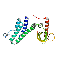
Crystal structure of DNMT3a ADD domain E545R mutant bound to H3T3ph peptide
Descriptor: DNA (cytosine-5)-methyltransferase 3A, Histone H3, SULFATE ION, ...
Authors:Wang, H, Li, H.
Deposit date:2014-05-08
Release date:2015-05-13
Method:X-RAY DIFFRACTION (1.8 Å)
Cite:Engineering of a histone-recognition domain in a de novo DNA methyltransferase alters the epigenetic landscape of ESCs
To be Published
6W6G
DownloadVisualize
BU of 6w6g by Molmil
The Mycobacterium tuberculosis ClpB disaggregase hexamer structure in conformation I in the presence of DnaK chaperone and a model substrate
Descriptor: ADENOSINE-5'-DIPHOSPHATE, Chaperone protein ClpB, PHOSPHOTHIOPHOSPHORIC ACID-ADENYLATE ESTER, ...
Authors:Yin, Y, Li, H.
Deposit date:2020-03-16
Release date:2021-03-17
Last modified:2024-05-29
Method:ELECTRON MICROSCOPY (3.1 Å)
Cite:Structural basis for aggregate dissolution and refolding by the Mycobacterium tuberculosis ClpB-DnaK bi-chaperone system.
Cell Rep, 35, 2021
6W6H
DownloadVisualize
BU of 6w6h by Molmil
The Mycobacterium tuberculosis ClpB disaggregase hexamer structure in conformation II in the presence of DnaK chaperone and a model substrate
Descriptor: ADENOSINE-5'-DIPHOSPHATE, Chaperone protein ClpB, PHOSPHOTHIOPHOSPHORIC ACID-ADENYLATE ESTER, ...
Authors:Yin, Y, Li, H.
Deposit date:2020-03-16
Release date:2021-05-26
Last modified:2024-05-29
Method:ELECTRON MICROSCOPY (3.3 Å)
Cite:Structural basis for aggregate dissolution and refolding by the Mycobacterium tuberculosis ClpB-DnaK bi-chaperone system.
Cell Rep, 35, 2021
6W6I
DownloadVisualize
BU of 6w6i by Molmil
The Mycobacterium tuberculosis ClpB disaggregase hexamer structure in conformation T in the presence of DnaK chaperone and a model substrate
Descriptor: ADENOSINE-5'-DIPHOSPHATE, Chaperone protein ClpB, PHOSPHOTHIOPHOSPHORIC ACID-ADENYLATE ESTER, ...
Authors:Yin, Y, Li, H.
Deposit date:2020-03-17
Release date:2021-05-26
Last modified:2024-05-29
Method:ELECTRON MICROSCOPY (3.5 Å)
Cite:Structural basis for aggregate dissolution and refolding by the Mycobacterium tuberculosis ClpB-DnaK bi-chaperone system.
Cell Rep, 35, 2021
6W6E
DownloadVisualize
BU of 6w6e by Molmil
The Mycobacterium tuberculosis ClpB disaggregase hexamer structure with a locally refined ClpB middle domain and a DnaK nucleotide binding domain
Descriptor: ADENOSINE-5'-DIPHOSPHATE, Chaperone protein ClpB, Chaperone protein DnaK, ...
Authors:Yin, Y, Li, H.
Deposit date:2020-03-16
Release date:2021-05-26
Last modified:2024-05-29
Method:ELECTRON MICROSCOPY (3.7 Å)
Cite:Structural basis for aggregate dissolution and refolding by the Mycobacterium tuberculosis ClpB-DnaK bi-chaperone system.
Cell Rep, 35, 2021

221051

PDB entries from 2024-06-12

PDB statisticsPDBj update infoContact PDBjnumon