 Movie
Movie Controller


+ Open data

- Basic information
| Entry | Database: PDB / ID: 5i04 | ||||||||||||||||||||||||
|---|---|---|---|---|---|---|---|---|---|---|---|---|---|---|---|---|---|---|---|---|---|---|---|---|---|
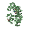
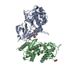
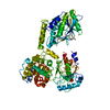
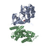
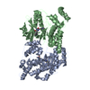
| Title | Crystal structure of the orphan region of human endoglin/CD105 | ||||||||||||||||||||||||
 Components Components | Maltose-binding periplasmic protein,Endoglin | ||||||||||||||||||||||||
 Keywords Keywords | SIGNALING PROTEIN / ORPHAN DOMAIN / ANGIOGENESIS / GLYCOPROTEIN / RECEPTOR | ||||||||||||||||||||||||
| Function / homology |  Function and homology information Function and homology informationatrioventricular canal morphogenesis / detection of hypoxia / endothelial microparticle / venous blood vessel morphogenesis / dorsal aorta morphogenesis / cell migration involved in endocardial cushion formation / vascular associated smooth muscle cell development / atrial cardiac muscle tissue morphogenesis / positive regulation of vascular associated smooth muscle cell differentiation / central nervous system vasculogenesis ...atrioventricular canal morphogenesis / detection of hypoxia / endothelial microparticle / venous blood vessel morphogenesis / dorsal aorta morphogenesis / cell migration involved in endocardial cushion formation / vascular associated smooth muscle cell development / atrial cardiac muscle tissue morphogenesis / positive regulation of vascular associated smooth muscle cell differentiation / central nervous system vasculogenesis / epithelial to mesenchymal transition involved in endocardial cushion formation / positive regulation of epithelial to mesenchymal transition involved in endocardial cushion formation / cardiac ventricle morphogenesis / regulation of transforming growth factor beta receptor signaling pathway / galactose binding / cardiac atrium morphogenesis / smooth muscle tissue development / type II transforming growth factor beta receptor binding / activin binding / type I transforming growth factor beta receptor binding / glycosaminoglycan binding / ventricular trabecula myocardium morphogenesis / positive regulation of BMP signaling pathway / outflow tract septum morphogenesis / transforming growth factor beta binding / artery morphogenesis / endocardial cushion morphogenesis / branching involved in blood vessel morphogenesis / detection of maltose stimulus / negative regulation of endothelial cell proliferation / heart looping / maltose transport complex / negative regulation of SMAD protein signal transduction / positive regulation of systemic arterial blood pressure / carbohydrate transport / signaling receptor activator activity / positive regulation of SMAD protein signal transduction / extracellular matrix disassembly / carbohydrate transmembrane transporter activity / maltose binding / maltose transport / maltodextrin transmembrane transport / BMP signaling pathway / vasculogenesis / regulation of cell adhesion / coreceptor activity / ATP-binding cassette (ABC) transporter complex, substrate-binding subunit-containing / transforming growth factor beta receptor signaling pathway / ATP-binding cassette (ABC) transporter complex / negative regulation of cell migration / cell chemotaxis / cell motility / negative regulation of transforming growth factor beta receptor signaling pathway / wound healing / positive regulation of angiogenesis / transmembrane signaling receptor activity / cell migration / regulation of cell population proliferation / outer membrane-bounded periplasmic space / response to hypoxia / periplasmic space / receptor complex / positive regulation of phosphatidylinositol 3-kinase/protein kinase B signal transduction / cell adhesion / nuclear body / negative regulation of gene expression / external side of plasma membrane / focal adhesion / DNA damage response / regulation of DNA-templated transcription / cell surface / negative regulation of transcription by RNA polymerase II / protein homodimerization activity / positive regulation of transcription by RNA polymerase II / extracellular space / identical protein binding / membrane / plasma membrane Similarity search - Function | ||||||||||||||||||||||||
| Biological species |   Homo sapiens (human) Homo sapiens (human) | ||||||||||||||||||||||||
| Method |  X-RAY DIFFRACTION / SYNCHROTRON / MOLECULAR REPLACEMENT / Resolution: 2.42 Å X-RAY DIFFRACTION / SYNCHROTRON / MOLECULAR REPLACEMENT / Resolution: 2.42 Å | ||||||||||||||||||||||||
 Authors Authors | Saito, T. / Bokhove, M. / de Sanctis, D. / Jovine, L. | ||||||||||||||||||||||||
| Funding support |  Sweden, 7items Sweden, 7items
| ||||||||||||||||||||||||
 Citation Citation | Journal: Cell Rep / Year: 2017 Title: Structural Basis of the Human Endoglin-BMP9 Interaction: Insights into BMP Signaling and HHT1. Authors: Saito, T. / Bokhove, M. / Croci, R. / Zamora-Caballero, S. / Han, L. / Letarte, M. / de Sanctis, D. / Jovine, L. #1: Journal: J. Biol. Chem. / Year: 1990 Title: Primary structure of endoglin, an RGD-containing glycoprotein of human endothelial cells. Authors: Gougos, A. / Letarte, M. #2: Journal: J. Biol. Chem. / Year: 2011 Title: Soluble endoglin specifically binds bone morphogenetic proteins 9 and 10 via its orphan domain, inhibits blood vessel formation, and suppresses tumor growth. Authors: Castonguay, R. / Werner, E.D. / Matthews, R.G. / Presman, E. / Mulivor, A.W. / Solban, N. / Sako, D. / Pearsall, R.S. / Underwood, K.W. / Seehra, J. / Kumar, R. / Grinberg, A.V. #3: Journal: PLoS ONE / Year: 2012 Title: Structural and functional insights into endoglin ligand recognition and binding. Authors: Alt, A. / Miguel-Romero, L. / Donderis, J. / Aristorena, M. / Blanco, F.J. / Round, A. / Rubio, V. / Bernabeu, C. / Marina, A. #4: Journal: PLoS ONE / Year: 2012 Title: Endoglin requirement for BMP9 signaling in endothelial cells reveals new mechanism of action for selective anti-endoglin antibodies. Authors: Nolan-Stevaux, O. / Zhong, W. / Culp, S. / Shaffer, K. / Hoover, J. / Wickramasinghe, D. / Ruefli-Brasse, A. | ||||||||||||||||||||||||
| History |
|
- Structure visualization
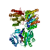
| Structure viewer | Molecule: MolmilJmol/JSmol |
|---|
- Downloads & links
-Download
| PDBx/mmCIF format | 5i04.cif.gz | 275.7 KB | Display | PDBx/mmCIF format |
|---|---|---|---|---|
| PDB format | pdb5i04.ent.gz | 221.1 KB | Display | PDB format |
| PDBx/mmJSON format | 5i04.json.gz | Tree view | PDBx/mmJSON format | |
| Others |  Other downloads Other downloads |
-Validation report
| Arichive directory | https://data.pdbj.org/pub/pdb/validation_reports/i0/5i04ftp://data.pdbj.org/pub/pdb/validation_reports/i0/5i04 | HTTPS FTP |
|---|
-Related structure data
| Related structure data |  5hzvC 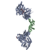 5hzwC  5i05C 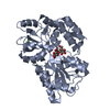 3setS 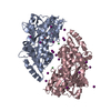 3sexS 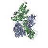 4wrnS C: citing same article ( S: Starting model for refinement |
|---|---|
| Similar structure data |


-Links
 PDBj
PDBj


- Assembly
| Deposited unit | 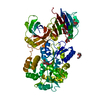
| ||||||||
|---|---|---|---|---|---|---|---|---|---|
| 1 |
| ||||||||
| Unit cell |
|
-Components
| #1: Protein | Mass: 75254.336 Da / Num. of mol.: 1 Mutation: I57T, D137A, K138A, E227A, N228A, A270H, K274H, K294A, A367V, I372V, E414A, E417A, D418A, R422N,I57T, D137A, K138A, E227A, N228A, A270H, K274H, K294A, A367V, I372V, E414A, E417A, D418A, ...Mutation: I57T, D137A, K138A, E227A, N228A, A270H, K274H, K294A, A367V, I372V, E414A, E417A, D418A, R422N,I57T, D137A, K138A, E227A, N228A, A270H, K274H, K294A, A367V, I372V, E414A, E417A, D418A, R422N,I57T, D137A, K138A, E227A, N228A, A270H, K274H, K294A, A367V, I372V, E414A, E417A, D418A, R422N,I57T, D137A, K138A, E227A, N228A, A270H, K274H, K294A, A367V, I372V, E414A, E417A, D418A, R422N,I57T, D137A, K138A, E227A, N228A, A270H, K274H, K294A, A367V, I372V, E414A, E417A, D418A, R422N,I57T, D137A, K138A, E227A, N228A, A270H, K274H, K294A, A367V, I372V, E414A, E417A, D418A, R422N,I57T, D137A, K138A, E227A, N228A, A270H, K274H, K294A, A367V, I372V, E414A, E417A, D418A, R422N,I57T, D137A, K138A, E227A, N228A, A270H, K274H, K294A, A367V, I372V, E414A, E417A, D418A, R422N,I57T, D137A, K138A, E227A, N228A, A270H, K274H, K294A, A367V, I372V, E414A, E417A, D418A, R422N,I57T, D137A, K138A, E227A, N228A, A270H, K274H, K294A, A367V, I372V, E414A, E417A, D418A, R422N,I57T, D137A, K138A, E227A, N228A, A270H, K274H, K294A, A367V, I372V, E414A, E417A, D418A, R422N,I57T, D137A, K138A, E227A, N228A, A270H, K274H, K294A, A367V, I372V, E414A, E417A, D418A, R422N,I57T, D137A, K138A, E227A, N228A, A270H, K274H, K294A, A367V, I372V, E414A, E417A, D418A, R422N,I57T, D137A, K138A, E227A, N228A, A270H, K274H, K294A, A367V, I372V, E414A, E417A, D418A, R422N,I57T, D137A, K138A, E227A, N228A, A270H, K274H, K294A, A367V, I372V, E414A, E417A, D418A, R422N,I57T, D137A, K138A, E227A, N228A, A270H, K274H, K294A, A367V, I372V, E414A, E417A, D418A, R422N Source method: isolated from a genetically manipulated source Details: THIS PROTEIN IS A CHIMERA. RESIDUES 56-422 ARE FROM E. COLI MALTOSE BINDING PROTEIN (MBP), CORRESPOND TO RESIDUES 27-393 OF SWISS-PROT DATABASE ENTRY P0AEX9 AND CONTAIN MUTATIONS I57T, ...Details: THIS PROTEIN IS A CHIMERA. RESIDUES 56-422 ARE FROM E. COLI MALTOSE BINDING PROTEIN (MBP), CORRESPOND TO RESIDUES 27-393 OF SWISS-PROT DATABASE ENTRY P0AEX9 AND CONTAIN MUTATIONS I57T, D137A, K138A, E227A, N228A, A270H, K274H, K294A, A367V, I372V, E414A, E417A, D418A AND R422N (CORRESPONDING TO I28T, D108A, K109A, E198A, N199A, A241H, K245H, K265A, A338V, I343V, E385A, E388A, D389A AND R393N IN P0AEX9). RESIDUES 426-737 ARE FROM HUMAN ENDOGLIN PROTEIN AND CORRESPOND TO RESIDUES 26-337 OF SWISS-PROT DATABASE ENTRY P17813. SUBTRACTING 400 FROM THE PDB ENTRY RESIDUE NUMBERING RESULTS IN THE NUMBERING ACCORDING TO UNIPROT ENTRY P17813. Source: (gene. exp.) Homo sapiens (human)Cell: Endothelial / Gene: malE, b4034, JW3994, ENG, END / Plasmid: pHLsec / Cell line (production host): HEK293S / Production host: Homo sapiens (human) / References: UniProt: P0AEX9, UniProt: P17813 | ||||
|---|---|---|---|---|---|
| #2: Polysaccharide | alpha-D-glucopyranose-(1-4)-alpha-D-glucopyranose / alpha-maltose  Source method: isolated from a genetically manipulated source Details: oligosaccharide / References: alpha-maltose | ||||
| #3: Sugar | ChemComp-NAG /   Type: D-saccharide, beta linking / Mass: 221.208 Da / Num. of mol.: 1 Type: D-saccharide, beta linking / Mass: 221.208 Da / Num. of mol.: 1Source method: isolated from a genetically manipulated source Formula: C8H15NO6 | ||||
| #4: Chemical |   Mass: 150.173 Da / Num. of mol.: 3 / Source method: obtained synthetically / Formula: C6H14O4 Mass: 150.173 Da / Num. of mol.: 3 / Source method: obtained synthetically / Formula: C6H14O4#5: Water | ChemComp-HOH / |  Mass: 18.015 Da / Num. of mol.: 26 / Source method: isolated from a natural source / Formula: H2O Mass: 18.015 Da / Num. of mol.: 26 / Source method: isolated from a natural source / Formula: H2OHas protein modification | Y | |
-Experimental details
-Experiment
| Experiment | Method: X-RAY DIFFRACTION / Number of used crystals: 1 |
|---|
- Sample preparation
| Crystal | Density Matthews: 3.02 Å3/Da / Density % sol: 57.8 % / Description: Droplet |
|---|---|
| Crystal grow | Temperature: 293 K / Method: vapor diffusion, hanging drop / pH: 8.5 / Details: 30% PEG 1000, 0.1 M TRIS-HCL / PH range: 7.0-8.5 |
-Data collection
| Diffraction | Mean temperature: 100 K |
|---|---|
| Diffraction source | Source: SYNCHROTRON / Site: ESRF  / Beamline: ID29 / Wavelength: 0.97625 Å / Beamline: ID29 / Wavelength: 0.97625 Å |
| Detector | Type: DECTRIS PILATUS 6M / Detector: PIXEL / Date: Sep 10, 2014 |
| Radiation | Monochromator: Si Single Crystal / Protocol: SINGLE WAVELENGTH / Monochromatic (M) / Laue (L): M / Scattering type: x-ray |
| Radiation wavelength | Wavelength: 0.97625 Å / Relative weight: 1 |
| Reflection | Resolution: 2.42→45.74 Å / Num. obs: 33737 / % possible obs: 99.1 % / Observed criterion σ(I): -3 / Redundancy: 3.4 % / Biso Wilson estimate: 70.03 Å2 / Rsym value: 0.046 / Net I/σ(I): 11.76 |
| Reflection shell | Resolution: 2.42→2.49 Å / Redundancy: 3.5 % / Rmerge(I) obs: 0.811 / Mean I/σ(I) obs: 1.25 / % possible all: 98.5 |
- Processing
| Software |
| ||||||||||||||||||||||||||||||||||||||||||||||||||||||||||||||||||||||||||||||||||||||||||||||||||||||||
|---|---|---|---|---|---|---|---|---|---|---|---|---|---|---|---|---|---|---|---|---|---|---|---|---|---|---|---|---|---|---|---|---|---|---|---|---|---|---|---|---|---|---|---|---|---|---|---|---|---|---|---|---|---|---|---|---|---|---|---|---|---|---|---|---|---|---|---|---|---|---|---|---|---|---|---|---|---|---|---|---|---|---|---|---|---|---|---|---|---|---|---|---|---|---|---|---|---|---|---|---|---|---|---|---|---|
| Refinement | Method to determine structure: MOLECULAR REPLACEMENT Starting model: 3SEX, 3SET, 4WRN Resolution: 2.42→45.737 Å / SU ML: 0.47 / Cross valid method: FREE R-VALUE / σ(F): 1.39 / Phase error: 35.15
| ||||||||||||||||||||||||||||||||||||||||||||||||||||||||||||||||||||||||||||||||||||||||||||||||||||||||
| Solvent computation | Shrinkage radii: 0.6 Å / VDW probe radii: 1 Å | ||||||||||||||||||||||||||||||||||||||||||||||||||||||||||||||||||||||||||||||||||||||||||||||||||||||||
| Displacement parameters | Biso mean: 76.6 Å2 | ||||||||||||||||||||||||||||||||||||||||||||||||||||||||||||||||||||||||||||||||||||||||||||||||||||||||
| Refinement step | Cycle: LAST / Resolution: 2.42→45.737 Å
| ||||||||||||||||||||||||||||||||||||||||||||||||||||||||||||||||||||||||||||||||||||||||||||||||||||||||
| Refine LS restraints |
| ||||||||||||||||||||||||||||||||||||||||||||||||||||||||||||||||||||||||||||||||||||||||||||||||||||||||
| LS refinement shell | Refine-ID: X-RAY DIFFRACTION
| ||||||||||||||||||||||||||||||||||||||||||||||||||||||||||||||||||||||||||||||||||||||||||||||||||||||||
| Refinement TLS params. | Method: refined / Refine-ID: X-RAY DIFFRACTION
| ||||||||||||||||||||||||||||||||||||||||||||||||||||||||||||||||||||||||||||||||||||||||||||||||||||||||
| Refinement TLS group |
|
