 Movie
Movie Controller

[English] 日本語
 Yorodumi
- PDB-4wrn: Crystal structure of the polymerization region of human uromoduli... -
+ Open data

- Basic information
| Entry | Database: PDB / ID: 4wrn | ||||||||||||||||||||||||
|---|---|---|---|---|---|---|---|---|---|---|---|---|---|---|---|---|---|---|---|---|---|---|---|---|---|
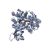
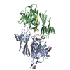
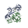
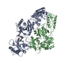
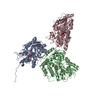
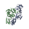
| Title | Crystal structure of the polymerization region of human uromodulin/Tamm-Horsfall protein | ||||||||||||||||||||||||
 Components Components | Maltose-binding periplasmic protein,Uromodulin | ||||||||||||||||||||||||
 Keywords Keywords | STRUCTURAL PROTEIN / ZP DOMAIN / EGF DOMAIN / EXTRACELLULAR MATRIX / GLYCOPROTEIN | ||||||||||||||||||||||||
| Function / homology |  Function and homology information Function and homology informationcitric acid secretion / protein localization to extracellular region / metanephric thick ascending limb development / metanephric distal convoluted tubule development / connective tissue replacement / protein transport into plasma membrane raft / Asparagine N-linked glycosylation / organ or tissue specific immune response / collecting duct development / urea transmembrane transport ...citric acid secretion / protein localization to extracellular region / metanephric thick ascending limb development / metanephric distal convoluted tubule development / connective tissue replacement / protein transport into plasma membrane raft / Asparagine N-linked glycosylation / organ or tissue specific immune response / collecting duct development / urea transmembrane transport / micturition / metanephric ascending thin limb development / protein localization to vacuole / regulation of protein transport / juxtaglomerular apparatus development / intracellular chloride ion homeostasis / renal urate salt excretion / glomerular filtration / antibacterial innate immune response / urate transport / renal sodium ion absorption / neutrophil migration / response to water deprivation / regulation of urine volume / endoplasmic reticulum organization / potassium ion homeostasis / intracellular phosphate ion homeostasis / intracellular sodium ion homeostasis / heterophilic cell-cell adhesion / IgG binding / extrinsic component of membrane / leukocyte cell-cell adhesion / ciliary membrane / detection of maltose stimulus / maltose transport complex / carbohydrate transport / cellular response to unfolded protein / multicellular organismal response to stress / carbohydrate transmembrane transporter activity / maltose binding / maltose transport / maltodextrin transmembrane transport / cellular defense response / renal water homeostasis / side of membrane / ATP-binding cassette (ABC) transporter complex, substrate-binding subunit-containing / ERAD pathway / ATP-binding cassette (ABC) transporter complex / RNA splicing / tumor necrosis factor-mediated signaling pathway / apoptotic signaling pathway / cell chemotaxis / lipid metabolic process / Golgi lumen / regulation of blood pressure / autophagy / intracellular calcium ion homeostasis / spindle pole / outer membrane-bounded periplasmic space / protein folding / gene expression / response to lipopolysaccharide / defense response to Gram-negative bacterium / basolateral plasma membrane / periplasmic space / cilium / apical plasma membrane / response to xenobiotic stimulus / inflammatory response / negative regulation of cell population proliferation / calcium ion binding / DNA damage response / cell surface / endoplasmic reticulum / : / extracellular exosome / membrane Similarity search - Function | ||||||||||||||||||||||||
| Biological species |   Homo sapiens (human) Homo sapiens (human) | ||||||||||||||||||||||||
| Method |  X-RAY DIFFRACTION / SYNCHROTRON / MOLECULAR REPLACEMENT / Resolution: 3.2 Å X-RAY DIFFRACTION / SYNCHROTRON / MOLECULAR REPLACEMENT / Resolution: 3.2 Å | ||||||||||||||||||||||||
 Authors Authors | Bokhove, M. / De Sanctis, D. / Jovine, L. | ||||||||||||||||||||||||
| Funding support |  Sweden, 7items Sweden, 7items
| ||||||||||||||||||||||||
 Citation Citation | Journal: Proc Natl Acad Sci U S A / Year: 2016 Title: A structured interdomain linker directs self-polymerization of human uromodulin. Authors: Marcel Bokhove / Kaoru Nishimura / Martina Brunati / Ling Han / Daniele de Sanctis / Luca Rampoldi / Luca Jovine /   Abstract: Uromodulin (UMOD)/Tamm-Horsfall protein, the most abundant human urinary protein, plays a key role in chronic kidney diseases and is a promising therapeutic target for hypertension. Via its bipartite ...Uromodulin (UMOD)/Tamm-Horsfall protein, the most abundant human urinary protein, plays a key role in chronic kidney diseases and is a promising therapeutic target for hypertension. Via its bipartite zona pellucida module (ZP-N/ZP-C), UMOD forms extracellular filaments that regulate kidney electrolyte balance and innate immunity, as well as protect against renal stones. Moreover, salt-dependent aggregation of UMOD filaments in the urine generates a soluble molecular net that captures uropathogenic bacteria and facilitates their clearance. Despite the functional importance of its homopolymers, no structural information is available on UMOD and how it self-assembles into filaments. Here, we report the crystal structures of polymerization regions of human UMOD and mouse ZP2, an essential sperm receptor protein that is structurally related to UMOD but forms heteropolymers. The structure of UMOD reveals that an extensive hydrophobic interface mediates ZP-N domain homodimerization. This arrangement is required for filament formation and is directed by an ordered ZP-N/ZP-C linker that is not observed in ZP2 but is conserved in the sequence of deafness/Crohn's disease-associated homopolymeric glycoproteins α-tectorin (TECTA) and glycoprotein 2 (GP2). Our data provide an example of how interdomain linker plasticity can modulate the function of structurally similar multidomain proteins. Moreover, the architecture of UMOD rationalizes numerous pathogenic mutations in both UMOD and TECTA genes. #1: Journal: Proc Soc Exp Biol Med / Year: 1950 Title: Characterization and separation of an inhibitor of viral hemagglutination present in urine. #2: Journal: J.Biol.Chem. / Year: 1991 Title: Isolation of the cDNA encoding glycoprotein-2 (GP-2), the major zymogen granule membrane protein. Homology to uromodulin/Tamm-Horsfall protein. Authors: Hoops, T.C. / Rindler, M.J. #3: Journal: Nat.Genet. / Year: 1998 Title: Mutations in the human alpha-tectorin gene cause autosomal dominant non-syndromic hearing impairment. Authors: Verhoeven, K. / Van Laer, L. / Kirschhofer, K. / Legan, P.K. / Hughes, D.C. / Schatteman, I. / Verstreken, M. / Van Hauwe, P. / Coucke, P. / Chen, A. / Smith, R.J. / Somers, T. / Offeciers, ...Authors: Verhoeven, K. / Van Laer, L. / Kirschhofer, K. / Legan, P.K. / Hughes, D.C. / Schatteman, I. / Verstreken, M. / Van Hauwe, P. / Coucke, P. / Chen, A. / Smith, R.J. / Somers, T. / Offeciers, F.E. / Van de Heyning, P. / Richardson, G.P. / Wachtler, F. / Kimberling, W.J. / Willems, P.J. / Govaerts, P.J. / Van Camp, G. #4: Journal: Nat Cell Biol / Year: 2002 Title: The ZP domain is a conserved module for polymerization of extracellular proteins. Authors: Luca Jovine / Huayu Qi / Zev Williams / Eveline Litscher / Paul M Wassarman /  Abstract: Many eukaryotic extracellular proteins share a sequence of unknown function, called the zona pellucida (ZP) domain. Among these proteins are the mammalian sperm receptors ZP2 and ZP3, non-mammalian ...Many eukaryotic extracellular proteins share a sequence of unknown function, called the zona pellucida (ZP) domain. Among these proteins are the mammalian sperm receptors ZP2 and ZP3, non-mammalian egg coat proteins, Tamm-Horsfall protein (THP), glycoprotein-2 (GP-2), alpha- and beta-tectorins, transforming growth factor (TGF)-beta receptor III and endoglin, DMBT-1 (deleted in malignant brain tumour-1), NompA (no-mechanoreceptor-potential-A), Dumpy and cuticlin-1 (refs 1,2). Here, we report that the ZP domain of ZP2, ZP3 and THP is responsible for polymerization of these proteins into filaments of similar supramolecular structure. Most ZP domain proteins are synthesized as precursors with carboxy-terminal transmembrane domains or glycosyl phosphatidylinositol (GPI) anchors. Our results demonstrate that the C-terminal transmembrane domain and short cytoplasmic tail of ZP2 and ZP3 are not required for secretion, but are essential for assembly. Finally, we suggest a molecular basis for dominant human hearing disorders caused by point mutations within the ZP domain of alpha-tectorin. #5: Journal: Am J Kidney Dis / Year: 2003 Title: Tamm-Horsfall glycoprotein: biology and clinical relevance. Authors: Franca Serafini-Cessi / Nadia Malagolini / Daniela Cavallone / Abstract: Tamm-Horsfall glycoprotein (THP) is the most abundant urinary protein in mammals. Urinary excretion occurs by proteolytic cleavage of the large ectodomain of the glycosyl phosphatidylinositol- ...Tamm-Horsfall glycoprotein (THP) is the most abundant urinary protein in mammals. Urinary excretion occurs by proteolytic cleavage of the large ectodomain of the glycosyl phosphatidylinositol-anchored counterpart exposed at the luminal cell surface of the thick ascending limb of Henle's loop. We describe the physical-chemical structure of human THP and its biosynthesis and interaction with other proteins and leukocytes. The clinical relevance of THP reported here includes: (1) involvement in the pathogenesis of cast nephropathy, urolithiasis, and tubulointerstitial nephritis; (2) abnormalities in urinary excretion in renal diseases; and (3) the recent finding that familial juvenile hyperuricemic nephropathy and autosomal dominant medullary cystic kidney disease 2 arise from mutations of the THP gene. We critically examine the literature on the physiological role and mechanism(s) that promote urinary excretion of THP. Some lines of research deal with the in vitro immunoregulatory activity of THP, termed uromodulin when isolated from urine of pregnant women. However, an immunoregulatory function in vivo has not yet been established. In the most recent literature, there is renewed interest in the capacity of urinary THP to compete efficiently with urothelial cell receptors, such as uroplakins, in adhering to type 1 fimbriated Escherichia coli. This property supports the notion that abundant THP excretion in urine is promoted in the host by selective pressure to obtain an efficient defense against urinary tract infections caused by uropathogenic bacteria. #6: Journal: Proc Natl Acad Sci U S A / Year: 2004 Title: A duplicated motif controls assembly of zona pellucida domain proteins. Authors: Luca Jovine / Huayu Qi / Zev Williams / Eveline S Litscher / Paul M Wassarman / Abstract: Many secreted eukaryotic glycoproteins that play fundamental roles in development, hearing, immunity, and cancer polymerize into filaments and extracellular matrices through zona pellucida (ZP) ...Many secreted eukaryotic glycoproteins that play fundamental roles in development, hearing, immunity, and cancer polymerize into filaments and extracellular matrices through zona pellucida (ZP) domains. ZP domain proteins are synthesized as precursors containing C-terminal propeptides that are cleaved at conserved sites. However, the consequences of this processing and the mechanism by which nascent proteins assemble are unclear. By microinjection of mutated DNA constructs into growing oocytes and mammalian cell transfection, we have identified a conserved duplicated motif [EHP (external hydrophobic patch)/IHP (internal hydrophobic patch)] regulating the assembly of mouse ZP proteins. Whereas the transmembrane domain (TMD) of ZP3 can be functionally replaced by an unrelated TMD, mutations in either EHP or IHP do not hinder secretion of full-length ZP3 but completely abolish its assembly. Because mutants truncated before the TMD are not processed, we conclude that the conserved TMD of mammalian ZP proteins does not engage them in specific interactions but is essential for C-terminal processing. Cleavage of ZP precursors results in loss of the EHP, thereby activating secreted polypeptides to assemble by using the IHP within the ZP domain. Taken together, these findings suggest a general mechanism for assembly of ZP domain proteins. #7: Journal: Annu Rev Biochem / Year: 2005 Title: Zona pellucida domain proteins. Authors: Luca Jovine / Costel C Darie / Eveline S Litscher / Paul M Wassarman / Abstract: Many eukaryotic proteins share a sequence designated as the zona pellucida (ZP) domain. This structural element, present in extracellular proteins from a wide variety of organisms, from nematodes to ...Many eukaryotic proteins share a sequence designated as the zona pellucida (ZP) domain. This structural element, present in extracellular proteins from a wide variety of organisms, from nematodes to mammals, consists of approximately 260 amino acids with eight conserved cysteine (Cys) residues and is located close to the C terminus of the polypeptide. ZP domain proteins are often glycosylated, modular structures consisting of multiple types of domains. Predictions can be made about some of the structural features of the ZP domain and ZP domain proteins. The functions of ZP domain proteins vary tremendously, from serving as structural components of egg coats, appendicularian mucous houses, and nematode dauer larvae, to serving as mechanotransducers in flies and receptors in mammals and nonmammals. Generally, ZP domain proteins are present in filaments and/or matrices, which is consistent with the role of the domain in protein polymerization. A general mechanism for assembly of ZP domain proteins has been presented. It is likely that the ZP domain plays a common role despite its presence in proteins of widely diverse functions. #8: Journal: Cell / Year: 2010Title: Insights into egg coat assembly and egg-sperm interaction from the X-ray structure of full-length ZP3. Authors: Ling Han / Magnus Monné / Hiroki Okumura / Thomas Schwend / Amy L Cherry / David Flot / Tsukasa Matsuda / Luca Jovine / Abstract: ZP3, a major component of the zona pellucida (ZP) matrix coating mammalian eggs, is essential for fertilization by acting as sperm receptor. By retaining a propeptide that contains a polymerization- ...ZP3, a major component of the zona pellucida (ZP) matrix coating mammalian eggs, is essential for fertilization by acting as sperm receptor. By retaining a propeptide that contains a polymerization-blocking external hydrophobic patch (EHP), we determined the crystal structure of an avian homolog of ZP3 at 2.0 Å resolution. The structure unveils the fold of a complete ZP domain module in a homodimeric arrangement required for secretion and reveals how EHP prevents premature incorporation of ZP3 into the ZP. This suggests mechanisms underlying polymerization and how local structural differences, reflected by alternative disulfide patterns, control the specificity of ZP subunit interaction. Close relative positioning of a conserved O-glycan important for sperm binding and the hypervariable, positively selected C-terminal region of ZP3 suggests a concerted role in the regulation of species-restricted gamete recognition. Alternative conformations of the area around the O-glycan indicate how sperm binding could trigger downstream events via intramolecular signaling. #9: Journal: Kidney Int / Year: 2011 Title: The rediscovery of uromodulin (Tamm-Horsfall protein): from tubulointerstitial nephropathy to chronic kidney disease. Authors: Luca Rampoldi / Francesco Scolari / Antonio Amoroso / Gianmarco Ghiggeri / Olivier Devuyst / Abstract: Uromodulin (Tamm-Horsfall protein) is the most abundant protein excreted in the urine under physiological conditions. It is exclusively produced in the kidney and secreted into the urine via ...Uromodulin (Tamm-Horsfall protein) is the most abundant protein excreted in the urine under physiological conditions. It is exclusively produced in the kidney and secreted into the urine via proteolytic cleavage. Its biological function is still not fully understood. Uromodulin has been linked to water/electrolyte balance and to kidney innate immunity. Also, studies in knockout mice demonstrated that it has a protective role against urinary tract infections and renal stone formation. Mutations in the gene encoding uromodulin lead to rare autosomal dominant diseases, collectively referred to as uromodulin-associated kidney diseases. They are characterized by progressive tubulointerstitial damage, impaired urinary concentrating ability, hyperuricemia, renal cysts, and progressive renal failure. Novel in vivo studies point at intracellular accumulation of mutant uromodulin as a key primary event in the disease pathogenesis. Recently, genome-wide association studies identified uromodulin as a risk factor for chronic kidney disease (CKD) and hypertension, and suggested that the level of uromodulin in the urine could represent a useful biomarker for the development of CKD. In this review, we summarize these recent investigations, ranging from invalidation studies in mouse to Mendelian disorders and genome-wide associations, which led to a rediscovery of uromodulin and boosted the scientific and clinical interest for this long discovered molecule. | ||||||||||||||||||||||||
| History |
|
- Structure visualization
| Structure viewer | Molecule: MolmilJmol/JSmol |
|---|
- Downloads & links
-Download
| PDBx/mmCIF format | 4wrn.cif.gz | 752.9 KB | Display | PDBx/mmCIF format |
|---|---|---|---|---|
| PDB format | pdb4wrn.ent.gz | 637 KB | Display | PDB format |
| PDBx/mmJSON format | 4wrn.json.gz | Tree view | PDBx/mmJSON format | |
| Others |  Other downloads Other downloads |
-Validation report
| Arichive directory | https://data.pdbj.org/pub/pdb/validation_reports/wr/4wrnftp://data.pdbj.org/pub/pdb/validation_reports/wr/4wrn | HTTPS FTP |
|---|
-Related structure data
| Related structure data | 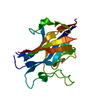 5bupC 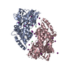 3sexS C: citing same article ( S: Starting model for refinement |
|---|---|
| Similar structure data |


-Links
 PDBj
PDBj


- Assembly
| Deposited unit | 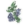
| ||||||||
|---|---|---|---|---|---|---|---|---|---|
| 1 |
| ||||||||
| Unit cell |
|
-Components
| #1: Protein | Mass: 76539.414 Da / Num. of mol.: 2 Mutation: I28T, D108A, K109A, E198A, N199A, A241H, K245H, K265A, A338V, I343V, E385A, E388A, D389A, R393N,R586A, R588A,I28T, D108A, K109A, E198A, N199A, A241H, K245H, K265A, A338V, I343V, E385A, ...Mutation: I28T, D108A, K109A, E198A, N199A, A241H, K245H, K265A, A338V, I343V, E385A, E388A, D389A, R393N,R586A, R588A,I28T, D108A, K109A, E198A, N199A, A241H, K245H, K265A, A338V, I343V, E385A, E388A, D389A, R393N,R586A, R588A Source method: isolated from a genetically manipulated source Details: THIS PROTEIN IS A CHIMERA. RESIDUES 25-391 ARE FROM E. COLI MALTOSE BINDING PROTEIN (MBP), CORRESPOND TO RESIDUES 27-393 OF SWISS-PROT DATABASE ENTRY P0AEX9 AND CONTAIN MUTATIONS I26T, ...Details: THIS PROTEIN IS A CHIMERA. RESIDUES 25-391 ARE FROM E. COLI MALTOSE BINDING PROTEIN (MBP), CORRESPOND TO RESIDUES 27-393 OF SWISS-PROT DATABASE ENTRY P0AEX9 AND CONTAIN MUTATIONS I26T, D106A, K107A, E196A, N197A, A239H, K243H, K263A, A336V, I341V, E383A, E386A, D387A AND R391N (CORRESPONDING TO I28T, D108A, K109A, E198A, N199A, A241H, K245H, K265A, A338V, I343V, E385A, E388A, D389A AND R393N IN P0AEX9). RESIDUES 395-710 ARE FROM HUMAN UROMODULIN PROTEIN AND CORRESPOND TO RESIDUES 295-610 OF SWISS-PROT DATABASE ENTRY P07911 AND CONTAIN MUTATIONS N613Q, R686A AND R688A (CORRESPONDING TO N513Q, R586A AND R588A). Source: (gene. exp.) Homo sapiens (human)Gene: malE, Z5632, ECs5017, UMOD / Plasmid: pHLSEC / Cell line (production host): HEK293S / Production host: Homo sapiens (human)References: UniProt: P0AEY0, UniProt: P07911, UniProt: P0AEX9*PLUS #2: Polysaccharide |   Source method: isolated from a genetically manipulated source Details: oligosaccharide / References: alpha-maltose #3: Sugar |   Type: D-saccharide, beta linking / Mass: 221.208 Da / Num. of mol.: 3 / Source method: obtained synthetically / Formula: C8H15NO6 Type: D-saccharide, beta linking / Mass: 221.208 Da / Num. of mol.: 3 / Source method: obtained synthetically / Formula: C8H15NO6#4: Chemical |   Mass: 65.409 Da / Num. of mol.: 2 / Source method: obtained synthetically / Formula: Zn Mass: 65.409 Da / Num. of mol.: 2 / Source method: obtained synthetically / Formula: ZnHas protein modification | Y | |
|---|
-Experimental details
-Experiment
| Experiment | Method: X-RAY DIFFRACTION |
|---|
- Sample preparation
| Crystal | Density Matthews: 4.91 Å3/Da / Density % sol: 75 % / Description: Rhombus |
|---|---|
| Crystal grow | Temperature: 293 K / Method: vapor diffusion, hanging drop / pH: 8.2 / Details: Sodium/potassium tartrate, Tris-HCl / PH range: 7.0 - 8.4 |
-Data collection
| Diffraction | Mean temperature: 100 K |
|---|---|
| Diffraction source | Source: SYNCHROTRON / Site: ESRF / Beamline: ID29 / Wavelength: 0.97625 Å |
| Detector | Type: DECTRIS PILATUS 6M-F / Detector: PIXEL / Date: Jan 24, 2013 |
| Radiation | Monochromator: Si Crystal / Protocol: SINGLE WAVELENGTH / Monochromatic (M) / Laue (L): M / Scattering type: x-ray |
| Radiation wavelength | Wavelength: 0.97625 Å / Relative weight: 1 |
| Reflection | Resolution: 3.2→35 Å / Num. all: 47837 / Num. obs: 47837 / % possible obs: 99.41 % / Redundancy: 6.3 % / Biso Wilson estimate: 108.3 Å2 / Rmerge(I) obs: 0.08846 / Net I/σ(I): 16.21 |
| Reflection shell | Resolution: 3.2→3.314 Å / Redundancy: 6.2 % / Rmerge(I) obs: 1.462 / Mean I/σ(I) obs: 1.29 / % possible all: 99.52 |
- Processing
| Software |
| |||||||||||||||||||||||||||||||||||||||||||||||||||||||||||||||||||||||||||||||||||||||||||||||||||||||||||||||||||||||||||||||||||||||||||||||||||||||||||||||||||||||||||||||
|---|---|---|---|---|---|---|---|---|---|---|---|---|---|---|---|---|---|---|---|---|---|---|---|---|---|---|---|---|---|---|---|---|---|---|---|---|---|---|---|---|---|---|---|---|---|---|---|---|---|---|---|---|---|---|---|---|---|---|---|---|---|---|---|---|---|---|---|---|---|---|---|---|---|---|---|---|---|---|---|---|---|---|---|---|---|---|---|---|---|---|---|---|---|---|---|---|---|---|---|---|---|---|---|---|---|---|---|---|---|---|---|---|---|---|---|---|---|---|---|---|---|---|---|---|---|---|---|---|---|---|---|---|---|---|---|---|---|---|---|---|---|---|---|---|---|---|---|---|---|---|---|---|---|---|---|---|---|---|---|---|---|---|---|---|---|---|---|---|---|---|---|---|---|---|---|---|
| Refinement | Method to determine structure: MOLECULAR REPLACEMENT Starting model: 3SEX Resolution: 3.2→34.976 Å / SU ML: 0.49 / Cross valid method: FREE R-VALUE / σ(F): 1.33 / Phase error: 28.21 / Stereochemistry target values: ML
| |||||||||||||||||||||||||||||||||||||||||||||||||||||||||||||||||||||||||||||||||||||||||||||||||||||||||||||||||||||||||||||||||||||||||||||||||||||||||||||||||||||||||||||||
| Solvent computation | Shrinkage radii: 0.9 Å / VDW probe radii: 1.11 Å / Solvent model: FLAT BULK SOLVENT MODEL | |||||||||||||||||||||||||||||||||||||||||||||||||||||||||||||||||||||||||||||||||||||||||||||||||||||||||||||||||||||||||||||||||||||||||||||||||||||||||||||||||||||||||||||||
| Displacement parameters | Biso mean: 141.3 Å2 | |||||||||||||||||||||||||||||||||||||||||||||||||||||||||||||||||||||||||||||||||||||||||||||||||||||||||||||||||||||||||||||||||||||||||||||||||||||||||||||||||||||||||||||||
| Refinement step | Cycle: LAST / Resolution: 3.2→34.976 Å
| |||||||||||||||||||||||||||||||||||||||||||||||||||||||||||||||||||||||||||||||||||||||||||||||||||||||||||||||||||||||||||||||||||||||||||||||||||||||||||||||||||||||||||||||
| Refine LS restraints |
| |||||||||||||||||||||||||||||||||||||||||||||||||||||||||||||||||||||||||||||||||||||||||||||||||||||||||||||||||||||||||||||||||||||||||||||||||||||||||||||||||||||||||||||||
| LS refinement shell | Refine-ID: X-RAY DIFFRACTION
| |||||||||||||||||||||||||||||||||||||||||||||||||||||||||||||||||||||||||||||||||||||||||||||||||||||||||||||||||||||||||||||||||||||||||||||||||||||||||||||||||||||||||||||||
| Refinement TLS params. | Method: refined / Refine-ID: X-RAY DIFFRACTION
| |||||||||||||||||||||||||||||||||||||||||||||||||||||||||||||||||||||||||||||||||||||||||||||||||||||||||||||||||||||||||||||||||||||||||||||||||||||||||||||||||||||||||||||||
| Refinement TLS group |
|
