 Movie
Movie Controller

[English] 日本語
 Yorodumi
- PDB-5hbm: Crystal Structure of a Dihydroxycoumarin RNase H Active-Site Inhi... -
+ Open data

- Basic information
| Entry | Database: PDB / ID: 5hbm | ||||||
|---|---|---|---|---|---|---|---|
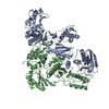
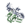
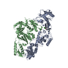
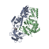
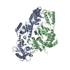
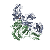
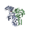
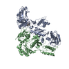
| Title | Crystal Structure of a Dihydroxycoumarin RNase H Active-Site Inhibitor in Complex with HIV-1 Reverse Transcriptase | ||||||
 Components Components |
| ||||||
 Keywords Keywords | TRANSFERASE / HYDROLASE/INHIBITOR / HYDROLASE / INHIBITOR / HYDROLASE-INHIBITOR complex | ||||||
| Function / homology |  Function and homology information Function and homology informationHIV-1 retropepsin / symbiont-mediated activation of host apoptosis / retroviral ribonuclease H / exoribonuclease H / exoribonuclease H activity / DNA integration / viral genome integration into host DNA / establishment of integrated proviral latency / RNA-directed DNA polymerase / RNA stem-loop binding ...HIV-1 retropepsin / symbiont-mediated activation of host apoptosis / retroviral ribonuclease H / exoribonuclease H / exoribonuclease H activity / DNA integration / viral genome integration into host DNA / establishment of integrated proviral latency / RNA-directed DNA polymerase / RNA stem-loop binding / viral penetration into host nucleus / host multivesicular body / RNA-directed DNA polymerase activity / RNA-DNA hybrid ribonuclease activity / Transferases; Transferring phosphorus-containing groups; Nucleotidyltransferases / host cell / viral nucleocapsid / DNA recombination / DNA-directed DNA polymerase / aspartic-type endopeptidase activity / Hydrolases; Acting on ester bonds / DNA-directed DNA polymerase activity / symbiont-mediated suppression of host gene expression / viral translational frameshifting / symbiont entry into host cell / lipid binding / host cell nucleus / host cell plasma membrane / virion membrane / structural molecule activity / proteolysis / DNA binding / zinc ion binding Similarity search - Function | ||||||
| Biological species |  Human immunodeficiency virus type 1 BH10 Human immunodeficiency virus type 1 BH10 | ||||||
| Method |  X-RAY DIFFRACTION / SYNCHROTRON / MOLECULAR REPLACEMENT / molecular replacement / Resolution: 3.043 Å X-RAY DIFFRACTION / SYNCHROTRON / MOLECULAR REPLACEMENT / molecular replacement / Resolution: 3.043 Å | ||||||
 Authors Authors | Kirby, K.A. / Sarafianos, S.G. | ||||||
| Funding support |  United States, 1items United States, 1items
| ||||||
 Citation Citation | Journal: To Be Published Title: Crystal Structure of a Dihydroxycoumarin RNase H Active-Site Inhibitor in Complex with HIV-1 Reverse Transcriptase Authors: Kirby, K.A. / Sarafianos, S.G. | ||||||
| History |
|
- Structure visualization
| Structure viewer | Molecule: MolmilJmol/JSmol |
|---|
- Downloads & links
-Download
| PDBx/mmCIF format | 5hbm.cif.gz | 209.7 KB | Display | PDBx/mmCIF format |
|---|---|---|---|---|
| PDB format | pdb5hbm.ent.gz | 163.7 KB | Display | PDB format |
| PDBx/mmJSON format | 5hbm.json.gz | Tree view | PDBx/mmJSON format | |
| Others |  Other downloads Other downloads |
-Validation report
| Arichive directory | https://data.pdbj.org/pub/pdb/validation_reports/hb/5hbmftp://data.pdbj.org/pub/pdb/validation_reports/hb/5hbm | HTTPS FTP |
|---|
-Related structure data
| Related structure data |  3qipS S: Starting model for refinement |
|---|---|
| Similar structure data |

-Links
 PDBj
PDBj


- Assembly
| Deposited unit | 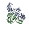
| ||||||||
|---|---|---|---|---|---|---|---|---|---|
| 1 |
| ||||||||
| Unit cell |
|
-Components
-Protein , 2 types, 2 molecules AB
| #1: Protein | Mass: 63989.238 Da / Num. of mol.: 1 / Fragment: p66 domain, residues 600-1153 / Mutation: K172A, K173A, C280S Source method: isolated from a genetically manipulated source Source: (gene. exp.) Human immunodeficiency virus type 1 BH10Strain: isolate BH10 / Gene: gag-pol / Plasmid: pET52a / Production host:  References: UniProt: P03366, RNA-directed DNA polymerase, DNA-directed DNA polymerase, retroviral ribonuclease H, exoribonuclease H |
|---|---|
| #2: Protein | Mass: 50096.539 Da / Num. of mol.: 1 / Fragment: p51 domain, residues 604-1027 / Mutation: C280S Source method: isolated from a genetically manipulated source Source: (gene. exp.) Human immunodeficiency virus type 1 BH10Strain: isolate BH10 / Gene: gag-pol / Plasmid: pET52a / Production host: |
-Non-polymers , 4 types, 6 molecules 


| #3: Chemical | ChemComp-NVP /  Mass: 266.298 Da / Num. of mol.: 1 / Source method: obtained synthetically / Formula: C15H14N4O / Comment: medication, antiretroviral*YM Mass: 266.298 Da / Num. of mol.: 1 / Source method: obtained synthetically / Formula: C15H14N4O / Comment: medication, antiretroviral*YM | ||
|---|---|---|---|
| #4: Chemical | ChemComp-F95 / ( Mass: 236.178 Da / Num. of mol.: 1 / Source method: obtained synthetically / Formula: C11H8O6 Mass: 236.178 Da / Num. of mol.: 1 / Source method: obtained synthetically / Formula: C11H8O6 | ||
| #5: Chemical |  Mass: 54.938 Da / Num. of mol.: 2 / Source method: obtained synthetically / Formula: Mn Mass: 54.938 Da / Num. of mol.: 2 / Source method: obtained synthetically / Formula: Mn#6: Water | ChemComp-HOH / | Mass: 18.015 Da / Num. of mol.: 2 / Source method: isolated from a natural source / Formula: H2O |
-Experimental details
-Experiment
| Experiment | Method: X-RAY DIFFRACTION / Number of used crystals: 1 |
|---|
- Sample preparation
| Crystal | Density Matthews: 3.43 Å3/Da / Density % sol: 64.17 % |
|---|---|
| Crystal grow | Temperature: 277 K / Method: vapor diffusion, hanging drop / pH: 6.6 / Details: PEG 8000, (NH4)2SO4, GLYCEROL, BIS TRIS PROPANE / PH range: 6.4-6.8 |
-Data collection
| Diffraction | Mean temperature: 100 K | |||||||||||||||||||||||||||
|---|---|---|---|---|---|---|---|---|---|---|---|---|---|---|---|---|---|---|---|---|---|---|---|---|---|---|---|---|
| Diffraction source | Source: SYNCHROTRON / Site: ALS / Beamline: 4.2.2 / Wavelength: 1 Å | |||||||||||||||||||||||||||
| Detector | Type: NOIR-1 / Detector: CCD / Date: Jul 25, 2012 | |||||||||||||||||||||||||||
| Radiation | Monochromator: ROSENBAUM-ROCK Si(111) SAGITALLY FOCUSED MONOCHROMATOR Protocol: SINGLE WAVELENGTH / Monochromatic (M) / Laue (L): M / Scattering type: x-ray | |||||||||||||||||||||||||||
| Radiation wavelength | Wavelength: 1 Å / Relative weight: 1 | |||||||||||||||||||||||||||
| Reflection | Resolution: 3.04→57.45 Å / Num. obs: 29497 / % possible obs: 98.9 % / Redundancy: 3.6 % / Biso Wilson estimate: 75.72 Å2 / CC1/2: 0.998 / Rmerge(I) obs: 0.065 / Rpim(I) all: 0.04 / Net I/σ(I): 16.7 / Num. measured all: 106171 / Scaling rejects: 19 | |||||||||||||||||||||||||||
| Reflection shell | Diffraction-ID: 1 / Rejects: _
|
-Phasing
| Phasing | Method: molecular replacement |
|---|
- Processing
| Software |
| ||||||||||||||||||||||||||||||||||||||||||||||||||||||||||||||||||||||||||||||||||||
|---|---|---|---|---|---|---|---|---|---|---|---|---|---|---|---|---|---|---|---|---|---|---|---|---|---|---|---|---|---|---|---|---|---|---|---|---|---|---|---|---|---|---|---|---|---|---|---|---|---|---|---|---|---|---|---|---|---|---|---|---|---|---|---|---|---|---|---|---|---|---|---|---|---|---|---|---|---|---|---|---|---|---|---|---|---|
| Refinement | Method to determine structure: MOLECULAR REPLACEMENT Starting model: 3QIP Resolution: 3.043→39.36 Å / SU ML: 0.46 / Cross valid method: FREE R-VALUE / σ(F): 1.35 / Phase error: 30.06 / Stereochemistry target values: ML
| ||||||||||||||||||||||||||||||||||||||||||||||||||||||||||||||||||||||||||||||||||||
| Solvent computation | Shrinkage radii: 0.9 Å / VDW probe radii: 1.11 Å / Solvent model: FLAT BULK SOLVENT MODEL | ||||||||||||||||||||||||||||||||||||||||||||||||||||||||||||||||||||||||||||||||||||
| Displacement parameters | Biso max: 194.13 Å2 / Biso mean: 73.8711 Å2 / Biso min: 51.14 Å2 | ||||||||||||||||||||||||||||||||||||||||||||||||||||||||||||||||||||||||||||||||||||
| Refinement step | Cycle: final / Resolution: 3.043→39.36 Å
| ||||||||||||||||||||||||||||||||||||||||||||||||||||||||||||||||||||||||||||||||||||
| Refine LS restraints |
| ||||||||||||||||||||||||||||||||||||||||||||||||||||||||||||||||||||||||||||||||||||
| LS refinement shell | Refine-ID: X-RAY DIFFRACTION / Total num. of bins used: 11
|
