 Movie
Movie Controller

[English] 日本語
 Yorodumi
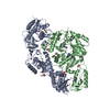
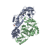
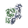
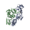
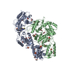
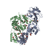
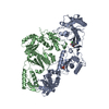
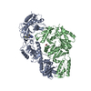
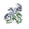
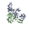
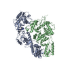
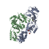
- PDB-3is9: Crystal structure of the HIV-1 reverse transcriptase (RT) in comp... -
+ Open data

- Basic information
| Entry | Database: PDB / ID: 3is9 | ||||||
|---|---|---|---|---|---|---|---|
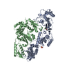
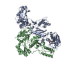
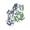
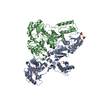
| Title | Crystal structure of the HIV-1 reverse transcriptase (RT) in complex with the alkenyldiarylmethane (ADAM) Non-nucleoside RT Inhibitor dimethyl 3,3'-(6-methoxy-6-oxohex-1-ene-1,1-diyl)bis(5-cyano-6-methoxybenzoate). | ||||||
 Components Components |
| ||||||
 Keywords Keywords | transferase/hydrolase / NNRTI / NONNUCLEOSIDE INHIBITOR / AIDS / HIV / P51/P66 / ADAM / Aspartyl protease / Cell membrane / Cytoplasm / DNA integration / DNA recombination / DNA-directed DNA polymerase / Endonuclease / Hydrolase / Lipoprotein / Membrane / Metal-binding / Multifunctional enzyme / Myristate / Nuclease / Nucleotidyltransferase / Nucleus / Phosphoprotein / Protease / RNA-binding / RNA-directed DNA polymerase / Transferase / Viral nucleoprotein / Virion / Zinc / Zinc-finger / transferase-hydrolase COMPLEX | ||||||
| Function / homology |  Function and homology information Function and homology informationHIV-1 retropepsin / symbiont-mediated activation of host apoptosis / retroviral ribonuclease H / exoribonuclease H / exoribonuclease H activity / DNA integration / viral genome integration into host DNA / establishment of integrated proviral latency / RNA-directed DNA polymerase / RNA stem-loop binding ...HIV-1 retropepsin / symbiont-mediated activation of host apoptosis / retroviral ribonuclease H / exoribonuclease H / exoribonuclease H activity / DNA integration / viral genome integration into host DNA / establishment of integrated proviral latency / RNA-directed DNA polymerase / RNA stem-loop binding / viral penetration into host nucleus / host multivesicular body / RNA-directed DNA polymerase activity / RNA-DNA hybrid ribonuclease activity / Transferases; Transferring phosphorus-containing groups; Nucleotidyltransferases / host cell / viral nucleocapsid / DNA recombination / DNA-directed DNA polymerase / aspartic-type endopeptidase activity / Hydrolases; Acting on ester bonds / DNA-directed DNA polymerase activity / symbiont-mediated suppression of host gene expression / viral translational frameshifting / symbiont entry into host cell / lipid binding / host cell nucleus / host cell plasma membrane / virion membrane / structural molecule activity / proteolysis / DNA binding / zinc ion binding Similarity search - Function | ||||||
| Biological species |  Human immunodeficiency virus type 1 BH10 Human immunodeficiency virus type 1 BH10 | ||||||
| Method |  X-RAY DIFFRACTION / SYNCHROTRON / MOLECULAR REPLACEMENT / Resolution: 2.55 Å X-RAY DIFFRACTION / SYNCHROTRON / MOLECULAR REPLACEMENT / Resolution: 2.55 Å | ||||||
 Authors Authors | Ho, W.C. / Bauman, J.D. / Das, K. / Arnold, E. | ||||||
 Citation Citation | Journal: J.Med.Chem. / Year: 2009 Title: Crystallographic study of a novel subnanomolar inhibitor provides insight on the binding interactions of alkenyldiarylmethanes with human immunodeficiency virus-1 reverse transcriptase. Authors: Cullen, M.D. / Ho, W.C. / Bauman, J.D. / Das, K. / Arnold, E. / Hartman, T.L. / Watson, K.M. / Buckheit, R.W. / Pannecouque, C. / De Clercq, E. / Cushman, M. | ||||||
| History |
|
- Structure visualization
| Structure viewer | Molecule: MolmilJmol/JSmol |
|---|
- Downloads & links
-Download
| PDBx/mmCIF format | 3is9.cif.gz | 212.6 KB | Display | PDBx/mmCIF format |
|---|---|---|---|---|
| PDB format | pdb3is9.ent.gz | 167.8 KB | Display | PDB format |
| PDBx/mmJSON format | 3is9.json.gz | Tree view | PDBx/mmJSON format | |
| Others |  Other downloads Other downloads |
-Validation report
| Arichive directory | https://data.pdbj.org/pub/pdb/validation_reports/is/3is9ftp://data.pdbj.org/pub/pdb/validation_reports/is/3is9 | HTTPS FTP |
|---|
-Related structure data
| Related structure data |  3irxC  2zd1S C: citing same article ( S: Starting model for refinement |
|---|---|
| Similar structure data |


-Links
 PDBj
PDBj


- Assembly
| Deposited unit | 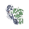
| ||||||||
|---|---|---|---|---|---|---|---|---|---|
| 1 |
| ||||||||
| Unit cell |
|
-Components
| #1: Protein | Mass: 64046.293 Da / Num. of mol.: 1 / Mutation: C879S, K771A, K772A Source method: isolated from a genetically manipulated source Source: (gene. exp.) Human immunodeficiency virus type 1 BH10Gene: gag-pol / Production host:  References: UniProt: P03366, RNA-directed DNA polymerase, DNA-directed DNA polymerase, ribonuclease H |
|---|---|
| #2: Protein | Mass: 50039.488 Da / Num. of mol.: 1 / Mutation: C879S Source method: isolated from a genetically manipulated source Source: (gene. exp.) Human immunodeficiency virus type 1 BH10Gene: gag-pol / Production host: References: UniProt: P03366, RNA-directed DNA polymerase, DNA-directed DNA polymerase |
| #3: Chemical | ChemComp-AC7 / 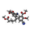  Mass: 506.504 Da / Num. of mol.: 1 / Source method: obtained synthetically / Formula: C27H26N2O8 Mass: 506.504 Da / Num. of mol.: 1 / Source method: obtained synthetically / Formula: C27H26N2O8 |
| #4: Water | ChemComp-HOH /  Mass: 18.015 Da / Num. of mol.: 124 / Source method: isolated from a natural source / Formula: H2O Mass: 18.015 Da / Num. of mol.: 124 / Source method: isolated from a natural source / Formula: H2O |
-Experimental details
-Experiment
| Experiment | Method: X-RAY DIFFRACTION / Number of used crystals: 1 |
|---|
- Sample preparation
| Crystal | Density Matthews: 2.82 Å3/Da / Density % sol: 56.4 % |
|---|---|
| Crystal grow | Temperature: 277 K / pH: 6.4 Details: 11% PEG8000, 15 mM MgSO4, 10 mM Spermine, 100 mM Ammonium sulphate, 50 mM imidazole pH 6.4., temperature 277K |
-Data collection
| Diffraction | Mean temperature: 93 K |
|---|---|
| Diffraction source | Source: SYNCHROTRON / Site: CHESS  / Beamline: F1 / Wavelength: 0.916 Å / Beamline: F1 / Wavelength: 0.916 Å |
| Detector | Type: ADSC QUANTUM 270 / Detector: CCD / Date: May 27, 2006 |
| Radiation | Protocol: SINGLE WAVELENGTH / Monochromatic (M) / Laue (L): M / Scattering type: x-ray |
| Radiation wavelength | Wavelength: 0.916 Å / Relative weight: 1 |
| Reflection | Resolution: 2.55→40 Å / Num. all: 41647 / Num. obs: 41074 / % possible obs: 98.6 % / Redundancy: 11.67 % / Rmerge(I) obs: 0.086 / Net I/σ(I): 18.3 |
| Reflection shell | Highest resolution: 2.55 Å / Rmerge(I) obs: 0.512 / Mean I/σ(I) obs: 3 |
- Processing
| Software |
| ||||||||||||||||||||
|---|---|---|---|---|---|---|---|---|---|---|---|---|---|---|---|---|---|---|---|---|---|
| Refinement | Method to determine structure: MOLECULAR REPLACEMENT Starting model: 2ZD1 Resolution: 2.55→40 Å / Stereochemistry target values: Engh & Huber
| ||||||||||||||||||||
| Displacement parameters | Biso mean: 80.8 Å2 | ||||||||||||||||||||
| Refinement step | Cycle: LAST / Resolution: 2.55→40 Å
|
