histone H1-4S187 kinase activity / P-TEFb complex / Interactions of Tat with host cellular proteins / nucleus localization / 7SK snRNA binding / cyclin/CDK positive transcription elongation factor complex / positive regulation of protein localization to chromatin / regulation of muscle cell differentiation / regulation of mRNA 3'-end processing / regulation of cyclin-dependent protein serine/threonine kinase activity ...histone H1-4S187 kinase activity / P-TEFb complex / Interactions of Tat with host cellular proteins / nucleus localization / 7SK snRNA binding / cyclin/CDK positive transcription elongation factor complex / positive regulation of protein localization to chromatin / regulation of muscle cell differentiation / regulation of mRNA 3'-end processing / regulation of cyclin-dependent protein serine/threonine kinase activity / cyclin-dependent protein serine/threonine kinase activator activity / transcription elongation-coupled chromatin remodeling / host-mediated activation of viral transcription / RNA polymerase binding / positive regulation of DNA-templated transcription, elongation / transcription elongation factor activity / [RNA-polymerase]-subunit kinase / cellular response to cytokine stimulus / RNA polymerase II transcribes snRNA genes / replication fork processing / Pausing and recovery of Tat-mediated HIV elongation / Tat-mediated HIV elongation arrest and recovery / HIV elongation arrest and recovery / Pausing and recovery of HIV elongation / cyclin-dependent kinase / cyclin-dependent protein serine/threonine kinase activity / Tat-mediated elongation of the HIV-1 transcript / regulation of DNA repair / Formation of HIV-1 elongation complex containing HIV-1 Tat / Formation of HIV elongation complex in the absence of HIV Tat / RNA Polymerase II Transcription Elongation / Formation of RNA Pol II elongation complex / negative regulation of protein localization to chromatin / RNA polymerase II CTD heptapeptide repeat kinase activity / RNA Polymerase II Pre-transcription Events / transcription elongation factor complex / TP53 Regulates Transcription of DNA Repair Genes / transcription initiation at RNA polymerase II promoter / positive regulation of transcription elongation by RNA polymerase II / molecular condensate scaffold activity / transcription elongation by RNA polymerase II / SMAD2/SMAD3:SMAD4 heterotrimer regulates transcription / PML body / transcription coactivator binding / kinase activity / cytoplasmic ribonucleoprotein granule / transcription by RNA polymerase II / Estrogen-dependent gene expression / DNA-binding transcription factor binding / protein phosphorylation / protein kinase activity / cell population proliferation / regulation of cell cycle / transcription cis-regulatory region binding / RNA polymerase II cis-regulatory region sequence-specific DNA binding / response to xenobiotic stimulus / protein serine kinase activity / cell division / DNA repair / protein serine/threonine kinase activity / chromatin binding / protein kinase binding / positive regulation of transcription by RNA polymerase II / DNA binding / nucleoplasm / ATP binding / membrane / nucleus / cytosol Similarity search - Function
: / Cyclin-T2-like, C-terminal domain / Cyclin-like / Cyclin/Cyclin-like subunit Ssn8 / Cyclin A; domain 1 / Cyclin, N-terminal / Cyclin, N-terminal domain / Cyclin-like / domain present in cyclins, TFIIB and Retinoblastoma / Cyclin-like superfamily ...: / Cyclin-T2-like, C-terminal domain / Cyclin-like / Cyclin/Cyclin-like subunit Ssn8 / Cyclin A; domain 1 / Cyclin, N-terminal / Cyclin, N-terminal domain / Cyclin-like / domain present in cyclins, TFIIB and Retinoblastoma / Cyclin-like superfamily / : / Phosphorylase Kinase; domain 1 / Phosphorylase Kinase; domain 1 / Transferase(Phosphotransferase) domain 1 / Transferase(Phosphotransferase); domain 1 / Serine/threonine-protein kinase, active site / Serine/Threonine protein kinases active-site signature. / Protein kinase domain / Serine/Threonine protein kinases, catalytic domain / Protein kinase, ATP binding site / Protein kinases ATP-binding region signature. / Protein kinase domain profile. / Protein kinase domain / Protein kinase-like domain superfamily / 2-Layer Sandwich / Orthogonal Bundle / Mainly Alpha / Alpha Beta Similarity search - Domain/homology
In the structure databanks used in Yorodumi, some data are registered as the other names, "COVID-19 virus" and "2019-nCoV". Here are the details of the virus and the list of structure data.
Jan 31, 2019. EMDB accession codes are about to change! (news from PDBe EMDB page)
EMDB accession codes are about to change! (news from PDBe EMDB page)
The allocation of 4 digits for EMDB accession codes will soon come to an end. Whilst these codes will remain in use, new EMDB accession codes will include an additional digit and will expand incrementally as the available range of codes is exhausted. The current 4-digit format prefixed with “EMD-” (i.e. EMD-XXXX) will advance to a 5-digit format (i.e. EMD-XXXXX), and so on. It is currently estimated that the 4-digit codes will be depleted around Spring 2019, at which point the 5-digit format will come into force.
The EM Navigator/Yorodumi systems omit the EMD- prefix.
Related info.:Q: What is EMD? / ID/Accession-code notation in Yorodumi/EM Navigator
Yorodumi is a browser for structure data from EMDB, PDB, SASBDB, etc.
This page is also the successor to EM Navigator detail page, and also detail information page/front-end page for Omokage search.
The word "yorodu" (or yorozu) is an old Japanese word meaning "ten thousand". "mi" (miru) is to see.
Related info.:EMDB / PDB / SASBDB / Comparison of 3 databanks / Yorodumi Search / Aug 31, 2016. New EM Navigator & Yorodumi / Yorodumi Papers / Jmol/JSmol / Function and homology information / Changes in new EM Navigator and Yorodumi
 Movie
Movie Controller


 Open data
 Basic information
 Components
 Keywords
 Function and homology information
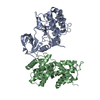
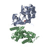
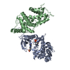
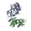
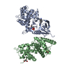
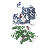
 Homo sapiens (human)
 X-RAY DIFFRACTION /
  Authors
 Citation
 Structure visualization
 Downloads & links
 Other downloads


 PDBj
PDBj


 Assembly
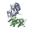

 Spodoptera frugiperda (fall armyworm) / Strain (production host): SF9


 Mass: 94.971 Da / Num. of mol.: 1 / Source method: obtained synthetically / Formula: PO4
 Mass: 92.094 Da / Num. of mol.: 5 / Source method: obtained synthetically / Formula: C3H8O3
 Mass: 319.141 Da / Num. of mol.: 1 / Source method: obtained synthetically / Formula: C12H12Cl2N2O4
 Sample preparation
 / Beamline: ID23-1 / Wavelength: 1.0055 Å
 Processing